40-50 % of diabetics are at risk, with a higher incidence in women.

- Diabetic foot
- Symptoms, stages, forms
- Possible causes of leg pain
- Poor circulation
- Specificity of pain in women and children
- Possible complications
- prognosis after surgery
- Diabetic foot care
- Prevention of diabetic foot
- foot ulcers
- Diabetic foot
- What a diabetic foot looks like, symptoms
- Examination in the Paracelsus Medical Center
- Why is smoking cessation recommended for diabetic foot ulcers?
- bibliography
- This is what a trophic ulceration looks like
- How trophic ulcers arise
- Possible complications of diabetic foot
- prognosis and prevention
Diabetic foot
platelet-rich plasma – A new highly effective treatment for diabetic ulcers.
Diabetic foot syndrome (diabetic foot) – is a syndrome of anatomical and functional changes developing on the background of diabetic neuropathy, micro- and macroangiopathy and osteoarthropathy. All these pathologies contribute to increased traumatization and infection of the soft tissues of the foot, the development of a purulent-arthritic process and, in advanced cases, amputation.
If you have had diabetes for several years, and even more so if you have not managed your diabetes well during this period, there is a significant risk of developing serious complications.
There is a significant risk of developing serious complications of diabetes if:
It is well known that all wounds and injuries in diabetics take a long time to heal. Even a small injury can become infected, and if not properly cared for, gangrene can develop. This condition is difficult to treat and often ends in amputation. To avoid complications, you should carefully read and follow the foot care instructions below.
Symptoms, stages, forms
Symptoms and treatment depend on the shape and stage of the diabetic foot. The process is divided into 6 stages:
- 0 – risk group. No ulceration, but feet are already deformed, pale and cold, with blisters.
- stage 1. Superficial ulcers appear, but they do not affect the deeper layers of the epidermis.
- stage 2 .. The ulcer develops deep in the foot and affects not only the skin but also the muscles, tendons and fibers.
- stage 3 .. The ulcer penetrates deep into the foot and reaches the bone.
- stage 4 .. The diabetic foot turns black (gangrene) with well-defined borders.
- Stage 5 .. The area of necrosis increases. The gangrene continues to increase and takes on larger and larger lesions. If left untreated, it can lead to complete loss of the leg.
Possible causes of leg pain
High blood sugar interferes with the transmission of nerve impulses and impairs the blood supply to the veins and arteries of the limbs. The combination of several pathological symptoms associated with impaired trophism, innervation and blood circulation is commonly referred to as 'diabetic foot syndrome'. But it is not always just this part of the leg that hurts - depending on the cause, the ankle, calf and lower leg can also be affected.
Poor circulation
If the blood flow to the lower limbs is normal, the tissues have sufficient nutrients and oxygen. High blood sugar levels cause the blood to clot and become sticky. This affects the permeability and elasticity of the arteries, veins and capillaries. If a person already has atherosclerosis or has just started it, it progresses dramatically. This explains why the lumen of the blood vessels narrows dramatically and certain parts of the leg with diabetes are no longer adequately supplied with blood. This damage to the blood vessels is called diabetic angiopathy.
The first symptoms of this pathological condition are:
- increased tiredness in the legs;
- numbness in certain areas of the skin;
- excessive sweating;
- Increasing pain, first on exertion and then at rest;
- limping when walking ;
- Feeling cold in the legs, even when the ambient temperature is pleasant.

As the diabetes complication progresses, covering the legs with a blanket can be painful.
With severe forms of angiopathy, all these ailments worsen and become a constant companion of the affected person. The person suffers from cramps and the skin on the feet changes color (gradually turning yellow and then blue). Burning, aching pain and numbness spread to the entire foot. If left untreated, trophic ulcers form on the feet, which can eventually lead to gangrene. Patients should have regular check-ups and seek immediate medical attention if there is even the slightest suspicion to avoid amputation.
Specificity of pain in women and children
In women, foot pain in diabetes is most often due to these pathological conditions:
- Arthropathy against the background of hormonal changes in the body (for example, during menopause);
- frequent blistering and ingrown toenails caused by wearing uncomfortable, tight, high-heeled shoes;
- Thrombophlebitis or high blood viscosity (these can be caused by pregnancy, use of oral contraceptives or metabolic disorders).
In children with diabetes, legs may ache after physical exertion or when there are sudden fluctuations in blood sugar levels. The discomfort in the child's lower limbs is also often caused by neuropathy. Therefore, in children, in addition to continuous monitoring by an endocrinologist, regular examinations by a neurologist and a vascular surgeon are very important. Timely diagnostic examinations can prevent foot problems before the first symptoms appear.

Sugar fluctuations in children should be monitored more frequently than in adults because age-related complications of diabetes can develop more quickly in children.
Possible complications
Foot phlegm surgery can be complicated by both local and systemic issues. Local complications include bleeding and persistent infection in the foot. Systemic complications include sepsis, pulmonary embolism, and perioperative infarction.
Because patients with diabetic foot phlegm often have multiple comorbidities, the risk of systemic complications is high. However, this risk is not related to the technique of the intervention, but to the reason for the intervention.
Patients with diabetic phlegm are patients who need intensive therapy, modern antibiotics and the fastest possible restoration of blood circulation.
prognosis after surgery
Predicting the outcome of diabetic phlegm surgery is difficult. It depends on many factors - general health, immune system, ability to restore blood flow, wound closure.
We know from our practice that if the phlegm is opened in good time and blood flow is restored at 75 %, the diabetic's accessory limb can be saved. The mortality rate is about 5 % and is associated with worsening of comorbidities.
After the operation, the treatment process is only beginning. The patient is given active antibiotic therapy based on an isolated bacterial flora, and blood flow is restored (angioplasty and stenting).
The final recovery process takes a lot of time. Often the patient is discharged on an outpatient basis and then re-admitted to the hospital for reconstructive surgery.
Diabetic foot care
According to people who have healed their diabetic foot, self-treatment should not be the first concern. Patients who made mistakes during treatment with folk remedies found that the disease had progressed further. Grandmother's prescriptions are not suitable for the treatment of psoriasis, in addition, not all drugs from the pharmacy can be used.
- Myorelaxants and antispasmodics are used to relieve pain.
- Drugs to improve blood circulation and metabolism are prescribed.
- If a bacterial flora is detected in the culture, the optimal dose of antibiotics is selected.
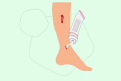
- Topical treatment consists of appropriate wound care and choice of dressings.
- Bandages help maintain social activity, but this type of therapy is indicated only for small wounds and in the absence of infection.
- Particular attention is paid to the correction of DM.
Diabetic foot care can be performed at home after patient training. An indispensable indication for hospitalization is the presence of severe infection and critical ischemia of the lower limbs. After discharge from the hospital, many patients relapse after some time. Even if you know how to care for your diabetic foot, you should see your doctor. He can adapt your therapy according to the examination results and the instrumental diagnosis and thus avoid treatment errors.

Prevention of diabetic foot
The most important measures to prevent AS is to correct the underlying disease. Patients should be aware that in addition to diabetic foot ulcers, other diseases such as B. cardiovascular diseases can occur. The risk increases with age, unhealthy habits, low activity level and obesity.
- Constantly check the condition of your feet. If this is difficult for you because of age, poor eyesight or musculoskeletal problems, ask your family to check your feet daily. Avoid wounds, blisters, tears and burns. Use a moisturizing cream and dress wounds as your doctor taught you - for example with a 0.05%igen chlorhexidine solution. Do not use green, iodine, alcohol or manganese products.
- If you buy closed-toe shoes, use the insole from an old shoe, not a fit. Shoes for the diabetic foot should fit loosely and be one size larger. When shopping for sandals, opt for soft, non-chafing materials. However, you are not immune to chafing, so wear shoes with socks or use band-aids. The shoes should not only be loose at the toes, but also at the cuff. Don't wear barefoot shoes and don't walk barefoot. If you have flat feet, choose blunt-toed shoes.
- Wash your feet daily in warm water at 36-37 degrees. Don't steam your feet. Only warm foot baths lasting no more than 5 minutes are allowed. After washing, wipe the soles of your feet and the spaces between your toes dry.
- Change your socks/stockings daily, choose knitwear with a loose elastic band and no rough seams.
- Trim your nails at the right time to avoid injury. Make sure the nails are not ingrown.
- Only remove calluses with a fine pumice stone or a special foot file. Do not cut rough nails with a blade. Do not steam the sole of the foot before treatment: use the device on dry skin.
- Make time for physical activity - 5 times a day for 30 minutes each time. Talk to your doctor about the intensity of the exercise.
- Enjoy foot massages. If you have trouble bending over, ask a family member or nurse for help.
foot ulcers
Foot ulcers are most common in diabetes, venous insufficiency of the lower limbs and after serious injury.

The ulcer makes walking difficult or impossible. The extensive skin defect is a gateway for infection and the inflammation of the foot can lead to serious complications such as necrosis and gangrene of the foot. The main treatment for foot ulcers is surgery.
Proper footwear allows pressure to be redistributed under the foot, reducing stress in the area of the ulcer and shifting it to areas with intact skin and softening the sole in overstressed areas.

The ulcers most commonly appear on the dorsal surface of the deformed toes and on the soles of the feet near the heads of the metatarsal bones.
Perseus Center shoes and insoles to relieve or relieve biomechanical pressure in the area of the ulcer:

Diabetic foot
Diabetic foot or diabetic foot syndrome (hereinafter DFS) is one of the late and most severe complications of diabetes in the form of purulent nerve processes, ulcers and osteoarthritic changes caused by peripheral nerve, vascular, skin and soft tissue, bone and joint injuries occur; Infections, ulcerations and/or deep tissue destruction associated with neurological disorders, reduction of the main blood flow in the arteries of the lower limbs of varying degrees) occur on the legs of people with type 1 and type 2 diabetes. Up to 15 % of diabetics are at risk of developing leg ulcers.
When the nervous system is damaged, a diabetic can no longer feel their feet properly. The healthy, normal secretion of sweat and sebum, which lubricate the skin of the foot, is disrupted. Together, these factors can cause abnormal pressure on the skin, bones, and joints of the foot when walking, and the skin on the foot to break down. Ulcers, including AS, may develop.
The damage to blood vessels and weakening of the immune system caused by diabetes make these wounds difficult to treat. Bacterial infections of the skin, connective tissue, muscles and bones can occur. From these infections, gangrene can develop (necrosis of certain organs and tissues of the living organism, which is black or very dark in color and spreads directly or through anatomical channels connected to the external environment, in the skin, lungs, intestines etc.) which can lead to the development of gangrene. The dark color is due to ferrous sulfide, which forms from iron and hemoglobin with hydrogen sulfide). Due to the poor blood circulation, the antibiotics have difficulty reaching the focus of infection. Often the only treatment for gangrene is amputation of the foot or the whole leg. If the infection spreads into the bloodstream, the process can be very life-threatening.
What a diabetic foot looks like, symptoms
All patients diagnosed with diabetes must have their feet checked daily to detect the disease at an early stage. According to Wagner's classification, there are 6 stages of diabetic foot syndrome:

- changes in bone structure.
- Appearance of ulcers on the skin.
- destruction of soft tissues.
- inflammation of bone tissue.
- gangrene of the toes.
- Gangrene of the foot and lower leg.
Patients with AS complain of long-lasting, non-healing wounds on the lower limbs. As can be seen in the patients' photographs, ulcerations form on the skin, extending in depth and around the periphery of the foot. Bacterial and fungal infections occur. The wounds affect tendons and joints. In the later stages, bones and joints are affected and the tissue dies.
Examination in the Paracelsus Medical Center
Patients with diabetes and AS symptoms can be examined by an endocrinologist in Sergiev Posad at the Paracelsus Clinic.
The center is equipped with a special ultrasound machine. The in-house laboratory delivers precise results quickly – on the same day as the examination.
The examinations are carried out by highly qualified endocrinologists who have over 11 years of experience in this field. In the endocrinology department, treatment is carried out by a doctor with 37 years of experience in this field.
Patients at high risk of developing diabetic foot disease should be examined not only by an endocrinologist, but also by a vascular surgeon and an orthopedic traumatologist. It is important to perform a diagnostic self-examination to identify the first signs of diabetic foot: change in skin color, appearance of dryness, swelling and pain, crooked toes, fungal infection.
The Paracelsus Clinic offers a comprehensive diagnosis, based on which the doctor selects a treatment method. In the clinic, you can undergo an examination of the arteries and veins, vascular lesions are detected at an early stage, and blood flow is assessed.
Appropriate treatment by our doctors can prevent the development of complications. Your doctor will help you adjust your lifestyle and diet.
Why is smoking cessation recommended for diabetic foot ulcers?
Smoking increases the risk of atherosclerosis of the blood vessels of the lower limbs, in which cholesterol deposits form on the vessel walls
and narrow their lumen. This leads to a reduction in blood supply to the tissues.
Dermatovenereologist, head of the DCD for the St. Petersburg City Paid Clinic for Dermatovenereology, St. Petersburg

Trophic ulcers are long-lasting, non-healing wounds that develop as a result of tissue nutrition disorders. Can such wounds be treated and how?
How to recognize a trophic ulcer on the leg and what causes such a skin defect?

Characteristics and properties of povidone iodine. What is povidone iodine used for? Instructions for using Betadine ® solution, ointment, suppositories with povidone iodine.
bibliography
- Clinical Guidelines 'Diabetic Foot Syndrome'/Public Organization 'Russian Union of Endocrinologists', Moscow- 2015.
- Instructions for the medicinal use of chlorhexidine, RLS.
- Instructions for the Medical Use of Hydrogen Peroxide, RLS.
- Instructions for the medicinal use of potassium permanganate, RLS.
- Instructions for the medicinal use of Miramistin solution, RLS.
- Instructions for the medicinal use of Octinisept solution, RLS.
- Instructions for Medical Use of Lavacept Solution, RLS.
- Instructions for the medicinal use of eosin solution, РЛС.
- Instructions for use of Betadine ® (solution, ointment), RU P015282/03, P015282/02.
- Michalsky VV, Bogdanov AE, Zhilina SV et al. Use of Betadin® in the treatment of infected wounds // RMJ №29, 12/23/2010.
- Instructions for the medicinal use of Levomekol ointment, RLS.
- Instructions for the medical use of Dioxicol ® ointment. OINTMENT.
- Instructions for the medicinal use of Dermazine ointment, РЛС.
- Instructions for the medical use of Asetisorb® -DT, RLS.
- Instructions for use of the drug Phenystil®, RLS.
- Instructions for medicinal use of Psilo-Balsam ® , РЛС.
- Yaremchuk An. A et al. Justification of the composition of a multicomponent ointment for the treatment of purulent wounds at the first stage of the wound formation process. Pharmaceutical Bulletin No. 3 (57) 2012.
- Borisov IV Povidone-iodine - new possibilities for a well-known drug (literature review). wounds and wound infections. Journal of Prof. BM Kostyuchenok. 2021, 8(3):12-18.
- Bigliardi PL et al. Povidone iodine in wound healing: A review of current concepts and practices. Int J Surg. 2017; 44: 260-268.
If you experience an adverse reaction while using any product from the portfolio of EGIS-RUS LLC, please report this information using one of the convenient contact forms:
This is what a trophic ulceration looks like
Even before the appearance of the sores, precursors of the disease can be seen. Initially, the skin is dark in color due to the accumulation of decomposing hemoglobin. Then the skin becomes pale and shiny. Gradually, the skin becomes damaged and begins to itch. The ulcer itself looks like a non-healing wound of varying size and depth. It is found on the feet, ankles and lower legs.
Ulcers develop when blood and lymph flow is impaired and tissue nutrition is disrupted. The regeneration processes are impaired, and the tissue becomes necrotic due to the disrupted trophic. Open wounds that do not heal over a long period of time are a breeding ground for bacteria, which is why wounds must be carefully cared for. Mucus or pus may come out of the wound.
It is worth remembering that the symptoms of a trophic ulcer are only the tip of the iceberg. Rather, it is the result of the progression of the underlying disease, which has its own character and characteristics. It does not heal spontaneously and requires treatment under medical supervision.
Many patients are unaware of their diagnosis, do not know how to properly treat trophic leg ulcers, and try home remedies. They consult a specialist when all the methods tried have no effect and the general condition worsens.
How trophic ulcers arise
As already mentioned, ulcers appear against the background of various diseases and conditions. Attention should be paid to the causative factors. Wet ulcers can occur with:
- people who are overweight.
- People who move little and prefer passive activities.
- Working people with a sedentary or standing job or physically demanding work.
- In bedridden patients.
- In people with diseases of the veins and arteries.
- Elderly people.
People with athlete's foot or nail fungus should also take a closer look. Rapidly progressing erosions require careful investigation. All skin changes are particularly dangerous for diabetics.
Possible complications of diabetic foot
Diabetic foot syndrome is one of the most serious complications of late-onset diabetes. Left untreated, the disease worsens, which can lead to amputation and death.
Diabetic foot syndrome is accompanied by reduced sensitivity of the limbs, which in turn is due to poor blood circulation. In this case, the formation of an ulcer, hidden by a dry scab, can go unnoticed until the bottom of the wound reaches the bones and tendons.
Untimely treatment of the disease can lead to the following complications:
Amputation of the affected limb does not eliminate the cause of the pathology. On the contrary, the doctor's recommendations must be followed even more strictly. If the underlying disease is not treated after the amputation, half of the amputations can recur within five years, and 40% of the amputations can be fatal.
Even after the ulcer has healed, visits to the doctor should be continued, otherwise the probability of recurrence within the next five years is 70 %.
prognosis and prevention
A negative prognosis in diabetic foot syndrome is more likely when the following factors are present
- Severe circulatory disorders in the lower limbs;
- osteoarthritis, which increases the risk of recurrence by 20-30 %;
- necrotic and gangrenous manifestations;
- wound infection;
- Lack of qualified medical care.
To minimize the risk, diabetes should be diagnosed early and treatment started as soon as possible.
Amputations can be avoided. For this, preventive measures should be taken:
- Daily inspection of the surface of the foot, careful hygiene of the lower limbs, and then thorough drying of the skin;
- Avoid abrasions and blisters by wearing loose-fitting footwear and socks with the seams facing outward;
- Avoid cuts and never go barefoot;
- Avoid overheating as skin sensitivity is greatly reduced;
- Do not remove corns and calluses yourself;
- Do not remove corns yourself.
! This item is intended for patients who have received a medical diagnosis. It does not replace a visit to the doctor and should not be used for self-treatment or self-diagnosis.
Read more:- Diabetic foot ulcers.
- Why amputate legs for diabetes?.
- The reamputation is.
- X-ray of Charcot's foot.
- Indications for amputation.
- The toe formation.
- Life after a leg amputation.
- Metatarsal amputation.