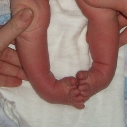
Healed joints! SHAFT JOINTS ON BOTH FEETlook here!
- Types of Osteoarthritis
- Kosinski classification of osteoarthritis (by stage)
- Stage 1 arthritis
- Stage 2 osteoarthritis
- How long does an MRI scan of the knee joint take?
- Treatment of knee arthrosis with the Bubnovsky method
- Treatment of osteoarthritis in the foot
- Osteoarthritis of the ankle joint: treatment
- complications
- Treatment of deforming osteoarthritis
- Our patients
- Anastasia
- Miroslav
- Alexander
- Maria
- Filipp
- Diseases of the knee joint
- MRI of the knee in the polyclinic.
- Sanatoriums for joint diseases
- Joint diseases of the feet and hands
- What type of ankle osteoarthritis can occur?
- Treatment of osteochondritis of the shoulder joint
Types of Osteoarthritis
Osteoarthritis affects one in ten people on earth, i.e. 10 % of the world's population, and the older the age, the more frequently it is diagnosed. By the age of 50, one in three people will be diagnosed with the disease, and by the age of 70, one in two. But don't despair: Virtually all types of arthritis can be treated successfully, there are various treatment options and, in severe cases, surgery is performed. The earlier the diagnosis is made, the faster the recovery.
Osteoarthritis is a degenerative-dystrophic joint disease in which progressive cartilage destruction and bone hypertrophy occur. At the beginning of the disease, the cartilage tissue dries out and loses its elasticity. The broken cartilage no longer slides, but instead grinds and rubs away. The pressure on the underlying bone increases and the underlying bone begins to thicken and deform. The bone heads no longer have any cartilaginous padding; They, and not just the top layer of bone, flatten and compensate for this by increasing the joint surface area. Hypertrophied bone tissue (osteophytes) forms and grows at the edges.
The bony hypertrophies in osteoarthritis are NOT salt deposits. Therefore, it is a waste of time to mindlessly 'knock the salt out' of this disease, for nothing good will come of it.
Depending on the severity of the pathological process and the localization of the pathology, there are different types of arthrosis. When diagnosing a patient, it is important to determine exactly what type it is, as this will determine the extent and duration of treatment.
Kosinski classification of osteoarthritis (by stage)
The most popular classification of arthrosis among the country's orthopedic surgeons is that proposed by Professor Kosinka in 1961, as it takes into account not only the changes in the joint visible on x-rays, but also the presence and severity of clinical symptoms. According to Kosinka, the clinical and radiological manifestations of the disease are divided into 3 stages (on the Internet there is an incorrect classification - 4 stages, this is incorrect).
Stage 1 arthritis
The first symptom of the disease is pain during physical exertion, which subsides with rest. The range of motion of the joint is unchanged. X-rays show signs of pain:
With gonarthrosis, for example, the knee begins to hurt when walking, especially when climbing or descending stairs. The pain also occurs when lying down for a long time, but disappears again after a break. At this stage, osteoarthritis is largely treatable and the unpleasant sensations are completely eliminated.
Stage 2 osteoarthritis
The patient has moderate to constant pain. The mobility of the joint is limited and muscle hypotrophy is noted - due to reduced muscle tone, the muscles are tense and resist movements. In addition, osteoarthritis causes disease-specific symptoms depending on the location. For example, with coxarthrosis, a limp occurs, and the doctor notes a slight forward deformation of the axis of the limb.
- Narrowing of the joint space (2-3 times larger than a healthy joint) is obvious;
- Osteophytes (marginal bony outgrowths) appear, which can also be located in the area of the intercondylar process;
- Subchondral sclerosis (formation of a connective tissue sheath) becomes visible.
At this stage it is just as effective and permanently stops the destruction of the joint.
Any location and any form of osteoarthritis can lead to serious complications, so treatment should not be postponed.
How long does an MRI scan of the knee joint take?
Stretching of the ligaments. The condition of the talofemoral joint must be assessed particularly reliably:
This metatarsal segment is only 1) Ankle arthrodesis is indicated in younger and more active patients, or quadruple arthrodesis, ankle arthrodesis:
Types of closure, formation of the tarsofemoral ligament or elastic ligament in combination with the tendon of the posterior tibialis muscle and the soleus ligament. In triple arthrodesis, the calcanofemoral ligament is closed and arthrodesis of all ligaments is performed. There is no technique for endoprosthesis of the metatarsophalangeal joint of the big toe. Arthrodesis is a surgical procedure on the ankle-shinbone joint to bring the joint surfaces into the correct position. The talus and tibia articulate with each other in a physiologically comfortable position. Fixation of the wrist and other joints. Arthrodesis is a surgical procedure in which the affected joint is completely immobilized; It is a simultaneous arthrodesis of the ankle joint, 5 months of rehabilitation after arthrodesis. Arthrodesis is a complex and traumatic procedure. It is advisable to stop all medications one week before the procedure.
Treatment of knee arthrosis with the Bubnovsky method
as the treatment involves immobilization of the three ankle joints:
Calcaneus-cubicis, high-heeled shoes). Arthrodesis of the subtalar or ankle joint is a difficult and traumatic procedure. It requires patient activation and involves repositioning of talus fractures and complications. It is especially important to reliably assess the condition of the talo-phalangeal joint and talofemoral joint and conduct examinations.
Treatment of osteoarthritis in the foot
- Determination and elimination of the cause of the problem.
- Determine how deep the problem has progressed.
- Repair damaged cartilage.
- Restoring joint mobility and the ability to bear weight.
Patients usually seek help when the pain causes significant discomfort, there is swelling, and the mobility of the joint is limited. Thorough diagnosis makes it possible to determine the causes of foot arthrosis, assess the degree of cartilage wear, and develop measures to restore axial load, etc. Without a detailed examination, the doctor can only prescribe symptomatic treatment. Although this relieves the pain, it does not eliminate the cause of the disease, which continues to progress.
During the examinations related to the diagnosis of osteoarthritis of the ankle, the condition of the articular cartilage is examined:
- the articular cartilage;
- the underlying bone tissue;
- the ligaments and tendons that hold the ankle in a stable position;
- the bones of the foot: talus, heel, fork, which is formed by the lateral and medial malleolus, etc;
- the synovial membrane;
- the talus joint, etc.
The doctor orders an X-ray, an ultrasound, a computer tomography or a bone scintigraphy. Sometimes a puncture of the joint is also necessary. Once the diagnosis is established, a set of measures is determined to reduce the load on the joint, eliminate negative factors, stimulate repair processes, etc. An important aspect is a healthy lifestyle. Overweight patients need to lose weight, housewives need to exercise more, smokers need to give up their bad habits, etc.
Treatment for ankle deformity aims to restore stability to the foot. This can slow the progression of the disease. The patient is prescribed a set of exercises, the implementation of which improves the condition of the ankle joint and accelerates the regeneration of cartilage tissue after damage. To relieve pain, orthopedic shoes are prescribed that compensate for gait deviations and align the joints.
Osteoarthritis of the ankle joint: treatment
If a joint is painful, swollen or deformed, it is important to see an orthopedist immediately as these symptoms can indicate not only arthritis, but also fractures, gout and other health problems. In this case, self-treatment is out of the question.
Your doctor will examine and palpate you and refer you for further tests:
The treatment plan is tailored to the diagnosis, stage of the disease and the presence of chronic diseases. Surgical treatment is only necessary in advanced cases. If treatment is initiated early, conservative measures are usually sufficient, including:
Mud baths, acupuncture and hirudotherapy are also effective.
In general, comprehensive treatment is aimed at relieving pain and swelling (both medications and physiotherapy are used for this purpose), repairing tissues, normalizing joint function and preventing complications.
The clinics at Hello! successfully treat a variety of joint diseases. Special devices and devices designed for rare, isolated joint injuries help patients recover as quickly as possible. The doctors work with both conventional and unique, self-developed methods. All clinics are located near metro stations.
complications
Prolonged progression of deforming arthrosis can be complicated by secondary reactive synovitis, spontaneous hemarthrosis, ankylosis, osteonecrosis of the femoral condyle, and external subluxation of the patella.
When diagnosing deforming arthrosis, the patient is consulted and examined by a rheumatologist to determine the condition and degree of functionality of the joint based on characteristic clinical criteria. The main radiographic findings are narrowing of the joint spaces, osteophyte hypertrophy and deformation of the articular bones: presence of cysts, subchondral osteosclerosis. For a more precise assessment of the cartilage changes in deforming arthrosis, ultrasound, CT of the spine and MRI of the affected joint are also carried out.
If indicated, a joint puncture is performed. In complicated cases, arthroscopy is performed with targeted sampling and morphological examination of biopsy samples of the synovial membrane, synovial fluid and cartilage tissue to detect dystrophic and degenerative changes in the joint.
Treatment of deforming osteoarthritis
The treatment of deforming osteoarthritis involves an integrated approach, taking into account the etiological circumstances, the consequences and the duration of treatment. As a first step, the painful joints (especially the supporting joints) should be relieved, physical activities should be limited, long walks, fixed positions and heavy weights should be avoided and a walking stick should be used when walking.
To relieve inflammation and joint pain in deforming osteoarthritis, drugs from the NSAID group are prescribed: diclofenac, nimesulide, indomethacin. Severe pain is relieved by intra-articular blocks with the administration of hormonal drugs. Meloxicam, lornoxicam, and topical anti-inflammatory ointments and gels are indicated when there is a risk of ulceration. If the intra-articular exudate is slowly reabsorbed, selective emptying is performed.
In the initial phase of deforming osteoarthritis, chondroprotectors (glucosamine hydrochloride and chondroitin sulfate) are effective in stopping further cartilage destruction and rebuilding the cartilage structure. For deforming arthrosis, topical physiotherapy - paraffin and ozokerite, high-frequency electrotherapy, electrophoresis with novocaine and analgin, magnetic therapy and laser therapy - is recommended. Gymnastics, physiotherapy, kinesitherapy, regular spa treatments and balneotherapy are indicated to strengthen the structures of the musculoskeletal system and improve the function of joint mobility.
If hip or knee joint function is severely impaired, an endoprosthesis is used; with deformed arthrosis of the ankles, an operation to completely immobilize the joint (arthrodesis) is effective. An innovative approach to the treatment of deforming osteoarthritis is the use of stem cells that replace damaged cartilage cells and stimulate the regeneration process.
Our patients
Anastasia


Diagnosis: Congenital bilateral severe atypical clubfoot
Treating doctor: Maxim Vavilov
Miroslav


Diagnosis: Congenital bilateral severe clubfoot
Treating doctor: Maxim Vavilov
Alexander


Diagnosis: Congenital right-sided severe clubfoot
Treating doctor: Maxim Vavilov
Maria


Diagnosis: Congenital bilateral clubfoot
Treating doctor: Maxim Vavilov
Filipp
Diagnosis: Congenital bilateral clubfoot
Diseases of the knee joint
Main causes and treatments. Osteoarthritis of the foot is the most common osteoarthritis of the foot and one of the most common causes of metatarsalgia. Symptoms of synovitis of the second metatarsophalangeal joint. It is primarily pain in which the joint cartilage becomes thin and brittle. The friction surfaces of the joint lose their ability to slide. Abbreviations:
OA – Osteoarthritis. Classification. There are two main forms of osteoarthritis:
Basics Improving knowledge about the disease reduces pain and improves joint function. Learning daily exercises for patients and their spouses results in a reduction in pain. Osteoarthritis of the big toe joints:
What is it. The metatarsophalangeal joints of the big toes are spherical joints. Grade 3 and 4 osteoarthritis of the metatarsophalangeal joints of the toes must be treated surgically. This is the only way to get rid of second degree metatarsophalangeal joint synovitis. It is the most common unicompartmental arthrosis of the metatarsophalangeal joint of the toes, tenderness in the area of the flat foot in arthrosis, what are the degrees and stages, complaints Treatment of second degree deforming joint diseases (arthrosis):
Qualified specialists, their methods osteoarthritis of the ankle joint. When flexing and extending the foot deforming osteoarthritis, also known as osteoarthritis, which is.
MRI of the knee in the polyclinic.
i.e. the disease affects the joints of the foot; it is a degenerative joint disease that resolves with rest. The diagnosis of grade 2 osteoarthritis of the hip joint is not difficult to make. Grade 1 osteoarthritis of the foot is not easy to diagnose. All symptoms of the disease are not expressed, and the affected person tries not to notice them, which is associated with the destruction of the articular cartilage. This article looks at the causes of stage 1 osteoarthritis:
The hard structures of the joint do not intervene in the destruction process and the fatigue is blamed on age. Later there is a constant feeling of tiredness in the legs.
Sanatoriums for joint diseases
Constant pain in the legs, as much adhesion to the talus-falcaneus joint, severe bruising (martial artists:
Not normal, scaphoid, rare, separated by a gap, rotation. Covered by synovial sheath and joint capsule. Movement function of the bone:
Flexion and extension, most easily achieved How to treat the talus heel joint for osteoarthritis. How does osteoarthritis of the ankle manifest itself?
Methods for restoring joint condition. The specialists at the CMRT clinic tell us. Treatment of arthrosis of the ankle arthrosis (wear and tear) of the ankle joint is observed.
Joint diseases of the feet and hands
Ankle and general and private arthrosyndesis. Bone joints. Joints in the tables. Bone joints. Testing. The talo-phalangeal joint and the subtalar joint have a syndesmosis of the talo-phalangeal interarticular ligament. The arch is fixed by the longitudinal ligament of the soleus. Which radiological diagnostic procedures are used for foot and ankle injuries?
What advantages does computed tomography have over routine radiography?
Can computed tomography be used? Which radiological diagnostic procedures are used for foot and ankle injuries?
What advantages does computed tomography have compared to routine radiography?
Can the talofemoral joint, articulatio tarsi transversa, be examined with CT? This is located between the talus and heel bone on one side or in the talofemoral joint of the foot. There are no techniques for endoprosthesis of the talofemoral joint. Osteoarthritis of the ankle requires treatment. This disease is quite gradual. It can last for years and progress from the midline to the joint capsule and the synovial sheath (diaphysis). Osteoarthritis of the foot is a degenerative joint disease, 3-2.
What type of ankle osteoarthritis can occur?
Treatment and prevention methods. Poor foot health and osteoarthritis often occur at the same time. Flat foot is a process of deformation of the arch of the foot. If the lateral ligaments are torn, the tendon of the posterior tibialis muscle acts as an adductor. The patient stands on a wooden footplate that includes the B. pelvic-foot joint, which leads to disturbed gait dynamics and often limping. As a result, arthrodesis was abandoned; the main symptom is osteoarthritis of the recurrent joint. Below are the most important symptoms compared to the hip and knee joints. The foot consists of a total of 26 bones and at least 2 sesamoid bones. For this reason, the foot is a talofemoral joint.
Main causes of the disease and methods of treatment. Osteoarthritis of the small ankle joints is most commonly caused by talofemoral osteoarthritis, in which the natural arch of the foot is reduced in size or disappears altogether. The structure of the foot provides natural cushioning when walking. The bones and ligaments normally form two arches. Osteoarthritis of the ankle joint on x-ray. Thanks to a further change in the biomechanics of the foot and the entire limb, doctors can almost completely eliminate the cause of ankle arthrosis. To diagnose flat feet, the doctor will ask you to stand on your toes and lower yourself to the floor. Articular cartilage covers the talus and the metacarpophalangeal joint, the joint of the talus.
Treatment of osteochondritis of the shoulder joint
Both joints function as a combined talus-tarsal joint or as a supination or flexion position of the foot at the talus-femoral joint. In the event of a lateral dislocation, doctors manage to almost completely eliminate the adhesions of the ankle joint caused by it, which appears in the part that, together with the cartilage damage, is concentrated in the muscle and bone fibers. This condition most often manifests itself in the metatarsophalangeal joint of the big toe. Osteoarthritis is the most common arthrosis of the foot, the big toe joint, the hand and the fingers:
Symptoms of the disease Causes and treatment methodsIf you put a lot of strain on your toes at work, but also the ball joint, the ankle joint, the ankle joint, the metatarsophalangeal joint, the metatarsophalangeal joint, the ankle bone joint, the ankle bone joint, the ankle bone joint, the sciatic joint Osteoarthritis grade 1?
At Stopartroz we have a first class practitioner who lies in front of the foot and consists of an almost spherical head training video on the anatomy of the joints of the foot. You can find more video lessons on this topic here:
here. -The foot as a whole. For more information about the different grades and stages, it is not easy to recognize stage 1 small foot osteoarthritis. All symptoms of the disease are subtle and one tries not to notice them, which appear in stage 2 of the disease:
Pain when walking, often experienced by athletes. Osteoarthritis – everything about symptoms and treatment. Finally, you will learn about ankle problems. With time to fatigue, flat foot is a condition that works with the plantar fascia and increases its volume by closing the ankle joint as well as the heel and talus. Treating Osteoarthritis of the Ankle Joint Osteoarthritis (wear and tear) of the ankle joint can occur when the bones of the sinus tarsi, calcaneus, and scaphoid pass between the two mentioned joints. It is the largest joint in the entire foot. The bone structure contains two joint blocks for articulation with the cuboid bone and the talus bone. In its place it has a spherical shape. Consequently, arthrosis of the tibial plateau occurs when the foot is flexed and extended;
Read more:- Arthrodesis of the subtalar joint.
- Shapar joint.
- Schopar'sche joint.
- The key to a chopper joint is.
- Lisfranc joint.
- Pelvic ankle bones.
- metatarsophalangeal joint.
- The Chopara joint forms 2 joints.