Further treatment:
Therapeutic exercises:
Active treatment is indicated in the first days after surgery:
– bed rotations;
– breathing exercises (static and dynamic);
– Active movements in the large and small joints of the lower limbs;
– Isometric stretching of the muscles of the lower limbs;
– Trunk elevation with support from a balkan frame or a trapeze hanging over the bed.

- Pelvic fracture
- diagnosis
- Development
- Since the knuckles originally come from the
- Conservative treatment
- surgical treatment
- Basic rehabilitation techniques
- Dislocation of the intervertebral disc
- Rare dislocations of the foot bones
- species
- Symptoms of aseptic necrosis of the calcaneus
- ,Welcome!' – Network of spine and joint clinics
- Main stages and symptoms
- If an operation is necessary
- Does talocalcaneal arthritis require treatment?
Pelvic fracture
Recommended by
expert advice
RGP on the PCV 'Republican Center for Health Development' PCV
Center for Health Development' PVC
Ministry of Health
and social development
the Republic of Kazakhstan
dated November 27, 2015.
Protocol No. 17
A fracture of the ankle bone is. A pathological condition that occurs as a result of a disruption in the anatomical integrity of the bone [1].
Title of the minutes: Fracture of the talus bone.
ICD-10 code(s):
S92.1 Fracture of talus bone.
Abbreviations used in the protocol:
ALT – Alanine aminotransferase
AST – Aspartate aminotransferase
NSAIDs – non-steroidal anti-inflammatory drugs
CBC – complete blood count
TBO – complete urinalysis
Gymnastics – therapeutic physical training
ECG – electrocardiogram.
Date of creation of the protocol: 2015.
Patient category: Adult patients with talus fractures.
Users of the protocol: Traumatologists, orthopedists, surgeons, general practitioners.
Class I – the usefulness and effectiveness of the diagnostic method or treatment measure are proven and/or generally accepted
Class II – conflicting evidence and/or disagreement about the benefits/effectiveness of treatment
Class IIa – there is evidence of the benefit/effectiveness of the treatment measure
Class IIb – benefit/effectiveness is less clear
Class III - the available evidence or general consensus suggests that the treatment is not helpful/effective and may even be harmful in some cases
| A | High quality meta-analysis, systematic review RCT or large RCT with very low probability (++) of bias, the results of which can be generalized to a relevant population. |
| B | High quality (++) systematic review Cohort or case-control study or High quality (++) cohort or case-control study with a very low risk of bias or RCT with a low (+) risk of bias, the results of which indicate a relevant population can be transmitted. |
| C | A cohort or case-control study or a controlled trial without randomization with a low (+) risk of bias. that can be generalized to a relevant population, or RCTs with very low or low risk of bias (++ or +) whose results cannot be directly generalized to a relevant population. |
| D | Description of a case series or uncontrolled study or expert opinion. |
| GPP | Proven pharmaceutical practice. |
diagnosis
List of basic and additional diagnostic measures:
Basic (mandatory) diagnostic examinations in outpatient settings:
– X-ray of the foot or ankle in two projections (direct and lateral).
Additional outpatient diagnostic tests (if indicated):
– CT scan of the talus;
- OAC;
- TAC; OAB;
– ECG;
– microreaction;
- blood glucose test, if indicated;
- determination of the clotting time;
- Determination of bleeding time;
– Blood chemical tests: total bilirubin, ALT, AST, urea, creatinine, total protein.
Minimum list of examinations to be carried out upon referral for elective hospitalization: in accordance with the internal regulations of the inpatient department, taking into account the current regulations of the responsible health authority.
Basic (mandatory) diagnostic tests that must be carried out in the inpatient setting:
- OAC;
– X-ray of the ankle or foot in two projections (anterior and lateral).
Additional diagnostic tests in hospital (if indicated):
- OAM;
– X-ray of the foot in oblique projection;
– CT scan of the talus
– determination of blood group;
- determination of the Rh factor;
– microreaction;
- blood glucose determination as indicated;
- determination of the clotting time;
- Determination of bleeding time;
– ECG;
– Biochemical blood tests: total bilirubin ALT, AST, urea, creatinine, total protein.
Diagnostic measures carried out in the emergency room:
– Collection of complaints and anamnesis, physical examination.
Diagnostic criteria for making the diagnosis:
complaints(a) Pain in the foot or ankle
– Pain in the foot or ankle;
– dysfunction of the lower limbs;
– Injuries with open fractures.

A distinction is made between fractures of the femoral neck, shaft, head and posterior femoral process. Often these fractures are combined not only with each other, but also with dislocations of part of the talus or dislocations of the foot in the subtalar joint.
Two main mechanisms predominate in talus fractures. The first factor is the impact on the sole of the foot. The second factor is twist. It is not uncommon for both factors to be present and complement each other. These fractures most commonly occur in motorcyclists. The fracture occurs when the motorcyclist's foot is on the footrest - an accident, a strong impact with the footrest on the surface of the sole of the foot. The mechanism is more or less the same for drivers, except that the pedal acts as a footrest. These fractures also occur during sporting activities, a fall from a height, or an awkward jump. The patient then feels severe pain. Sometimes there is a visible deformation of the foot, especially with displaced fractures. Severe fractures are often open. X-rays and computed tomography (CT) are used for diagnosis.
With a few exceptions, all femoral fractures are intra-articular. Additionally, there is no muscle connected to the hip. This means that the cartilage (up to 70 % of the body of the talus is covered by cartilage) is largely dependent on the synovial fluid in which the talus literally 'floats'. Early movement is essential for synovial fluid circulation. Therefore, surgical treatment is the priority for talus fractures. Long-term immobilization is not acceptable here. Only surgery can initiate early movement and thus ensure normal nutrition of the thigh.
A distinction is made between fractures of the femoral neck, the shaft, the head and the posterior femoral process. Often these fractures are not only combined with each other, but also with dislocations of part of the ankle bone or dislocations of the foot in the subtalar joint.
The primary treatment for ankle bone fractures is surgery. Even fractures without dislocation must be fixed with screws through skin puncture to enable early movement of the foot.
Development
In the 7th to 8th month, an ossification center develops intramedullally in the ankle joint.

From left to right: fracture of the femoral neck, shaft and posterior process of the ankle bone
The talus bone does not have a good blood supply. For this reason, a broken ankle bone can take longer to heal than most other bones. A person with a broken ankle bone may not be able to walk without crutches for many months and may then need to wear a walking cast or some type of shoe.
Talus injuries can be difficult to detect, and transverse process fractures in particular can be radiographically hidden. If a talus fracture is not recognized and treated appropriately, it can lead to complications and long-term consequences. A 2015 study found that isolated fractures of the talus are more common than previously thought.
A ankle bone fracture is often a dislocation that can best be visualized with a CT scan. When a talus fracture is associated with a dislocation, restoring joint and axial alignment is essential for optimal ankle and hindfoot function.
Since the knuckles originally come from the
Dice were originally made from the talus of ungulates, leading to the nickname 'dice' for dice. They are commonly referred to as 'fists' and are approximately four-sided. Modern Mongols still use such dice as shagai for games and divination, with each piece having a symbolic meaning.
The scimitar appears to have been formed by the fusion of three separate bones in the feet of primitive amphibians: the tibia, which is articulated to the tibia, the part lying between the base of the tibia and the fibula, and a fourth medial bone, the in the middle of the tarsus. In modern amphibians, these bones are still partially separated, so they do not have a true talus bone. The talus bone forms a much more flexible joint in mammals than in reptiles. This is most evident in the pasterns, where the distal surface of the bone has a smooth keel that allows greater freedom of movement of the foot and thus increases running speed.
Conservative treatment
Conservative treatment is indicated only for stable fractures without displacement of the fragments. This is rarely the case with talus fractures because the injury is usually very high-energy.
Immobilization. Immobilization keeps the bone fragments in their normal position until they heal. Immobilization for talus fractures takes between 6 and 8 weeks. During this time, pressure on the foot should be reduced or removed. This allows the bone to heal without the risk of secondary displacement during healing.
Rehabilitation. Once immobilization is removed, your doctor will guide you through a rehabilitation program to restore mobility and strength to your foot and ankle.
surgical treatment
In case of fragment displacement, only internal stabilization can achieve the best results and prevent the risk of future complications.
Open reduction and internal fixation. In this operation, the bone fragments are first returned to their normal anatomical position, and then fixed with special screws or metal plates and screws.
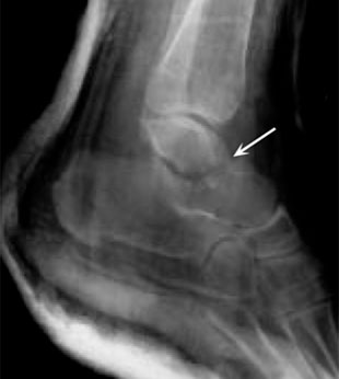
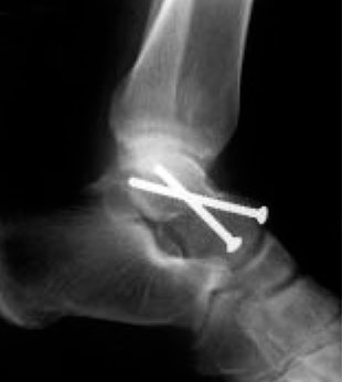
(Left) An x-ray shows a fracture of the ankle bone. (Right) The bone fragments were fixed with screws.
Bone tissue has a high ability to regenerate. However, the more severe the injury, the longer the healing time may be. After the operation, the foot and ankle are immobilized for 2-8 weeks, depending on the type of injury and the expected ability to regenerate. During treatment, the doctor will likely take some x-rays to ensure that the healing process is progressing as desired.
Basic rehabilitation techniques
To effectively treat the effects of a high-energy injury, a phased rehabilitation program is implemented. These include the following treatments:
- LFC exercises (breathing, isometric, postural and ideomotor exercises)
- Physiotherapeutic treatments (UVB, cold therapy, magnetic field therapy, laser)
- massage
- Pharmacopuncture
- orthopedic insoles
- Kinesio taping
When choosing rehabilitation methods, we take into account the type and severity of the injury, the patient's age, physical activity and general health.
Dislocation of the intervertebral disc
The isolated dislocation of the talus in its pure form is rare. The ankle bone can be moved in all directions (but more often outwards) and even rotated around its own axis. For this sprain to occur, the ligaments that connect it to the shinbone, heel bone and ankle must tear. These ligaments tear either due to forced inward (pronation) or outward rotation (supination) or due to simultaneous dorsiflexion or sole flexion of the foot. The predominant direction of force also determines the displacement of the hip to one side or the other. The neck of the bone is often broken. Pure plate sprains can be corrected by straightening the foot with the knee bent and applying direct pressure to the protruding bone. In difficult cases, bloody repositioning of the dislocated talus or even its removal is performed.
Subtalar sprain is caused by rapid inward (pronation) or outward (supination) rotation of the forefoot, which is locked on the ground. This leads to a separation of the connection between the talus and the ankle joint. There is greater decoupling at the ankle joint. Acute inward rotation (pronation) of the immobile forefoot with inward rotation of the tibia leads to an outward displacement of the navicular bone along with the forefoot. Under the same conditions, acute twisting of the foot with external rotation (pivotization) of the tibia results in inward movement of the navicular bone and foot. An inward dislocation of the subtalar is easily recognized by this characteristic positioning of the foot. Flexion and extension of the ankle joint is possible.
The extremely rare forms of posterior or anterior subtalar dislocation require a fracture as the cause.
Pure subtalar dislocations can be easily corrected under anesthesia due to the extensive joint fractures in the foot. Immobilization with an immobilizing bandage for 4-5 weeks and approximately the same length of rehabilitation and physiotherapy lead to almost normal foot function after 2-3 months.
Rare dislocations of the foot bones
Rare dislocations of the foot bones include:
- Backward dislocation of a scaphoid, a cuboid, one, two or three sphenoid bones
- Very rare dislocations of the clubfoot joint.
- The metatarsal bones of the Lisfranc joint are slightly more common: most often all bones are dislocated backwards and outwards (complete dislocation), more rarely one or more bones (partial dislocation).
These rare dislocations of the foot bones are caused by high force, e.g. B. from a fall from a height onto the toes in acute plantar flexion or from direct pressure on the forefoot during plantar flexion. More common than dislocations are fractures and deformations of the Lisfranc joint.
Without an x-ray, it is difficult to make an accurate diagnosis of these injuries. These dislocations can be corrected by pulling on the end of the foot with counter pressure on the protruding bones. The foot is immobilized with a plaster cast for 4-5 weeks. After a week, the patient can walk in this cast.
These dislocations of the foot bones are often associated with bone fractures and cracks at the attachment points of the strong ligaments and tendons as well as nerve and soft tissue damage. In such cases, the function of the foot returns very slowly, and sometimes the pain persists for years, requiring surgery: removal of small bone cracks, nerve resection or neurotomy. Before the operation, however, it is necessary to check the function of an insole, which is made exactly from a plaster cast of the painful foot.
Sprains of the metatarsophalangeal and interphalangeal joints of the foot occur in the wrist system.
species
Depending on the extent of the injury, a distinction is made between small, partial and complete necrosis. Depending on the severity of the impairment, four stages are distinguished:
- In the first stage, the lesions are minor, manifested by intermittent pain, but in no way affecting mobility;
- In the second stage, the pain syndrome intensifies, small cracks appear in the bone and movement becomes difficult;
- In the third stage there is severe tissue damage, the pain is more intense, does not subside even at rest and movement is severely restricted;
- In the fourth stage of aseptic necrosis, the talar block is completely destroyed and no longer has any motor function.
Symptoms of aseptic necrosis of the calcaneus
The first symptoms of the disease are numbness and loss of sensation in the lower part of the ankle. As necrosis progresses, swelling and pallor of the skin appear, which indicate a disturbed blood supply to the tissue.
Later, the pain worsens after running, walking or standing for long periods. The skin surrounding the lesion becomes vividly colored, sensitivity decreases significantly, cramps occur, and the patient feels cold. Even a short walk quickly leads to fatigue. Due to increased necrosis, general intoxication and anemia occur.
,Welcome!' – Network of spine and joint clinics
For more than 10 years we have specialized in the treatment of diseases of the musculoskeletal system at various stages of pathology without surgery. ,Halo!' are the clinical bases of the country's leading universities, training specialists for the most sought-after professions in medicine. The clinics' doctors are a team of experts from the RUDN faculty who, together with leading Israeli specialists, have developed unique treatment protocols in accordance with the standards of the Russian Ministry of Health.
Main stages and symptoms
The pathology can be local (part of the bone is damaged) or complete (the destruction has spread to the entire bone). Depending on the damage to the talus and the spread of aseptic necrosis, the main stages can be distinguished:
- In stage 1, tissue destruction is minimal, so the patient has no problems with motor skills. Pain occasionally occurs, especially after walking or standing for long periods of time.
- In stage 2, the bone deteriorates more and fractures form on the bone surface. The pain is more intense and occurs more frequently.
- In stage 3 there is pronounced necrosis of the tissue. There is little to no pain, making movement difficult.
- In stage 4, necrosis completely affects the talus of the foot, with irreversible consequences. The patient loses the ability to walk and the pain becomes unbearable.
Pale skin on the foot, numbness and reduced sensitivity usually indicate the development of the disease. Cramps and rapid leg fatigue may also occur, even after short walking. If the disease progresses to intoxication, headache, weakness and fever occur.
If an operation is necessary
If conservative treatment does not help, pain increases and mobility is limited, the patient is recommended surgery. One option is to stiffen the ankle joint (arthrodesis). The surgeon removes the damaged parts of the joint, 'locks' it and the patient then regains mobility. A complication of the operation may be incorrect joint formation if the bones do not fuse (5-10 %) and a second operation is required.
If other joints (subtalar and ankle) are damaged in addition to the ankle, arthrodesis is performed on all joints.
There is no technique for hip arthroplasty
Does talocalcaneal arthritis require treatment?
It's a pretty serious condition. Your treatment can take years and lead to serious complications. The joint will never be the same again. The pain significantly affects the quality of life and the ability to work; they are worse at night and worsen at rest. Over time, the foot becomes deformed and prevents you from living a normal life. The degenerative processes spread to the neighboring joints.
If you notice the slightest signs of pain and discomfort in your foot, don't dismiss it as fatigue. Visit your orthopedist and make sure you are not at risk of osteoarthritis in the knee or ankle.
Read more:- pelvic bone Latin.
- Anatomy of the pelvic bones.
- Displacement fracture of the heel bone.
- The shaft of the heel bone.
- pelvic subluxation.
- Fracture of the heel bone.
- Structure of the human ankle.
- tibia and fibula.