Pour 200 ml of boiling water over about a tablespoon of the grated herb and leave to steep for an hour. The resulting tincture is drunk as a tea.
- Functional disorders of the organs, movement disorders in diseases of the nervous system
- Paralysis, hemiparesis, hemiparesis, hemiparesis, hemiparesis, hemiparesis
- General information.
- Why there is an upper hemiparesis
- Amyotrophic Lateral Sclerosis
- Spinal anthrophia
- Multifocal motor neuropathy
- diagnosis
- Follow
- diagnosis
- Treatment
- Special features of the diagnosis
- Peculiarities in the development of pediatric hemiparesis
- Symptoms of Myelopathy
- examination and diagnosis
- treatment and prevention
- Traditional remedies
- symptoms
- Educate parents on how to heal their children
- Treatment of spastic tetraparesis
- Clinical Features
Functional disorders of the organs, movement disorders in diseases of the nervous system
Our clinic specializes in neurology and the diagnosis and treatment of organ and movement disorders. We focus on diagnosing and treating the cause of the disorder to give you the best possible treatment..
Disorders of the pelvic organs (pelvic organ disorders) are caused by damage to the connection between the brain and the pelvic organs (paralysis or paresis) or between the pelvic organs and the brain (sensory disturbances). Our main goal is to find and treat the cause of pelvic organ dysfunction.
The following symptoms may occur:
- Constipation.
- Fecal incontinence is often due to weakness of the anal sphincter.
- difficulty urinating.
- Urinary incontinence (we advocate conservative treatment of urinary incontinence in both men and women is possible).
- sexual dysfunction.
- Perineal nausea.

Speeding up recovery is helpful:
- Identifying and treating the underlying condition that has damaged the connection between the brain and pelvic organ.
- Drugs that stimulate neuromuscular transmission.
- Magnetic stimulation.
If the disturbance lasts for a long time – secondary infections of the urinary tract (cystitis, pyelonephritis) and inflammation of the lower intestine (sigmoiditis, proctitis) are possible. In these cases, we suggest a laboratory test for the causative microorganism with selection of the antibiotic. At the same time, we recommend immunomodulatory treatment to prevent re-infection.
Paralysis, hemiparesis, hemiparesis, hemiparesis, hemiparesis, hemiparesis
We treat paralysis, paresis, hemiparesis, hemiparesis and monoparesis. The primary aim is to find and treat the cause of the paresis (paralysis).. This is the basic requirement for success. As the nervous system regenerates, the muscles resume their function, obeying signals from the brain and spinal cord, and the paralysis gradually disappears. We have proven methods for treating paralysis, many of which you can do at home. These are.
Paralysis or paresis is a state of weakness of muscle groups, caused by the disruption of the connection between the muscles and the nervous system. Paresis can be caused by damage to the brain, spinal cord, or peripheral nerves and loss of nerve impulses from the brain to the muscles.
paresis – is a paralysis of the limbs of varying degrees of severity. The degree of paresis (paralysis) is determined using a score:
- 0 points. – no voluntary movements. Total paralysis (aka 'paralysis')
- 1 point - Degree of paralysis in which muscle contractions are present but hardly felt, no movement in the joints
- grade 2 – Movement is possible in the horizontal plane (no gravity required), movement in the joints is restricted
- 3 points – The movement of the joints is severely limited, but the muscles can counteract gravity (the limb can be lifted).
- 4 points. – The range of motion is full, but muscle strength is clearly limited
- 5 points – The range of motion is full, muscle strength is normal and complete.

Hemiparesis (hemiplegic paralysis) – Hemiparesis is a weakness in muscle tone on one (right or left) side of the body. A distinction is made between hemiparesis of the right and left extremities.
General information.
Hemiparesis of the upper limbs - decreased strength in the hands. It is usually asymmetrical and develops gradually. There are differentiated variants of this syndrome:
- limp. Muscle tension and reflexes are reduced, and hypotrophy is noted.
- Spastic. Muscle tension and reflexes are increased, hypotrophy is not noted. Characteristic are pathological reflexes and joint movements that are normally absent (when one limb performs a movement act, the other limb repeats the same movement in a reduced form).
- Mixed. The symptoms of the above types of paresis occur together.
The severity ranges from mild muscle weakness to complete loss of the ability to perform active movements. Viewed in isolation, upper paraparesis occurs much less frequently than lower paraparesis (muscle weakness in the legs). In many disorders, it precedes the development of inferior paraparesis or occurs some time after loss of leg muscle strength. When the thickening of the spinal cord in the cervical region (below the fourth cervical vertebra) is affected, the spastic superior paraparesis is combined with a flaccid spastic inferior paraparesis.
Why there is an upper hemiparesis
Amyotrophic Lateral Sclerosis
Upper hemiparesis is considered a feature of ALS that is primarily suspected at the time of onset, as it is less common in other disorders. In patients with ALS with cervical onset, the disorder is present at the time of onset, is flaccid, asymmetric, and is associated with pyramidal symptoms and hyperreflexia.
In ALS with progressive hemiplegia, dysphagia, dysarthria, fasciculations, and changes in speech atrophy precede the onset of these symptoms. Patients with ALS with lumbar onset develop flaccid inferior hemiparesis before the onset of asymmetric superior paresis. Pseudobulbar and bulbar syndromes develop later.
Spinal anthrophia
This group of diseases is inherited and results from the degeneration of motor neurons. Spinal atrophies differ significantly in their clinical manifestations, with both types of disease being characterized by a predominance of upper paresis. In distal Duchenne-Arant CMA, the onset of symptoms is more common at a young age. The arms become weak and have the appearance of a 'clawed paw'.
The lesions then spread to the proximal limbs. Lower paraparesis develops much later. Sometimes the disease begins with an upper paresis. Scapuloperoneal Vulpian SMA occurs in young or middle age. Unlike the previous amyotrophy, the shoulder girdle is affected, not the arms, resulting in weakness in the arms. The muscles in the lower legs and feet are also weakened.
Multifocal motor neuropathy
MMN is a form of polyneuropathy of autoimmune etiology. Upper extremity paresis is asymmetric and develops gradually. There is no pain or sensory disturbances. The hands are affected in 95 % of cases, and half of the patients experience fasciitis and muscle spasms. The gradient is progressive or wavy. Hypotrophy is absent or appears at a late stage.
diagnosis
The patient should be examined by a neurologist and an orthopedist. Hospitalization may be required to confirm the diagnosis.
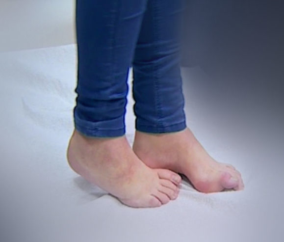
Spastic paresis in children can be diagnosed by the presence of Babinski's sign: when the upper part of the foot is stimulated by stroking movements, the big toe flexes outward instead of inward. This is a pathological symptom characteristic of the disease.
- Barre test - a person raises their limbs and holds them for 30 seconds;
- general and biochemical blood test;
- general urine examination;
- CT scan to assess the condition of the joints and spine;
- MRI to show abnormalities in the brain and spinal cord;
- Electromyography to diagnose neuromuscular transmission;
- Analysis of the cerebrospinal fluid;
- myelography to understand how the spinal cord works;
- blood tests for hepatitis, syphilis and HIV;
- EEG to assess the electrical activity of the brain;
- Determination of the B vitamins in the blood.
Based on the results of the comprehensive examination, the child's paraplegia is confirmed or disproved.
Follow
If the disease occurs early and without rehabilitation, it leads to difficulty walking and limits independence in adult life. Lack of mobility leads to motor deficits.
Abnormal body alignment leads to contractures, dislocations and joint subluxations. This leads to leg deformities, secondary muscle changes such as atrophy and fibrosis, and drastically reduced rehabilitation options.
A good rehabilitation program can prevent these consequences. A neurologist and an orthopedic surgeon develop a rehabilitation plan.
diagnosis
For a comprehensive diagnosis of paraparesis, the following examinations are carried out:
- General blood test (to detect infectious diseases)
- MRI of the brain (to detect tumors)
- Myography (to assess muscle function)
- EEG (to monitor brain activity).
- The EEG examination is considered to be the most effective type of examination.
Only a neurologist or osteopath can make an accurate diagnosis.
For further examinations, it may be necessary to consult specialized doctors - specialists in traumatology and orthopedics as well as neurology.
In children, the severity of the disease (moderate, severe, severe) is taken into account when prescribing treatment.
Treatment
Paraparesis can be treated with different methods: physiotherapy and medication.
For the treatment of paraparesis, therapies are used:
Massage therapy is often combined with physical therapy. When treating paraparesis, a special diet and nutrition plan is drawn up.
The therapy exercises should never be performed at home, but must be supervised by a doctor.
It is important to consult specialists in time to avoid paralysis (or paralysis).
Special features of the diagnosis
In most cases, the diagnosis is made by clinical methods alone, without the use of additional technology.
In the development of hemiparesis, the muscle strength of the left and right extremities is compared. Standardized resistance tests are carried out, but also a classic test procedure, the Barré test.
In this procedure, the limbs are kept under stress for a period of time. In the case of progressive paresis, the stretched limbs are slowly lowered after 20 seconds.
To determine the factors that lead to the development of paralysis of the limbs, the following devices and laboratory tests are used:
- CSF and brain tissue MRI and diagnostic CT;
- CSF analysis;
- electromyography;
- General blood count and biochemical examinations;
- electroneuromyography;
- Determination of the content of substance B12 and folic acid;
- Determination of genetic predisposition or possible tumors.
Peculiarities in the development of pediatric hemiparesis
If a child is developing normally overall, you may find them walking on tiptoe. This is of course a cause for great concern for parents. If the child walks independently, this does not mean that he suffers from paraparesis.
Tiptoe walking may be due to only certain groups of leg muscles being well tightened, which is not always a sign of paraparesis. In such cases, therapists often recommend massage and general therapeutic exercises.
When disorders occur, the repair processes in the child's nervous system are much more active than in the developed adult body due to its plasticity.
Therefore, the effects of significant disruption, hypoxia and birth trauma may soon no longer be visible.
If a child is diagnosed with paraparesis, continuous monitoring by a neurologist should be ensured. Monitoring should be supplemented with medication, physical therapy, and orthopedic examinations. Acupuncture can also have a positive effect.
Symptoms of Myelopathy
The spinal cord is the main 'cable' of the nervous system that connects the brain to the body. When this cable malfunctions, the connection between the brain and the body is disrupted, which can result in the following symptoms:
- Decreased muscle strength in the arms and/or legs (paralysis, paresis) below where the spinal cord was injured. Doesn't always go hand in hand with an emotional disorder.
- impairment of the senses Below the site of the spinal cord injury. It is possible that only one type of sensation (pain, touch, temperature, musculo-articular sensation) or all types of sensations are decreased or increased.
- Bladder and bowel disorders (pelvic floor disorders). Urinary incontinence or difficulty in emptying the bladder or bowel can occur.
- Muscle tension in the legs and sometimes in the arms (spasticity). This is caused by a disruption in the brain's control over the spinal cord. Nerve cells in the spinal cord tense the muscles of the limbs independently. In the legs, forced knee flexion and soleus flexion of the foot are characteristic. In the hands, there is a forced flexion of the elbow and fingers.
examination and diagnosis
MRI scan Provides a good visualization of the spinal cord and brain, intervertebral discs and spinal cord tumors. It is good for distinguishing areas of spinal hemorrhage.
Computed tomography (CT) With it, the bones of the spine can be well represented. CT angiography is a method of examining the blood vessels (including the spinal cord) by administering a contrast agent intravenously beforehand.
electromyography (EMG, ENMG, somatosensory evoked potentials, magnetic stimulation studies) assesses the flow of electrical stimulation through the spinal cord and peripheral nerves.
blood tests We use blood tests primarily to diagnose infectious, metabolic, and autoimmune diseases of the spinal cord. The examination also provides important information about circulatory disorders in the spinal cord, which are associated with increased blood viscosity.
treatment and prevention
A holistic approach is always required when treating hemiparesis of the upper and lower limbs. Treating the underlying condition causing the muscle weakness is just as essential as treating the symptoms intensively.
Gymnastic and massage treatments are intended to help the muscle tissue contract better. Over time, the exercises require more activity and increase the stress on the muscles. Each activity must be monitored by a doctor in a strict order.
Further treatment steps include physical therapies, water treatments, physiotherapy as well as electromyostimulation and magnetic field applications, acupuncture, individual massage therapy and acupuncture.
The use of all kinds of psychological techniques and various trainings to improve the mental state of patients continues to give remarkable results.
Operations are rarely used. Most of these measures are required to treat conditions that cause paralysis.
Water treatment is considered one of the most effective ways to prevent paralysis in the upper and lower limbs.
Traditional remedies
Traditional medicine is widely used to treat this condition. Herbal remedies always help to restore strength and strengthen the body's defenses.
symptoms
Note the main symptoms that are typical of this diagnosis.
- weakness in the muscles.
- Unsteady gait of the child.
- Pain and swelling in the lower limbs.
- Difficulty flexing and extending the lower leg and thigh.
- inability to step on the heel.
Ask Nikolay Borisovich Nikonov, the author of the treatment method, a question through the feedback form. It is also useful to schedule a follow-up appointment with a myologist.
Educate parents on how to heal their children
From my point of view (that of Nikolai Nikonov) it is the inability of muscle cells to release excess protein inside them when they start to form (from the 6th week of fetal life).
My point of view is based on:
In 2017, the Nobel Prize in Medicine was awarded to American geneticists. Outstanding scientists were honored for discovering the molecular mechanisms that control circadian rhythms. The discovery of the 2017 Nobel Prize winners confirms my view of the process of protein swelling in children with cerebral palsy. You can read more about this discovery at: /articles/nobelevskie-laureates-2017-goda-objasnivshie-vozniknovenie-dtsp.
- On the discovery of excess protein in muscle cells, the so-called 'protein dump', by 2016 Nobel Prize winner Yoshinori Ohsumi. More information about the discovery at: /articles/podderzhka-nobelevskogo-laureate.
- Using genetic engineering data, Professor Kiyotoshi Sekiguchi discovered that the lack of a polydomain protein means that lymph vessels don't grow as fast as muscle cells. Professor Kiyotoshi Sekiguchi studied the location of the polydome on embryos and discovered that the protein forms lymphatic vessels during embryonic development.
According to the World Health Organization, the primary cause of atonic-astatic tetraparesis and other forms of cerebral palsy is damage or abnormal development of the fetal brain in utero.
Treatment of spastic tetraparesis
As I write this article ya write, it is my experience that there is only one way to bring a child back. The only method I have developed myself is the Nikonov method. With this method, the swollen muscles are worked with the hand and the working muscle is slightly fixed.
neurologists Neurologists believe that Children with this diagnosis cannot recover and suggest that spastic tetraparesis (double hemiplegia) should be treated with a combination of conservative, orthopedic, and rehabilitative measures. The main goals of therapy are to eliminate the sequelae of the disease, such as spasticity, restricted motor function and abnormal motor stereotypies.
Conservative treatment includes pharmacotherapy with baclofen and botulinum toxin, which reduce spasticity, increase impulse conduction and improve metabolism in the brain, which should lead to an increase in passive movements in the limbs.
Conservative treatment does not result in an increase in passive movements, let alone active movements.
Neurologists recommend physical therapywhich includes a series of exercises, postures and movements to strengthen muscles.
How can spastic muscles be strengthened? Try to get them out of the way!
use neurologists Hirudotherapy, acupuncture, massage and exercise therapy, kinesitherapy.
Only the appearance of an improvement is awakened.
neurologists provide surgical treatment of double hemiplegia. During the operation, muscles are stretched and contractures are corrected, tendons are repaired.
Clinical Features
The incidence of this form of cerebral palsy is 2 %.
K Musculoskeletal disorders include:
Bilateral spasticity equally in the upper and lower limbs (spastic tetraplegia or quadriplegia). However, there are also forms in which the spasticity only predominates in the hands. In spastic tetraparesis, secondary complications such as contractures, trunk and limb deformities appear early. Wrist and foot reflex abnormalities are noted.
Impaired mental and language developmentAbnormalities of the sense organs:
Cognitive impairments are evident in this case. Squinting, optic nerve atrophy, hearing loss, pseudobulbar syndrome and consequent dysarthria, dysphagia, dysphonia. Children with double hemiplegia usually speak slurred and nasally and are difficult for others to understand. Vowels are often mispronounced. The speed of speaking is changed. The child pronounces words and sounds either excessively loud and fast, or weakly and slowly. In the most severe form of childhood cerebral palsy, thinking is slow and sluggish and memory is impaired. Disinhibition, euphoria, apathy and abulism are not uncommon.
Other pathologies: Epilepsy, secondary microcephaly.
Decreased intellect.Pronunciation: pronunciation.
Read more:- Paraparesis - what is it?.
- Inferior paresis - what is it?.
- Lower spastic paraparesis.
- paresis of the lower body.
- paresis is.
- Toe movement in children.
- Spastic gait is.
- A wobbly gait means.