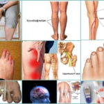
Syndactyly can be simple or compound, depending on the condition of the adjacent fingers. Simple syndactyly is a fusion of normally developed fingers (length, number of phalanxes, and range of joint movement). Complex syndactyly is the fusion of fingers with anomalies of the tendon-ligamentous and articular-bony apparatus (flexion contractures, adducted segments, sprains, clinodactyly, etc.).
- Polydactyly: Why are six fingers better than five?
- Premium care: What modern diapers can do
- Multiple fingers: good or bad?
- ICD-10
- Causes of syndactyly
- 3 techniques of toe treatment
- Classic pedicure
- European
- Insoles for foot treatment
- How is the diagnosis made?
- What helps prevent the disease?
- Classification of fractures
- Symptoms of a broken finger
- How to prevent this disease
- self-help and self-diagnosis
- Medical examination for paraesthesia of the toe
- Associated symptoms and diagnosis
- Treatment
- medication
- A nail falls off
- Paronychia – purulent inflammation in the nail area
- Acute paronychia
- Chronic paronychia
Polydactyly: Why are six fingers better than five?
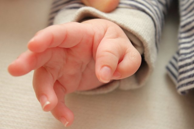
Polydactyly is a very common anomaly. It means that a person is born with extra fingers or toes, sometimes both. This can be an 'extra' finger or several fingers at the same time. Doctors have traditionally thought of this abnormality as a fetal malformation, but today scientists say moderate polydactyly can be beneficial. MedAboutMe tells you what the secret is.
Spoiler alert: It's not just the extra finger that gives you a better grip and hold of items!

Premium care: What modern diapers can do
Multiple fingers: good or bad?
It is estimated that one in 700 babies is born with polydactyly (from the Greek meaning 'many' and 'fingers'). The frequency is even 1 in 143 for Africans and Afro-Americans and 1 in 1400 for Europeans.
Polydactyly is the opposite of oligodactyly, in which there are fewer fingers than normal.
How many fingers can a human have? Here are the most surprising examples.
- Hong Hong, a Chinese boy who is 4 years old, has 31 fingers in total.
- The world record is held by Akshat Saxena from India. He is 10 years old and has 7 fingers on each hand and 10 toes. So he has 34 fingers in total!
Polydactyly is one of Mother Nature's usual jokes. In humans and animals, it occurs on one or more limbs. Supernumerary 'fingers' can occur in dogs, cats, mice, horses, pigs and even reptiles and birds. Most often it is a small piece of tissue that can be easily removed. Bones are rarely found in the mass, but no joints. It is even rarer to find a fully developed, functional finger.
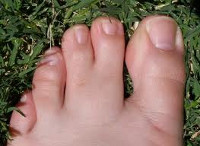
Most often, the little finger is additionally fused, less often the second thumb. The other fingers—index, middle, or ring finger—are even rarer.
Nine times out of 10 these fingers are on the feet and only 10 % on the hand.
If polydactyly is detected, genetic testing is required: this anomaly can be associated with certain syndromes. This association is found in about 6 % of the cases. Genetic testing is also required if polydactyly runs in families, which is the case in 14 % of cases.
Thirty-nine different genetic mutations, possibly more, are responsible for the occurrence of supernumerary toes: all mutations are not yet known.
Studies show that this anomaly occurs most frequently in the first 4 weeks of embryonic development, during rapid limb development.
ICD-10
Syndactyly is an anomaly of the fingers resulting from their separation during the embryonic period. Syndactyly accounts for about half of all congenital malformations of the upper limb and occurs with a frequency of 1 in 2000 to 3000 newborns. Syndactyly of a child's hand or foot may be an independent defect or may occur in combination with other limb malformations: polyphalangy and polydactyly, hypoplasia of fingers, brachydactyly, ectrodactyly, cleft hand, radial or ulnar clubfoot, humeral synostosis, etc. 60 % of children with syndactyly have at the same time a congenital pathology of the musculoskeletal system (pseudarthrosis, varus or valgus foot, clubfoot, etc.).
Causes of syndactyly
The presence of syndactyly in a child may be due to hereditary factors. In this case there is an autosomal dominant inheritance type and the syndactyly is familial. More than 20 % of syndactyly is hereditary.
In the absence of familial syndactyly, it is necessary to take into account the formation and differentiation of the fetal limbs during embryogenesis under the influence of various unfavorable factors. The positioning of the hands occurs at 4 and 5 weeks of pregnancy; physiological syndactyly is characteristic of the fetus at this stage. With normal development, the fingers form in the 7th to 8th week with rapid growth of the finger radii and slower growth of the interdigital spaces. With an abnormal shortening of the interdigital septa, the fingers are not separated, that is, syndactyly develops.
Factors affecting the normal development of the fetal limbs can be toxic influences on the pregnant woman's body (drugs, alcohol, occupational risks, unfavorable environmental conditions, etc.), X-rays, infectious diseases during pregnancy (flu, syphilis, tuberculosis, etc.). It is not uncommon for the cause of the birth of a child with syndactyly to be unknown.
Syndactyly is a common malformation and belongs to genetic syndromes (Smith-Lemli-Opitz, Aper, cryptophthalmia) and chromosomal syndromes (Cat's Cry, Edwards, etc.).
Acquired syndactyly, caused by thermal and chemical burns to the hand, is less common in children.
3 techniques of toe treatment
Every pedicure has advantages and disadvantages. You have to know them to feel comfortable and to ensure the safety of customers.
Modern cosmetology offers various options and types of toe treatments for women who constantly strive for perfection.
Classic pedicure
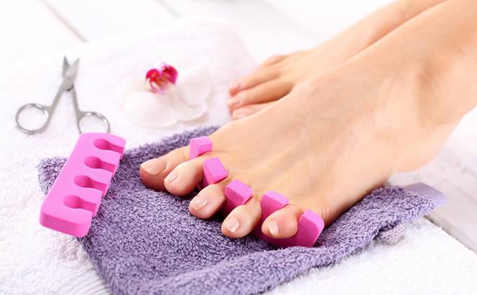
This type of pedicure can be done at a beauty salon or you can do it yourself.
- softening of the skin on the feet by steaming in a steam bath;
- Cutting nails, removing dead skin with a pumice stone;
- filing the nail plates;
- Cuticle care.
This type of pedicure is the most popular in beauty salons because it is simple, quick and inexpensive.
European
This type of pedicure is very similar to the classic nail scissors pedicure, but without the use of potentially dangerous tools and steaming your feet in the bathtub.
The European pedicure consists of the following steps:
- A special preparation is applied to the cuticles to soften them, and then the cuticles are pushed to the edge of the nail; this treatment is safer than trimming, and regular repetition of this treatment also slows down the growth of the cuticle;
- The desired shape of the nail plate is not changed by grinding, but with the help of scissors and tweezers;
- The dead skin on the toes and feet is removed with scrapers and erasers.
European pedicure has many advantages: safety, a long-lasting result and a beautiful appearance of the feet. However, there is also a downside. The result can be seen not only in one session, but also at regular intervals; a truly well-groomed appearance is achieved after six sessions.
Insoles for foot treatment
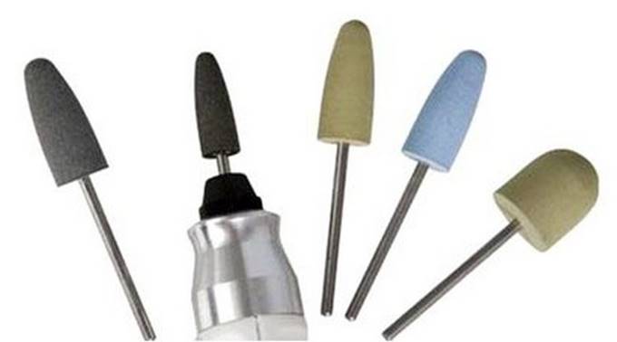
The inserts for foot treatment are all different and vary in shape, diameter and grit. A skilled craftsman always understands the purpose of each cutter - a sign of his high skill and a guarantee of choosing the right consumable. Let's take a closer look at what cutters there are and what they are made of.
Ceramic cutters They are rightly considered the least dangerous and can be used at home even by those who are just beginning to learn the art of pedicure. Ceramic burs are used in a wide variety of areas: to treat the cuticles, but also to remove rough cuticles and polish the nail plate.
We recommend.
The burrs with the coarsest grit are suitable for removing rough skin, e.g. B. heels or corns. The composition of this grit is diamond grit.
The skin can be treated with a medium grit nozzle. The finest grit is found on the cutters used to grind fingertips and nails.
The main advantage of these nozzles is their low hardness, which greatly reduces the risk of injury, especially to the delicate nail plate and surrounding skin.
But ceramic also has a major disadvantage: it lasts much shorter than others.
There are many variations of these cutters in shape, from cylindrical to oval. A grain nozzle is easily identified by the color of the stripe on the nozzle tip: blue means low, white means medium, and black means high.
diamond nozzles are used for the roughest parts of the skin and the free part of the nail plate.
Diamond-tipped milling cutters have different levels of abrasiveness:
- super coarse. They can be recognized by the black stripe and are intended for very rough skin.
- Extra coarse. The stripe on this router is green; its function is to remove roughness on the nail.
- Middle. Features a blue stripe and a streamlined shape. Designed for gentle treatments on sensitive areas: on the nail fiber and cuticle.
- Small. The red stripe burr is needed to pass the cuticle itself and the lateral dermal shafts.
- Extra fine. The nozzle has a yellow stripe and is intended for nail shape correction.
How is the diagnosis made?
The first stage of diagnosis consists of examining the foot externally and asking whether the pain has been present and for how long. An experienced doctor will already be able to reject part of the diagnosis at this stage. He will then either initiate treatment or order examinations: X-rays, less frequently blood tests, even more rarely an MRI or an ultrasound examination of the joints.
Treatment depends on the cause of the condition.
The ingrown nail is removed, and then the foot needs to be immobilized, bandaged, and pain relieved for a few days. After that, a visit to the podiatrist is necessary: the problem can be caused by a misalignment of the foot. If this is not corrected, the nail will keep growing back.
Calluses also need to be removed, and if they have already caused pain in the big toe joint, this should be done by a dermatologist or podiatrist. It is important to find out what is causing the callus. They can be caused by ill-fitting shoes, flat feet, or warts underneath. In all these cases, the treatment will be different. The condition of the feet can be corrected with cosmetic surgery if the doctor agrees.
Orthopedic treatment is necessary for flat feet and valgus deformities and can provide relief for patients with arthritis and osteoarthritis. Diabetes, gout and autoimmune diseases require a systemic approach, while joint pain is treated symptomatically.
What helps prevent the disease?
The most important prevention is suitable footwear. At the first symptoms, when the bones in the feet in the area of the big toe just start to hurt, you should take a close look at your shoes. are they comfortable Does the pain go away when you take them off?
The second preventive measure is a pedicure. If, despite good care, the nail is trying to grow back or blistering excessively, you should see your doctor.
Third, fight obesity and standing work. Both can overload the foot and joint problems can arise if overloaded.
The fourth point is early treatment. If your joints start to hurt, don't delay seeing a doctor. The sooner the changes in the body are corrected, the sooner relief occurs.
The Healthy Spine Hello! consists of certified doctors with many years of experience. These include doctors and candidates of medical sciences, professors and doctoral students of the highest qualification category, lecturers of the Faculty of Osteopathy and Manual Therapy of the Peoples' Friendship University of Russia. All of them have extensive experience of inpatient work in leading medical and preventive institutions in Moscow and form the strongest team of experts in the diagnosis and treatment of diseases of the musculoskeletal system.
Comprehensive treatment in the network's clinics is carried out by doctors from specialist areas such as traumatology and orthopaedics, chiropractic and osteopathy, neurology and therapy, physiology and reflexology, as well as specialists from other popular medical professions (cardiology, surgery, dietetics). Unique methods and innovative devices make it possible to quickly and accurately diagnose, and most importantly, to prescribe adequate and effective therapy.
Classification of fractures
There are many types of finger fractures and they are all different. Sometimes a single phalanx is damaged. Sometimes multiple fingers are broken. Diagnosing a fracture is fairly easy, but it should only be made by a doctor.
- Open and closed. It is difficult to determine whether a person has a closed fracture of a finger without the help of a doctor. An open fracture is easy to spot because it is almost always accompanied by bleeding. In an open fracture, the integrity of the bone and skin is compromised. Closed fractures are not only complete but also incomplete with fractures.
- With or without displacement. Treatment of fractures with displacement is very difficult, since the bone is badly shattered and has a large number of different fragments. One can tell by certain signs that a person has a displaced fracture. For example, the deformed area of the finger is shortened. The affected person can no longer move their fingers and suffers from a severe pain syndrome.
- edge fracture . This type of injury can be caused in a number of ways. A fracture of the little finger can result in a partial or complete fracture of the phalanx. This injury can be caused by a strong impact or falling with a heavy object on the hand or leg. It is not uncommon for patients to suffer a marginal fracture due to various pathologies. For example, trauma can occur as a result of a disease that causes bone fragility. The marginal fractures can be primary fractures, nail fractures, medial fractures and compound fractures.
- fracture of the index finger. If someone has broken their index finger, they should see a doctor immediately. The doctor can quickly put the dislocated bone back into place. If the finger is dislocated, the doctor may place a cast or splint over the injured area. The doctor will certainly prescribe medication for pain relief. In most cases, a broken index finger is not dangerous if you seek immediate medical attention.
- Fracture of the thumb on the hand. This injury is often caused by a strong blow. Due to the special position of the bone, some difficulties can arise during treatment. The broken finger needs to be restored by a doctor. This requires various devices that have a stretching effect. After straightening the injured finger, the doctor applies a plaster cast. The cast allows the finger to be in a specific position. The bones are immobilized until the treatment is complete.
Symptoms of a broken finger
The very first and foremost symptom of a broken finger is severe, unbearable pain. The intensity of the pain syndrome depends on the severity of the injury. Severe swelling or bruising follows the pain. This reaction is quite natural when a person suffers an injury.
- Sharp and strong pain that begins to increase with movement;
- The finger turns blue and gradually swells from bleeding internally;
- The injured area is deformed;
- The finger can shorten;
- Strange mobility of the phalanx that was absent before the injury;
- inability to make a full fist or relax the hand completely;
- A bruise forms under the nail, causing severe pain;
- An abnormal crunch is heard when moving the finger.
An open fracture of the finger causes shock with pain and bleeding. Do not try to straighten the bone yourself, as this can lead to serious consequences. This should only be done by an experienced traumatologist or surgeon in the hospital.
How to prevent this disease
Elementary prevention will help avoid swollen toes. You simply have to pay attention to your body and try to avoid possible injuries. The key to good foot health is personal hygiene, which should be a regular part of everyone's life.
Particular attention should be paid to choosing comfortable, high-quality shoes, which are made of natural materials, should have a low heel and a comfortable sole. Special orthopedic shoes or insoles can also be used. If fungal or other diseases are detected, they should be treated immediately.
You should also pay attention to your diet to ensure it is nutritious and contains all the necessary dietary micronutrients. Salt intake should be limited as it prevents excess water from leaving the body, which causes swelling. It is also better to avoid alcohol or keep it to a minimum.
Toe edema is easy to treat and often goes away completely, but only if treated on time. Basic principles can prevent swelling and keep you healthy.
self-help and self-diagnosis
To answer the question of what to do when toes go numb, it is important to understand the causes of this condition.
Loss of feeling in the toes, localized fever, swelling and limited mobility can indicate a disease of the small joints: arthritis, osteoarthritis or gout. Osteoarthritis and arthritis are characterized by a slow and gradual development of symptoms. With gout, the toes are numb and hot, swollen and uncomfortable, accompanied by a fever. If you have these symptoms, you should see an orthopedist or rheumatologist.
What to do if toes go numb because of uncomfortable shoes
The above measures will relieve the discomfort, but keep in mind that constant wearing of unsuitable shoes often leads to the development of osteoarthritis in the lower limbs.
Osteochondrosis, spondylosis, osteoarthritis of the spine, herniated and displaced discs, and tumors of the spine and soft tissue can also cause numbness in the fingers. All of these conditions can lead to poor circulation, nerve compression, and spinal cord damage. The following symptoms are typical of numbness in the fingers due to spinal abnormalities
- limited mobility
- impairment of movement coordination;
- crunching, cracking when moving;
- pain in the back and chest;
- Headache;
- changes in hearing and vision;
- dizziness;
- occasional 'disobedience' of the limbs;
- weakness without reason;
- rise in blood pressure.
It is important to see a neurologist or orthopedist as soon as possible if you experience these symptoms. Without treatment, dangerous complications such as paralysis, spinal cord damage or cerebral hemorrhage can occur.
With foot injuries, numbness and pain, as well as changes in the shape and color of the toe and swelling, may not appear immediately, but may appear several hours later or in the morning after sleep. If these symptoms are detected, urgent treatment by a trauma surgeon is required.
Medical examination for paraesthesia of the toe
Even if the symptoms are not very annoying, a medical examination is necessary. Initially mild discomfort can eventually lead to paralysis and other serious complications. If you start treatment early, both the cost and the duration of treatment will be much less than if you see a doctor late.
After a medical history and examination, the doctor will certainly refer the patient for further examinations. Various diagnostic methods may be necessary to determine the cause of your symptoms, e.g. B:
- X-rays and MRI scans of the joints and spine;
- computed tomography of the head;
- Ultrasound examination of blood vessels;
- Laboratory tests (blood and urine tests, aspiration of synovial fluid or cerebrospinal fluid).
Associated symptoms and diagnosis
Patients' complaints are rarely limited to numbness in the fingers. As a rule, a characteristic picture emerges, consisting of several clinical signs:
- Fatigue in the feet is a specific harbinger of numbness in the near future. This feeling can occur hours or days before the onset of numbness in the toes;
- paresthesia - an uncomfortable feeling in the sole area described by sufferers as 'standing on a ball' or 'walking on a puffy rice';
- Pain – occurs later in life and is an indication of a chronic condition. The pain is usually nagging, burning, or throbbing;
- Weak pulse – detected in the presence of vascular anomalies in the legs;
- Trophic abnormalities - are detected in the final stages when fingers freeze and turn blue, sores and ulcers appear on the skin, brittle nails and hair loss are observed.
You should see your GP for a diagnosis. Your doctor will listen to your concerns and refer you to one of the specialists below:
Depending on the suspected illness, a consultation appointment will be arranged. Treatment is decided after x-rays, computed tomography (CT) and magnetic resonance imaging (MRI).
Treatment
Consider how to treat toe numbness. The choice of treatment program depends on the characteristics of the clinical picture. Pharmacological treatment has special features.
medication
What to do if the numbness in the toes is caused by a systemic disease? When toes become numb, it is necessary to find an effective medication for the symptom. Prescribing medication is necessary not only to restore feeling in the lower limbs, but also to treat the underlying disease.
- Analgesics (Naise, Diclofenac) are indicated to reduce the pain and inflammation associated with the underlying disease. Topical ointments are best suited for limited effects - numbness of the big toe, numbness of the second or middle finger, ring finger, or numbness of the pinky finger;
- Nootropics (glycine, piracetam) – prescribed for brain damage (stroke, sclerosis, tumors, etc.). These drugs restore blood circulation in the head area and strengthen blood vessels;
- Anticoagulants (heparin and its derivatives) - If the cause of the numbness in the feet and toes is varicose veins, thrombosis or other diseases, blood-thinning medications are indicated. They reduce blood viscosity, increase blood flow to local tissues and improve cell nutrition;
- Vasodilators (nitroglycerin, isosorbide dinitrate) - are prescribed as an adjunct to previous treatment. These drugs expand the arteries and veins and improve tissue trophism;
- Vitamins – to strengthen the body with certain physiological abnormalities or to identify a leg disease at an early stage. Your doctor may prescribe tablets or capsules, as well as intravenous fluids, to get a quick effect.
Read more:A word of warning!!!
Depending on the cause of the numbness in the toes, the specialist doctor can prescribe special drugs to treat the pathology - these funds, as a rule, are prescribed only after a thorough examination.
A nail falls off
If the nail begins to peel and fall off as a result of an injury to the toe, this is normal. However, if the nail separates from the nail bed for no apparent reason, you should take notice. The most harmless cause of a nail prolapse is the excessive use of manicures, especially sharp tools to clean the space under the nails.
In rarer cases, the nail may fall off as a result of the following conditions:
- fungal infection of the nail;
- nail psoriasis;
- warts growing in clusters around the nail;
- hyperthyroidism;
- Sarcoidosis - a disease in which small clusters of cells form in organs and tissues;
- amyloidosis - a build-up of proteins in organs;
- Changes in the fibers of the connective tissue that support the body's organs and tissues;
- circulatory disorders, e.g. B. caused by smoking or Raynaud's disease (which causes the skin on your fingers to turn white when it's cold);
- an allergic reaction to medication (usually antibiotics) or nail cosmetics.
Destruction of the nail plate is possible for the following reasons:
- trauma, including nail biting;
- skin diseases such as psoriasis or tetanus;
- Growths in the surrounding tissue, which are usually harmless (e.g. common or keratotic warts) but can also be cancerous.
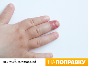
Paronychia – purulent inflammation in the nail area
Paronychia is inflammation of the perineal folds, skin, and soft tissues that surround and support the nail and is a form of panniculitis. Paronychia is caused by infection while trauma is a predisposing factor. The disease is about three times more common in women. Occasionally, paronychia is caused by a chronic skin condition, such as eczema or psoriasis, or another condition, such as diabetes or HIV.
Paronychia can be acute, meaning symptoms appear within a few hours, or chronic, meaning it lasts longer than six weeks.
Acute paronychia
Acute paronychia usually develops as a result of a minor injury to the periungual fold, e.g. B. with a manicure or with people who like to bite their nails. The affected area becomes red, hot, painful to the touch, and swollen. After some time, pus may appear and accumulate around the nail.
The most common cause of acute paronychia is Staphylococcus aureus, but any other microbe can also cause inflammation. In rare cases, the cause of a perianal infection is a herpes virus; in this case one speaks of a herpetic paronychia. The early stages of bacterial paronychia can be treated with antibiotics, while surgical removal of the pustules is more common. If left untreated, the process can progress to a chronic stage.
Chronic paronychia
Chronic paronychia progresses more slowly and can be more difficult to treat. It is most common in people whose hands come into frequent contact with water or chemicals, e.g. B. cleaning staff, bartenders, kitchen workers or fishmongers. The disease can start on one nail but then affect multiple nails. The affected perineal folds are swollen and can sometimes become red and sore, often after contact with water. The nail plate gradually thickens, furrows appear, and the nail may also turn yellow or green and become brittle.
- straightening of the fingers.
- Gum disease on feet.
- Why are a teenager's toes crooked?.
- What is another name for an orthopedist?.
- Why are the big toes crooked?.
- Why are the fingers curved?.
- ectrodactyly.
- Heels for girls 12.