How can diastasis be surgically removed if conservative treatment is unsuccessful? There are several effective methods:
- types of skin cancer.
- basal cell carcinoma of the skin
- symptoms and signs
- Fascial training
- abnormalities of the fascia
- When and which doctor to see
- diagnosis
- complications
- Intertrochanteric syndesmosis
- Achilles tendon (AC)
- Take the quiz and rate your knowledge: how well did you absorb the material?
- Precautions
- Types and characteristics of molars
- Features of the maxillary premolars
- opinions
- last comments
- A personal experience
types of skin cancer.
There are 3 types of common malignant skin tumors. They differ both in terms of frequency (ie, the likelihood of contracting the disease) and the degree of life-threatening nature - such as e.g. B. Basal cell carcinoma, squamous cell carcinoma and melanoma.
melanoma – is one of the rare and dangerous types of skin cancer. Melanoma accounts for only 4 % of all malignant skin cancers, but accounts for nearly 80 % of deaths at this site. You can find out more about melanoma here.
basal cell carcinoma of the skin
basalioma – is the most common but also the safest form of skin cancer. Death from a basal cell carcinoma is only possible in very advanced cases or in aggressive (basasquamous) forms of the cancer. The favorable course of basal cell carcinoma is due to the fact that it almost never forms metastases (only 0.5 % of cases).
symptoms and signs
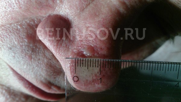
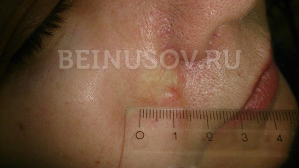
Basal cell carcinoma occurs most frequently on the skin of the nose, slightly less frequently on the face and much less frequently on other parts of the body.
The highest incidence is over 40 years of age. The youngest patient diagnosed histologically with basal cell carcinoma was 39 years old.
What basal cell carcinoma of the skin looks like depends on its shape:
- nodular form (equivalent to 'knotty). The tumor appears as a nodule. It is distinguished from other skin growths by an increased number of vessels on the surface, a waxy sheen, and small bluish-gray inclusions. All these signs can be seen in the photo.
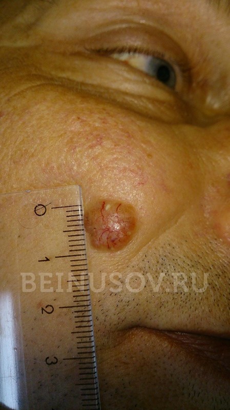
In addition, another characteristic feature may appear on the surface of a nodular basalioma: ulceration.
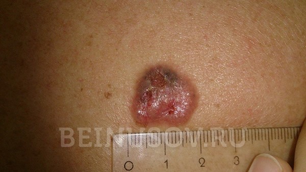
- Superficial shape In most cases, the basal cell carcinoma appears as a reddened area on the skin. Peeling elements and the already mentioned waxy shine are also possible.
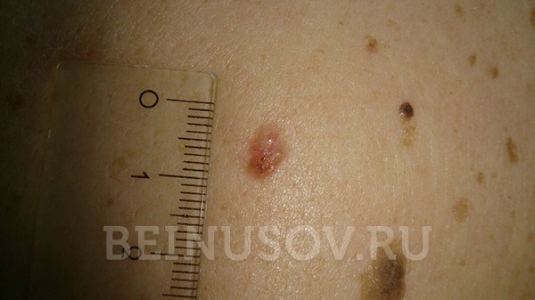
- The scleroderma-like form is very rare and often difficult to diagnose. It is characterized by a lighter and firmer thickening in relation to the surrounding skin.
Fascial training
The superficial sheets of fascia of the various anatomical areas together form the common fascia of the body. The fascial tissue connects the different areas of the musculo-articular system and makes them highly effective. Musculo-fascial trigger therapy targets the fascia that surrounds the sternocleidomastoid muscle when it contracts and causes it to relax. In the long term, the connective tissue becomes softer and muscle damage is reduced. To achieve the best effect, special rollers or balls are used and specific exercises are performed to stretch the fascia in a three-dimensional direction.
The aim of fascia training is to improve and stretch the properties of our thin fascia, the connective tissue covering that surrounds our muscles, tendons and bones. The training increases the metabolism in the tissue, improves blood circulation and the fascia and muscles remain soft and elastic. It is important that you feel a pleasant tension during the training; you may feel some muscle soreness, but no severe pain. Movements should be slow and gradual, repetitive and reaching a maximum.
abnormalities of the fascia
Fascial anomalies can result from open or closed trauma. Malformations can occur. Diseases are rare. The most common fascial pathology is Dupuytren's contracture.
The sliding fascial system plays a large role in the biomechanics of the body. The fascia wraps itself around our muscles like a thin net, sometimes very tightly, and literally sticks to them. Healthy and elastic fascia creates all the conditions for good muscle function by compensating for imbalances in certain fibers. The fascial training of this soft base improves the integrity of the body and strengthens physical fitness. In pathology, this thin network acts as a barrier. Well-trained fascial tissue helps maintain health and responsiveness, and protects muscles from damage.
When and which doctor to see
If you have pain in the ankle area, depending on the possible causes, you should consult a traumatologist, rheumatologist or neurologist. There are a number of medical conditions that require immediate medical attention. These include the following diseases:
- inability to move comfortably due to pain in the joint (on one or both sides);
- An injury that causes the ankle to deform;
- joint pain that occurs at night or after periods of rest;
- Persistent pain that does not go away or decreases and is poorly controlled with medication;
- inability to move the ankle;
- swelling of the joint, lower leg and foot;
- Signs of inflammation - rise in temperature, reddening of the skin, tenderness;
- any other symptoms lasting more than a day.
diagnosis
The diagnosis of ankle injuries is based on the patient's complaints and the symptoms present. However, it is also important to determine the severity of the injury and the extent of damage to the joint. For this purpose, various procedures are prescribed: ultrasound, X-ray, and in complicated cases, computed tomography or MRI of the joint. If gout, rheumatoid arthritis and certain other diseases are suspected, a number of blood tests are required - PCR for infections, determination of rheumatic markers, plasma uric acid levels, coagulation parameters and biochemistry.
Therapy depends on the specific diagnosis, but there are some general procedures that are recommended for all patients with ankle pain. Crutches, restraining bandages, immobilization, and limiting activity will help.
Cold compresses to reduce swelling and pain. Ice packs, cold compresses, and pads for 15-20 minutes up to three times a day or more may help relieve pain.
Above all, relief and rest are necessary to reduce acute inflammation and discomfort. Compression helps immobilize the joint and keep it stable for healing. To improve blood and lymph circulation, an elevated position is helpful; this helps reduce discomfort and promote healing. Crutches, walkers, canes, splints or joint splints, a cast, or orthotics can help mobility.
Supervised physiotherapy and physical therapy are helpful, and massage can be used when acute symptoms resolve.
Non-steroidal anti-inflammatory drugs in various forms - injections, capsules, tablets - are used to relieve pain syndrome. They suppress inflammatory reactions, reduce pain and swelling and help alleviate symptoms. It is taken in an age-appropriate dose for an acute period (up to 5-7 days) or according to a schedule prescribed by the doctor. Dialrapid is considered to be one of the most effective drugs to relieve ankle pain.
In severe cases, glucocorticoids, including intra-articular injections, can be used.
- Only the rectus muscle diverges;
- the lower lateral areas lose tension;
- widening of the ribs, saddle-likeness appears;
- curvature of the waist occurs.
In men, the disease is asymptomatic in the early stages. Clinical manifestations gradually increase. In women, the disease begins abruptly, some time after child birth.
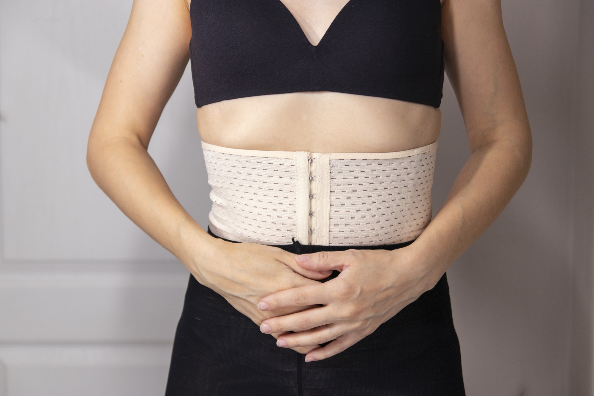
The first visible symptom is a characteristic bulge in the middle of the abdomen in a vertical and rounded shape. When you contract your abdominal muscles, you can clearly see the inner edges of the rectus abdominis muscle.
As the disease progresses, bowel function becomes impaired and many women experience pelvic muscle relaxation, resulting in incontinence during vigorous coughing, sneezing, and other activities that require abdominal contraction.
complications
In most cases, this pathology does not pose a major health risk. However, if left untreated, atrophy of the rectus muscle gradually develops, as a result of which the aponeurosis stretches even more.
The main unpleasant consequence of diastasis is the psychological discomfort and complexes that a person develops. It's embarrassing to go to the pool, sauna or any other place where you have to wear a bathing suit. Some women with this defect no longer want to have children.
Untreated symptoms of gastrointestinal disorders come later. As the disease progresses, hernias form. Internal organs can prolapse, which often leads to severe functional disorders up to and including intestinal obstruction. In women, renal colic and urinary incontinence occur.
Intertrochanteric syndesmosis
The ligaments of the intertrochanteric syndesmosis are the intertrochanteric membrane of the lower leg, the anterior intertrochanteric ligament, the posterior intertrochanteric ligament, and the transverse ligament. The anterior longitudinal ligament of the syndesmosis is 3 times weaker than the posterior ligament.The front longitudinal band is three times weaker than the rear band, while the rear band can withstand a pulling force of up to 30.0±2.3 kg. However, the most stable structure of the syndesmosis is the interosseous membrane, the thickness of which is twice that of the anterior and posterior ligaments combined.
In the case of an injury to the interosseous syndesmosis, the clinical picture is characterized by pronounced and persistent swelling of the lower leg and foot; severe injuries require surgical immobilisation of the tibia due to tibia diastasis and talar instability.

Achilles tendon (AC)
The Achilles tendon (AT) is the strongest and most powerful tendon in the human body, capable of withstanding significant static and dynamic loads. It is the common tendon of the superficial calf muscles and the deep cambium.
AC fibers have a spiral course like a rope, which gives them high strength and at the same time the ability to stretch slightly under physical stress, straightening this spiral and thus cushioning the stress.
Although the strength of AC is considerable, it has its limits: it is around 50 N/mm2.
AC strain (elongation) under stress of 3-5 % is to be regarded as physiological, up to 8 % as harmful. AC strain greater than 8 % will inevitably lead to micro and macro cracks.
The number of vessels supplying the AS decreases proximal to the calcaneus and reaches a minimum at a distance of 4-5 cm from the calcaneal tuberosity, where nutrition occurs only by diffusion from the synovial fluid.
The arch of the foot, together with the plantar fascia and the AC, form a single functional system that absorbs the impact of running and jumping. With a significant flattening or enlargement of the longitudinal arch of the foot (cavus foot), spatial discrepancies in the metatarsal structure, hyperpronation or hypersupination of the foot, the shock-absorbing properties of the AC are reduced and the load on the AC increases accordingly, leading to wear and the development of chronic diseases.
The pathogenesis of the spontaneous cruciate ligament is multifactorial. It is caused by a number of exogenous and endogenous factors or a combination of these factors.
The most important endogenous factor are dystrophic-degenerative changes – in the tendon itself (tendinopathy), their covering (paratendinopathy) and in the tendon sacs (Achilles bursitis, deep and superficial bursitis).
Take the quiz and rate your knowledge: how well did you absorb the material?
Anatomy of the back muscles. What are back muscles and what are they used for?

Diagnoses patients in traumatology and orthopedics. Reads X-rays and performs conservative chiropractic treatments for osteochondrosis and bulging discs. other authors
Precautions

After a botulinum toxin injection, a few rules should be followed:
- Do not take a horizontal position for several hours;
- Visits to baths, solariums or swimming pools are not recommended for a week;
- It is best to avoid strenuous physical activity for a few days;
- It is better to abstain from alcohol for a week.
Botox has been widely used for more than 20 years. It is approved for cosmetic use between the ages of 18 and 65. If you frown all the time, it is an excellent preventive measure against deep wrinkles. After repeated injections, the habit of involuntary frowning also disappears.
'I was hesitant to have the procedure for a long time, but on the advice of a friend I finally decided to do it. I work in a private kindergarten and my job is to entertain the children. Due to my daily facial expressions, I had some wrinkles on my forehead that made it difficult for me to accept my appearance. During the consultation, the beautician advised me to have a Botox treatment. I would like to say that the treatment was completely painless and the effect was immediate - my wrinkles disappeared.'
'For the past year and a half I have been undergoing regular beauty treatments around my eyes. On the plus side I can record the absence of expression lines for about five months and the freedom from pain. The downside, of course, is that Botox takes some getting used to. Not physically, but emotionally. When you walk around young for a few months, you don't want to look older.
'To be honest I haven't met a woman who would dare to have botox at the same age. But I'm a working grandmother and I always want to look good in front of my friends. Due to my age I had a lot of wrinkles on my face and we couldn't do it in one session, I had to come back after 3 months. But the effect was already noticeable on the eighth day after the first injection. As I am very sensitive, the beautician numbed my face and I only felt a light touch. As I am allergic to certain products, I was concerned about the negative effects of the treatment. But fortunately there was none. I want to tell you that you don't have to be afraid to correct your appearance even if you are 60 years old, because beauty is beyond age!
Types and characteristics of molars
In humans, the premolars only break through in childhood with the complete change from milk teeth to permanent teeth between the ages of eight and twelve. The structure of the lower and upper molars is quite different: the upper molars have one root, while the lower molars often have two roots.
The first premolar in the upper jaw is considered the largest. Opposite him is a smaller tooth in the lower jaw. All of these teeth have a long gap on their chewing surfaces.
Features of the maxillary premolars
The coronal part of this type of tooth, located on the upper alveolar process, has a prismatic shape. Its buccal and palatal halves are characterized by a convex surface. There are some differences in dentition between the second and first premolars. They have a special crown and root shape.
The first small dentition on the upper jawbone is characterized by the fact that its palatal surface is dominated by the vestibular part. The contact surface of this unit is characterized by its rectangular shape. He has a cheekbone with two clearly defined bumps. It is the only type of premolar whose root is forked.
The buccal surface of the second representative of this group is more rounded and dominant compared to the palatal part. The main feature of this premolar is that its single root is usually conical in shape. Occasionally the small molars are split.
opinions
A deputy about a management company in Voronezh: 'They collect money but do not provide any services'.
'A neural network must be accompanied by a switch'.
Russian paleoanthropologist, editor of the Anthropogenesis.ru website
Why didn't the officers start with 'MYO!' talked about the new repair program?
After the transport 'reform', waiting for a bus and squeezing into it became a chore.
last comments
Voronezher pensioner lost more than 1.6 million rubles shopping
Dog owners should be forced to pay taxes
Center. parking garages. Evacuation. Revolutsii Prospect, deserted.
Center. parking garages. Evacuation. Revolutsii Prospect, deserted.
A personal experience
Dear readers! We would like to present you our new project 'New Buildings in Voronezh'. We have marked new apartment complexes on an interactive map of the city. If you click on a complex, you will get all the information about it: location, terms of delivery, floor plan, price per square meter, etc. Apartment in the heart of a metropolis or in a sleeping district? With or without furniture? The map provides answers to all these questions and many more. 'Novostroyka Voronezh' offers the opportunity to find out quickly and comprehensively about new residential complexes and to learn how to find one's way in the variety of apartments!
The online edition of 'MYO!
(translated as Moyo! The Straight Line)
Online edition, registered on 12/30/2014. Federal Service for Supervision of Communications, Information Technologies and Mass Media (Roskomnadzor)
EL Registration Certificate No. FS77-60431 dated 12/30/2014.
Founder: LLC Publishing House 'Svobodnaya pressa'.
Irina Bulgakova is the editor-in-chief of the editorial board of 'MYO!'.
Address of the editorial office: Address of the editorial office: 54 L. Ryabtseva Street, Voronezh, 394049
Opinions of the authors of articles published on MOO! Articles posted online, material posted in the Opinions and Popular News sections, and user comments on material posted on the Site do not necessarily reflect the opinions of the editorial staff of MOO!
Do you have any news that interests you?
Call: (473) 267-94-00, 264-93-98. Write to: [email protected], [email protected].
If you have any questions about advertising on the Site, please call: (473) 267-94-11, 267-94-08, 267-94-07, 267-94-06, 267-94-05
- The digital newspaper 'MYO! Plus'
- About Us
- Contact
- site plan
- all messages
- Attractions in Voronezh
- Useful Materials
- rules of communication
- data protection
- tibial fasciitis.
- shin fascia.
- Ankle strap at heel.
- is liquorice.
- longitudinal ectromelia.
- Anatomy of the foot x-ray.
- Anatomy of the joints of the foot.
- Longitudinal soleus muscle.