All patients report pain in the area of the second or third toe. With a neuroma the pain is 'burning and throbbing'. It may be accompanied by numbness in the fourth toe.
- Removal of Morton's neuroma
- Causes of the disease
- metatarsalgia
- Causes of metatarsalgia of the foot:
- causes
- Symptoms of Morton's Neuroma
- diagnosis
- Treatment of chondromyxoid fibroma
- SPONTANEOUS RESORPTION OF METACARIAL AND METATARUS BONES IN THE PRACTICE OF A PEDIATRIC RHEUMATOLOGIST
- Reviewed in.
- Do cytowania:
- To quote:
- Conservative treatment of Morton metatarsalgia (neuroma)
- Surgical removal - excision of the neuroma
- Prevention of Morton's neuroma
- QUESTIONS AND ANSWERS
- Can Morton's neuroma be treated without surgery?
- Which orthoses are required for Morton's neuroma?
- How can I make an appointment for a doctor's visit and surgery for Morton's neuroma?
- What does recovery look like after Morton's neuroma removal?
- Recovery and maintenance phase of metatarsalgia
- Causes of metatarsalgia of the foot
- How is metatarsalgia of the foot diagnosed?
Removal of Morton's neuroma
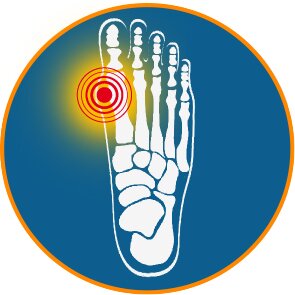
Morton's neuroma or Morton's metatarsalgia is a disease in which fibrous tissue forms around one of the branches of the supporting nerve. The nerve between the third and fourth fingers is most commonly affected, and less often between the second and third fingers. The pathological process develops as a result of irritation and compression of the nerve. The disease manifests itself as a burning, stabbing pain in the sole of the foot, tingling and numbness in the toes. It feels like a pebble has penetrated the shoe.
Statistically, one in three people suffers from Morton's neuroma. Women are affected 8-10 times more often than men - the cause is wearing uncomfortable high-heeled shoes.
The symptoms can often be managed with simple measures, e.g. B. by simply setting the heel to a lower height. However, in some cases, special treatment, including surgery, is required.
Causes of the disease
- shape of the foot. It most commonly affects people who have flat feet, high arches, and other deformities. This leads to instability in the toe joints.
- diseases of the foot. For example bunions, hallux valgus and hammer toe deformities.
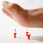
- Shoes that put pressure on the toes and balls of the feet: high heels (especially over 5 cm), tight shoes, wrong size, pointed toes.
- practice of certain sports. For example, when you run or play tennis, the balls of your feet feel increased pressure. And when skiing or climbing you have to wear uncomfortable, tight shoes.
- Foot injuries are also more common among athletes.
Patients often ask the doctors in our clinic this question. Some have heard or read that the nerve becomes 'enlarged' due to this pathology. Others are confused by the ending '-oma' - this is usually the name for benign and malignant tumors. Still others know that neuromas refer to nerve tumors in various parts of the body.
The name Morton's neuroma is misleading because the disease has nothing to do with real tumors. It develops according to the following pattern:

- A branch of the sooty nerve is often compressed and irritated;
- and a fibrous tissue forms around the nerve, a sort of scar;
- an inflammatory process begins.
metatarsalgia
Metatarsalgia is pain in the midfoot area (metatarsus); After a short period of time, patients return to normal activities of daily living, just without pain.
Metatarsalgia in the foot – is pain in the area of the metatarsus of the foot. Metatarsalgia can be mechanical or neurogenic.
Mechanical metatarsalgia is usually caused by overloading of the small metatarsal rays (can occur in hallux valgus, hallux rigidus and other foot diseases) or can be caused by anatomical peculiarities of the tarsal bones (too long second metatarsal bone, Freiberg's disease). A patient with metatarsalgia complains of discomfort in the sole of the foot. This is a fairly common complaint. The pain most commonly occurs under the second, third and fourth toes, and less commonly under the big toe. Metatarsalgia impairs walking and causes constant discomfort in the forefoot area.
Causes of metatarsalgia of the foot:
- damage to the interdigital nerves,
- Lesions of the big toe joints of the 2nd to 5th,
- hallux regidus,
- muscle fatigue,
- aseptic necrosis,
- frequent foot injuries,
- poor blood supply to the feet,
- neuroma,
- synovitis,
- overweight,
- tight shoes,
- augmentation of the metatarsal head,
- arthritis or other osteoarthritis,
- systemic diseases such as diabetes that can cause neuropathic foot pain
- aging, as the fatty layer of the foot thins or atrophies with age,
- Sports in which the arch of the foot is subjected to excessive impact loading (e.g. jogging).
As previously mentioned, the main symptom of metatarsalgia is arch pain, which is most noticeable when walking or exercising. Because the foot supports the core during all activities, chronic arch pain impairs the ability to perform even the simplest activities. Discomfort when wearing shoes and socks.
causes
Excessive stress on the forefoot is the main cause of Morton's neuroma. It can be caused by wearing high-heeled shoes, shoes that are too tight and/or uncomfortable, walking incorrectly, being overweight (e.g. obesity), walking for long periods of time, standing work and sports. Morton's neuroma can develop due to a foot deformity, most commonly flat feet (hallux valgus).
Various injuries to the foot (fractures, dislocations, bruises) can cause Morton's neuroma through direct nerve damage, hematoma compression, or post-traumatic transverse flatfoot. Other triggers are chronic infections of the foot, synovitis or tendinitis of the foot, obliterating atherosclerosis or obliterating arteritis of the lower limbs, the presence of a fatoma at the level of the metatarsal bone.
The above factors act as irritation or pressure on the common nerve of the toes. In response, local nerve sheath thickening and thickening, reactive fiber remodeling, and peri-neural connective tissue hypertrophy occur. Chronic trauma can cause inflammatory infiltration and lead to epineural fusion with surrounding musculoskeletal structures.
Symptoms of Morton's Neuroma
The most characteristic pain occurs in the distal parts of the foot, often in the 3rd and 4th toes. The pain is burning and is sometimes accompanied by shooting pain in the toes. In some cases, patients complain of discomfort and the feeling that a supposed foreign body is entering their shoes. At the onset of Morton's neuroma, the pain syndrome is closely associated with wearing shoes. Patients notice significant relief when the shoes are removed. Over time, these symptoms may subside and then reappear. An exacerbation is more often triggered by wearing tight shoes.
The progression of Morton's neuroma leads to a change in the pain syndrome. The pain becomes constant and increases when wearing shoes of any kind and does not subside, only decreasing when the shoes are removed. Numbness in the fingers occurs. The initially recurrent pain syndrome prompts patients to seek medical attention when the neuroma has reached an advanced stage and conservative treatment methods are ineffective.
diagnosis
The clinical picture is nonspecific and the diagnosis is made on the basis of further examinations. Radiographs show an exophytic mass with distinct festoon-like outlines, usually located in the metaphyseal area and growing toward the epiphysis. The tumor represents a well-defined focus of destruction that can be between 1 to 2 and 6 to 8 centimeters long. The cortical layer over the lesion is swollen and abraded. A sclerotic rim is found around the edge of the mass, and calcareous streaks and a trabecular pattern can be seen in the lesions. If the tumor is located subperiosteally, the cortical layer becomes irregular. Normally there is no periosteal reaction. Periosteal infiltrates are commonly observed in the spinal region.
The radiological appearance of chondromyxoid fibroma may resemble chondrosarcoma, enchondroma, benign chondroblastoma and chondroblastic variant of osteosarcoma, so bone trepanobiopsy is performed to make a definitive diagnosis. In the differential diagnosis, a highly aggressive malignant chondrosarcoma should first be excluded.
Macroscopically, the tumor tissue is a lobular mass with a dense, bluish-gray or grayish-white texture that is well demarcated from healthy tissue. It may resemble hyaline cartilage. Microscopic examination reveals a chondroid or mucous intercellular substance with elongated or stellate cells inside. Connective tissue areas are usually found in the interlobular spaces. Small vessels can also be found there. In addition to collagen fibers, the tumor tissue also contains osteoid and multinucleated giant cells.
The structure of the lobules can vary greatly within a single tumor. A variety of fibroblasts can be seen, both large and small, with some cells having 2 or 3 nuclei. These cells are located primarily at the periphery of the lobules. In one out of three cases, the cells of a benign chondromyxoid fibroma have an atypical structure and resemble those of a malignant chondrosarcoma. In 10 percent of cases, foci of necrosis are visible in the biopsy material.
Treatment of chondromyxoid fibroma
Treatment is exclusively surgical. The operation is routinely performed in the oncology department. Marginal bone resection followed by plasticization of the defect with autografts or allografts is the most effective surgical procedure to minimize recurrences.
In some cases, curettage (a surgery in which the tumor is scraped out with a curette, a spoon-like surgical instrument) is performed for this condition. However, this is not the preferred approach because it increases the likelihood that altered tumor cells will remain in the affected area and the chondromyxoid fibroma will recur over time.
SPONTANEOUS RESORPTION OF METACARIAL AND METATARUS BONES IN THE PRACTICE OF A PEDIATRIC RHEUMATOLOGIST
This article presents the observation of an 11-year-old boy diagnosed with spontaneous resorption of the metacarpal and metatarsal bones. The authors note the rarity of this pathology, cite literature data on spontaneous bone resorption and describe the features of the clinical picture and the need for a differential diagnosis with juvenile rheumatoid arthritis.
Keywords: Spontaneous metacarpal and metatarsal resorption, differential diagnosis.
(Vopros of contemporary paediatrics. – 2008; 7(3):113-116)
1) Eisenstein DM, Poznanski AK, Pachman LM Torg Osteolysis Syndrome. At the. J. Med. Genet. 1998; 16(80): 207-212.
2 Al Aqeel A, Al Sewairi W, Edress B, et al. Inherited multicentric osteolysis with arthritis: a variant resembling Torg syndrome in a Saudi family. At the. J. Med. Genet. 2000; 3 (93): 11-18.
3) Zankl A., Pachman L., Poznański A. et al. Torg syndrome is caused by inactivating mutations in MMP2 and is allelic to NAO and Winchester syndrome. J. Bone Miner Res. 2007; 22 (2): 329-333.
4) Rouzier C, Vanatka R, Bannwarth S et al. A new homozygous MMP2 mutation in a family with Winchester syndrome. Clin. Genet. 2006; 69 (3): 271-276.
5. Singh JA, Williams CB, McAlister WH Osteoliza kości łopatkowej, zapalenie błony maziowej and krótkie czwarte śródręcze u sióstr: nowy zespół? At the. J. Med. Genet. A. 2003; 30 (121): 118-125.
6. Faber MR, Verlaak R, Fiselier TJ et. al. Inherited multicentric osteolysis with carpal-tarsal localization, mimicking juvenile idiopathic arthritis. Eur. J. Pediatr. 2004; 163 (10): 612-618.
Reviewed in.
Do cytowania:
Zholobova E., Bobyleva V., Mikhaleva G. SPONTANEOUS RASTS OF METACARPAL AND METATARPAL BONES IN THE PRACTICE OF A CHILDREN'S REUMATOLOGIST. Issues in contemporary pediatrics.. 2008;7(3):113-116.
To quote:
Zholobova E., Bobyleva V., Mikhaleva G. SPONTANIC OSTEOLIZATION OF SUPERSTAR BONES IN THE PRACTICE OF A CHILDREN'S REUMATOLOGIST. Current Pediatrics. 2008;7(3):113-116.
Article appears in print
Conservative treatment of Morton metatarsalgia (neuroma)
Treatment of Morton metatarsal neuroma in the sole of the foot is individual and depends on the cause of the disease and the severity of the symptoms. In the initial stages, conservative treatment is recommended:
- Relieves pressure on the foot – avoid tight shoes in favor of comfortable or low-slung orthopedic shoes
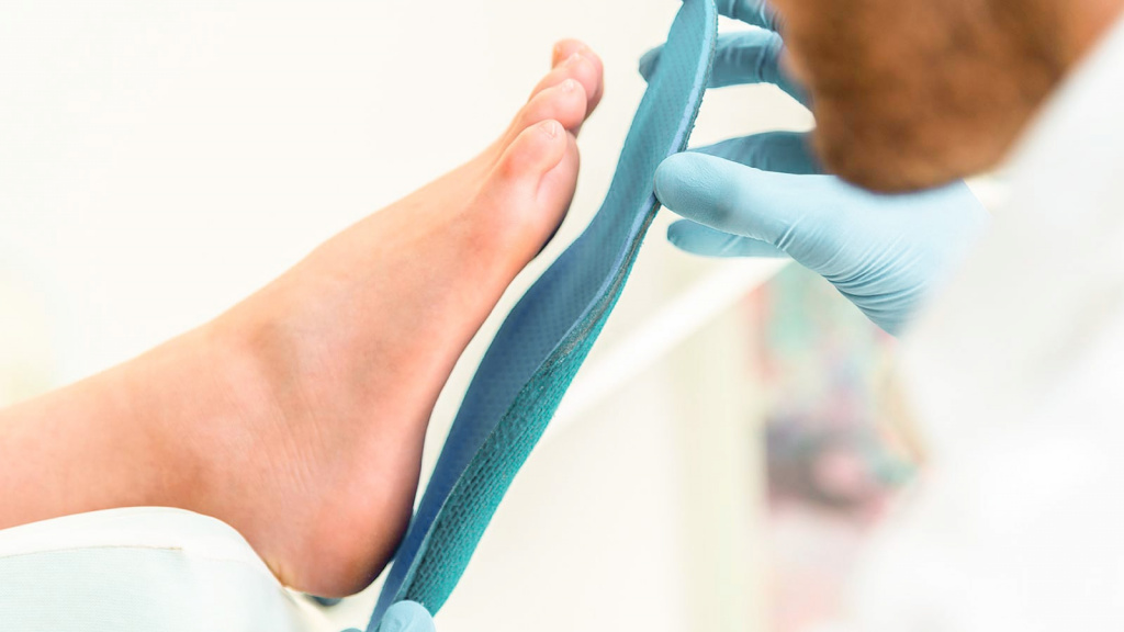
- Use of insoles and metatarsal lifters - orthopedic insoles placed under the ball of the foot and secured to the toes with elastic bands or silicone rings to spread the metatarsal bones and create an anatomically correct foot curvature
- Wearing shoes with heel support.
All of these methods have one goal: to reduce the pressure on the damaged nerve trunk. As a result, the further progression of the disease is stopped and the pain syndrome is reduced.
Even more effective is the constant wearing of orthoses specially made for the patient. Such a solution is capable of:
- to reduce the load on the forefoot and normalize the condition of the transverse arch;
- to reduce the pressure of bones and ligaments on the modified nerve trunk;
- to avoid or eliminate soft tissue inflammation, including nerve involvement;
- restore the anatomy of the foot to improve gait and bring it closer to normality.
Drugs used to treat Morton's neuroma do not have a clear therapeutic effect. Patients may be prescribed several compresses, NSAIDs, ointments or gels, but these do not solve the problem. Drug treatment improves the condition, but does not eliminate compression of the nerve.
Physiotherapeutic treatments also only treat the symptoms and the severity of the disease. They are only moderately effective:
Surgical removal - excision of the neuroma

Let's take a look at the surgical treatment of Morton's neuroma of the foot. The current level of development in orthopedics allows effective and rapid removal of masses at any stage of neuroblastoma formation.
The thickened nerve is severed under local anesthesia. The orthopedist decides individually whether access to the nerve should be from the back of the foot or from the sole side. An incision is then made into the soft tissue to visualize the nerve and the thickened part of the nerve is excised. The tissue is then sewn shut and covered with a sterile bandage.
The entire operation to remove neuroblastoma takes no more than 20 minutes. A recurrence of the disease is ruled out. The function of the foot and the sensitivity of the toes are completely restored. Just 2 weeks after the operation, patients are fully fit for everyday life again, and after 1.5 months they can do sports safely.
In the experience of practicing surgeons, patients are more likely to be anxious and search for information about the cure for Morton's neuroma on their own, rather than receiving expert advice and effective treatment. Surgical techniques have now improved so much that the disease can be eliminated quickly and with quality guarantees.
Prevention of Morton's neuroma
Morton's neuroma often develops in people who wear tight shoes with high heels and narrow toes. Therefore, tight and uncomfortable shoes as well as poor quality footwear should be avoided. The shoes should be made of natural materials, have good supination, fit in size and be comfortable to wear.
The development of a neuroma can be prevented by timely treatment of foot diseases:
It is also necessary to treat chronic diseases that can favor the development of a neuroma: obesity, diabetes, atherosclerosis, arteritis.
During heavy physical and standing work, which involves significant stress on the legs, regular foot massages, foot baths with herbal decoctions and sea salt are necessary.
QUESTIONS AND ANSWERS
Can Morton's neuroma be treated without surgery?
Morton's neuroma is a disease of the foot in which there is a thickening in the area of the longitudinal nerve. In the early stages of the pathological process, the disease can be treated without surgery using conservative methods: wearing special shoes and orthoses, exercise therapy, physical therapy, foot and lower leg massage and steroid injections. Indications for surgery for Morton's neuroma: failure of conservative treatment, persistent severe pain in the toe area. The main surgical treatments are neurolysis and neurectomy.
Which orthoses are required for Morton's neuroma?
In the case of Morton's neuroma, special insoles, orthoses and spinal insoles are recommended. These aids ensure even pressure distribution in the forefoot area, reduce the likelihood of blisters forming on the sole of the foot and prevent the development of a deformed foot. The following types of insoles are available: arch-supporting, arch-forming and pressure-relieving.
How can I make an appointment for a doctor's visit and surgery for Morton's neuroma?
To make an initial doctor's appointment, call the clinic's call center. The call center staff will then arrange a day and time for the consultation. During this visit, the orthopedist/traumatologist will determine whether surgery to remove Morton's neuroma is necessary. All necessary contacts (phone numbers, addresses) are listed on the clinic's website.
What does recovery look like after Morton's neuroma removal?
Rehabilitation after surgical treatment of Morton's neuroma usually proceeds without complications. After the operation, special footwear (forefoot relief, firm sole) should be worn for 1-2 weeks. It is advisable to keep your foot elevated most of the time. The sutures should be bandaged in a timely manner and infections should be avoided. It is advisable to start exercising immediately after surgery to restore mobility to the toes.
Recovery and maintenance phase of metatarsalgia
After a period of acute pain, the metatarsalgia patient should gradually return to more vigorous activity to allow the muscles and joints to heal and strengthen without causing further damage. The goal of this phase is to restore the normal biomechanics of the foot and relieve pressure and pain.
During this phase, the patient should perform a series of exercises designed by the physical therapist to improve the strength, stability and range of motion of the foot.
This phase can be difficult to maintain, especially in professional or highly motivated athletes who want to return to strenuous activity as quickly as possible. It is important that the relationship between patient and therapist is based on trust. Patients should understand that their body needs a period of reduced exertion.
become a member Pravda.ru Telegram channel with the opportunity to express your own opinion)
Add HealthPravda to your sources Yandex.News or News.Google
We also invite you to join our communities Zen, VKontakte, classmates, YouTube.
Causes of metatarsalgia of the foot
Not all causes of foot metatarsalgia are known. In addition to frequent walking, wearing uncomfortable shoes or high heels is also a cause. Being overweight also contributes to metatarsalgia. Rheumatoid arthritis, osteoarthritis or gout can also trigger metatarsalgia.
The main symptom of metatarsalgia is pain in the area of the metatarsal bone under the sole of the foot. Metatarsalgia may or may not be accompanied by bruising, swelling or inflammation. Symptoms may appear quickly or develop over time. They include:
- Pain in the sole of the foot. The pain can be stabbing, aching or burning. The pain may increase when running or walking.
- Numbness or tingling in the toes
- a pebble-like feeling in the shoe.
If you experience any of these persistent symptoms, you should see your doctor. Untreated metatarsalgia causes pounding in the toes and causes limping and pain in other parts of the body, including the lower back and thighs.
How is metatarsalgia of the foot diagnosed?
The podiatrist begins the examination of metatarsalgia by asking you about your symptoms. He or she will examine the foot. X-rays may be necessary to rule out fractures. In other cases, an MRI scan of the foot is recommended to evaluate the soft tissues surrounding the metatarsophalangeal joints.
Metatarsalgia can usually be treated without surgery. Your doctor will recommend a metatarsal pad, surgical shoes, or shoe inserts to relieve pressure on the painful part of the foot. Sports shoes or shoes with a wobbly sole may also be recommended. Other useful tips include:
- Choose shoes with a good sole, a wide toe box and a low heel
- Avoid walking barefoot.
- Soak your feet and use a pumice stone to remove corns.
If these remedies do not relieve metatarsalgia, an injection or surgery is necessary to address the underlying cause of the pain.
Read more:- metatarsalgia.
- Morton shoe inserts.
- Morton's Neuroma Ointment.
- Treatment of metatarsalgia of the foot.
- Cracked metatarsal.
- The tarsal and metatarsal bones.
- metatarsal bones.
- metatarsal and metacarpal bones.