An unhealed fracture is a pathological process characterized by a lack of bone marrow formation at the site of injury. There are three types of fractures: delayed consolidation, nonunion, and nonunion. These pathologies are considered stages of one and the same process and can proceed sequentially without timely treatment.

- Leg injuries (ankle, foot and toes)
- Acute leg injuries
- Pathological anatomy
- diagnosis
- TAYLOR DEFORMITY
- PHASES OF THE OPERATION
- An individual approach
- The main stages of rehabilitation massage:
- indications
- Surgical technique
- Diagnosis
- Treatment of delayed consolidation
- Fracture of the phalanx: causes and symptoms
- What complications can occur after a broken toe?
- causes
- Other possible causes of growth plate damage
- signs
- Treatment of a fracture of the 5th metatarsal bone
- Fracture of the 5th metatarsal, how long do you have to wear a cast?
Leg injuries (ankle, foot and toes)
Almost everyone suffers from minor injuries to their toes, feet, or ankles that cause pain or swelling.
Ankle, foot, or toe injuries most commonly occur during pregnancy:
In children, foot injuriesIn children, foot, leg and ankle injuries most commonly occur from falls or while playing sports. Foot injuries often occur in sports such as jumping, soccer or basketball that require faster and sharper changes in direction. Leg injuries in children require special attention because injuries to the bones surrounding the joint can affect the child's physical development as they grow.
The risk of leg fractures, ankle and foot injuries increases in older people. Natural aging weakens bone strength and muscle mass is lost. The risk of injury also increases due to loss of vision and the ability to maintain balance.
Most minor injuries to the ankle, foot, or toe are not serious and will heal on their own. Treating the injury at home can help relieve leg pain and other unpleasant symptoms until they go away.
Acute leg injuries
Acute leg injuries and trauma can result from a direct blow, a penetrating wound, or a fall, as well as from twisting, spraining, entrapment, or incorrect bending of the limb. In such a case, the leg pain can be severe and stabbing. Bruising and swelling may occur shortly after the injury. Acute injuries to the leg can take very different forms:
- Bruises (bruises) . After an ankle injury, the bruise can spread to the toes due to gravity.
- Puncture wounds. Puncture wounds on the leg can come from sharp objects - nails, buttons, knives, needles, animal teeth. A puncture wound to the leg poses an increased risk of infection because it is difficult to clean and the wound emits heat and high moisture - conditions that promote the growth of bacteria. A common possibility for infection is a foot wound where the puncture occurs through the sole of a shoe.
- Ligament injuries that support the joints;
- tendon injuries, of which rupture of the Achilles tendon is the most common;
- joint injuries, joint dislocations;
- Muscle strains. Muscles in the foot and ankle can be painful due to a sprain. A severe strain can lead to a torn muscle fiber;
- Broken bones. The most common fracture is a broken toe or ankle;
- Dislocation of a boneA dislocation is a condition in which a bone moves out of its normal position in relation to other bones. This can occur in the joints of the kneecap, hip, finger, elbow, or shoulder;
- severe traumaA severe trauma causing compression syndrome.
Pathological anatomy
The histological changes in Marchfoot are characterized by patchy resorption of bone substance and the formation of new bone structures. There is also a process of periosteal and less pronounced endosteal bone formation. The bone marrow is displaced by undifferentiated fibrous connective tissue. The reconstructed bone is rich in cellular elements but poor in minerals and has a 'loose' spongy substance. In the long term, a sclerotic, compact bone forms at the remodeling sites.
There are two forms of MS: the more common acute form, which usually occurs on the 2nd to 3rd day after walking or overexertion and has clear clinical signs, and the primary chronic form, which develops gradually. In both cases, without an acute forefoot injury, there is severe pain, the inability to strike the foot firmly, and limping. There is limited hard swelling and soft tissue edema on the dorsal surface of the foot over the diaphysis of the 2nd and 3rd and less commonly the 4th and 5th metatarsals (the 1st metatarsal is almost never affected). Metatarsal bone is almost never affected. The skin is usually unchanged, but hyperemia, cyanosis, and an increased venous pattern sometimes occur.
diagnosis
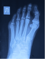
Radiological examination is crucial for the diagnosis of March's foot as well as for determining the course of the disease, distinguishing several phases of the disease. Depending on the stage, the radiological appearance varies from normal to severe bone remodeling (see loosening zone). The zone of metatarsal rearrangement is located in a typical area: the II-IV metatarsals usually in the middle or on the border of the middle and distal thirds of the diaphysis, and the V metatarsals on the border of the middle and proximal thirds. Remodeling of the bony structure of the metatarsal bones begins after the onset of pain, but it sometimes takes 4-8 weeks for visible changes to appear on the x-ray.
Radiological There are four different phases of March foot. The first phase is characterized by initial signs of remodeling of the diaphysis and surrounding periosteum. The entire diaphysis is crossed transversely or slightly obliquely by a 1 to 3 mm wide band of homogeneous calcification with indistinct contours. This zone, consisting of calcified osteoid tissue, gradually merges into the bone structure of the diaphysis and the surrounding periosteal layers. Simultaneously with the area of translucency around the diaphysis of the metatarsal bone, delicate fusiform periosteal layers appear, resembling ossified periostitis (see) or periosteal callus (see). These mucoid periosteal layers can be localized or cover almost the entire diaphysis. In the initial phase of the disease, the light area often extends into the periosteal layers. At this stage, the radiological appearance is very similar to a periosteal fracture.

TAYLOR DEFORMITY
Taylor deformity is a deformity of the fifth toe. This deformity is also known as 'shoemaker's foot' because cobblers sit cross-legged while working, leaning on the outer parts of their feet.
Taylor's foot deformity is a so-called bunion on the outside of the foot at the prominence of the head of the 5th.
As a rule, patients not only suffer from the cosmetic defect, but also from pain when wearing shoes and often develop bursitis, tenderness (redness) and hyperkeratosis (ossification) in the area of the 'hump'.
This deformity is caused by a hereditary predisposition based on hypermobile syndrome. As transverse flatfoot progresses, the 5th metatarsal tilts outwards and the little toe tilts inwards.
PHASES OF THE OPERATION
- A 2-3 cm long incision is made on the outer side surface of the foot.
- Exostomectomy (removal of the 'ankle').
- Osteotomy (sawing) of the 5th metatarsal bone. Different types of osteotomy are used (AUSTIN, SCARF, etc.). The type of osteotomy performed depends on the bone structure and the degree of Tayloristic deformity.
- Displacement of the bony fragments of the 5th metatarsal bone relative to each other.
- The immobilization of the bone fragments after the osteotomy is usually done with 1 titanium screw.
- Sewing the bag and seams to the skin.
- Sterile bandage with betadine and fixation bandage.
- First of all, a consultation with an orthopedist is required to clinically examine the feet, make a final diagnosis, select surgical treatment and obtain comprehensive information about surgery and rehabilitation. X-rays of the feet should be taken in 2 projections (straight and lateral).
- Standard examination before surgery (blood and urine test, chest x-ray, ultrasound of the veins of the lower extremities, consultation with the family doctor).
- The operation takes place on the day of admission to the hospital.
- Anesthesia: in most cases spinal anesthesia (epidural anesthesia).
- The duration of the procedure starts at 30 minutes. Up to 1 hour (for surgery on 2 feet).
- If Taylor deformity is present in both feet, it is advisable to operate on 2 feet at the same time.
- The titanium screws do not need to be removed! Titanium is an inert material for bones. The screws do not cause any complications, do not interfere and do not 'ring'.
- The hospital stay is only 1-2 days.
- The pain syndrome after surgery is negligible.
- Walking is allowed the day after surgery, but only in special postoperative orthopedic shoes.
- Wearing special postoperative orthopedic shoes for 3-4 weeks after surgery.
- Crutches and a cast are not required.
- Antibacterial, anti-inflammatory and anticoagulant treatment is mandatory after surgery.
- Post-surgery bandages are applied twice a week.
- The stitches are removed 14 days after the operation.
- To speed recovery, you should apply cold applications to your feet, limit walking, and elevate your feet in the first few weeks after surgery.
- Walk in normal footwear (non-slip shoes) for 4 weeks after surgery.
- Wearing orthoses (preferably custom-made) is mandatory for 4 weeks after surgery.
- Wearing shoes with heels no earlier than 3-4 months after the operation, with a recommended heel height of no more than 3-4 cm.
- Sporting activities are possible 4-5 months after the operation.
An individual approach
'A lot depends on the condition of a person's ligamentous-muscular apparatus,' says Maria Istifeeva. – In children, for example, the ligaments are more flexible, so if the injury is treated correctly, the rehabilitation phase will go faster than in most adults. The same applies to people who play sports and are well trained. In most cases 15 sessions are sufficient, in some cases 20 sessions, maximum 25′.
The number of massage sessions and their duration are determined individually, depending on the patient's age, the duration of the immobilization of the limbs and, of course, the type of injury.
The main stages of rehabilitation massage:
- Step 1: Drainage massage to reduce swelling is carried out for 2-3 days.
- Level 2: Massage to eliminate muscle tension and improve blood circulation.
- Level 3: Ligament radiation to improve ligament flexibility, both through massage and exercise.
CNMT is used for rehabilitation after fractures of the shoulder, forearm, fingers, wrist and elbow, rotator cuff tears, ligament and muscle tears, hip, shinbone and foot fractures.
indications
Osteosynthesis is used for complex fractures of the metatarsals (especially the first and fifth metatarsals) and fingers when conservative treatment is inadvisable or ineffective (including false joints). This is necessary if the fractures are severely displaced and the soft tissues between the fractures are at risk. An open fracture or risk of skin injury from bone fragments may also be an indication for surgery.
Preoperative preparation is a series of examinations to assess the damage, select the best method of surgical intervention, identify any contraindications and diagnose the condition of the body as a whole. The minimum number of examinations includes:
- X-rays of the bones of the foot and, if inconclusive, CT or MRI;
- Specialist consultations: surgeon, anesthesiologist, general practitioner;
- Laboratory tests: general blood and urine tests, blood chemistry, coagulation, infections and blood group tests;
- ECG and chest x-ray.
If necessary, doctors will order additional tests.
Surgical technique
Osteosynthesis is carried out under anesthesia. The doctor cuts the fracture site and exposes the bone. The bone fragments are then put back together and fixed with a wire. If necessary, the damaged tissue is sutured, the wound is sewn closed and covered with a sterile bandage. If necessary, a drain can be placed in the wound.
Take advantage of this unique opportunity for a free consultation about elective surgery. Learn more.

Diagnosis
The orthopedist and trauma surgeon diagnoses and treats these clinical pictures. First, the specialist clarifies the patient's specific concerns, determines the type of injury, develops a treatment plan, determines any limitations and carries out the necessary examinations. An essential prerequisite for a successful diagnosis is the availability of x-ray images, reports and statements.

Only 5-8 months after the injury can it be determined whether the patient has delayed fracture consolidation and nonunion. A nonunion is suspected at a later stage.
During the examination, the external condition of the hand/foot is checked, the temperature is checked, the presence of abnormal mobility and other symptoms are determined. In addition, the doctor assesses the mobility of the joints and determines the size of the paired limbs. Undoubtedly, a CT scan and an X-ray are necessary for a successful diagnosis. Based on the findings, the following problems are diagnosed:
- Delayed consolidation. The callus is barely visible. It serves as a ligamentous connection in fractures and slightly covers the fracture line. Areas without adhesion can be recognized by the thickening;
- Unhealed fracture. Sharp parts of the fracture have been ground down due to bone resorption. The bone marrow is not or only faintly visible. The fracture line is visible along its entire length;
- Pseudarthrosis. Thickening of the fracture ends and significant sclerotic abnormalities in the fracture area. The locking plates overlap the intramedullary canals.
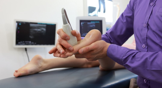
In addition, the patient may be referred for an X-ray and ultrasound examination of the vessels. Such diagnostics are necessary if there is suspicion of circulatory disorders.
Treatment of delayed consolidation
If delayed consolidation is diagnosed early, treatment with conservative methods can achieve the desired result. A plaster cast or orthopedic bandage is applied to stabilize the limb. The duration of application corresponds to the time the bone fragments need to heal. In addition, UWT, calcium electrophoresis, ultra-high frequency and electromagnetic therapy are recommended to aid consolidation. Doctors often use substances that promote anabolic processes in the body.

The significant advantages of conservative treatment include minimal risk of tissue damage and a safe rehabilitation period.
Disadvantages include prolonged immobilization, which can result in loss of muscle mass in the limb and reduced mobility.
If conservative treatment is unsuccessful or it is necessary to prevent the above consequences, surgical treatment is carried out. This consists of the following methods:
- Surgical repositioning of bone fragments. Various fixation constructs can be used: pins, screws, dynamic orthopedic plates, and external fixators. These are used when there is no noticeable displacement and axis shift;
- Bone grafts. Bone grafts taken from the patient are used. Most commonly, part of the hip bone is used. By transplantation of a cheekbone, the conversion of the callus into normal tissue is achieved;
- Activation of osteogenesis. This is done through tunneling and decoration. In the first case, tunnels are created to trigger bone formation. In the second case, an acetabulum is formed in the affected area, consisting of an impressive number of bone fragments connected to the periosteum.
Fracture of the phalanx: causes and symptoms
The toe bones of the phalanx are most often injured when dropping a heavy object, during a strong impact, or when the foot is accidentally turned upside down. In some cases, fractures are caused by conditions such as osteomyelitis (bone infection), diabetes, cancer and osteoporosis.
Most fractures are stress fractures: a microfracture in which the skin does not break and the bone is not displaced. Fragmentary fractures are less common: the bone breaks in several places. The diagnosis of an open fracture is simple: bony protrusions can be seen. The correct assessment of the severity of the injury determines the appropriate treatment.
The main symptoms of a broken toe include:
- visible swelling;
- unbearable pain;
- deformation of the toe bone;
- Bruise; Bruise;
- Crunching when you try to move your foot;
- tingling, cold, numbness;
- An open wound with bleeding.
What complications can occur after a broken toe?
Don't think that a toe injury is harmless. A number of problems can arise after an injury. If there is a hematoma, the nail can be removed. If there is irregular tissue fusion, surgical intervention is required: an osteotomy is performed to correct the joint and bone deformity.
There is also a risk of infection if the skin around the broken finger becomes inflamed. Redness, swelling, pus, soft tissue and fever are signs of infection. In this case, antibiotics are essential.
To avoid the consequences of a rupture, it is important to consult a qualified specialist. Not only podiatrists and orthopedists, but also osteopathic doctors and physiotherapists diagnose and treat injured limbs. Specialists make their diagnosis after examining and analyzing x-ray images. In some cases, CT, MRI, ultrasound and bone scans are also required.
causes
Although growth plate injuries are usually associated with acute trauma (fall or blow to the limb), chronic trauma resulting from excessive and frequent stress can also cause an injury. Such injuries to the growth plate can occur, for example, in athletes: gymnasts, track and field athletes, baseball players.
Some studies of childhood injuries indicate that growth plate injuries are caused by falls on the playground or from chairs. Sports such as football, athletics and gymnastics account for a third of all injuries. Other physical activities such as cycling, sledding, skiing and inline skating are responsible for a fifth of all growth plate fractures. Injuries from car, motorcycle and similar traffic accidents account for only a small percentage of growth plate fractures.
If a child has pain after an acute injury or overexertion that persists or decreases with a change in physical activity, or if localized pain occurs, medical advice is required. Under no circumstances should the child move with pain. Children who play sports often experience some discomfort when they have to perform new movements. In some cases, the occurrence of certain symptoms is quite predictable; Nevertheless, any complaint of a child deserves attention, since some injuries, if not properly treated, can lead to irreversible changes and interfere with the normal growth of the bones of the injured limb.
Although most growth plate damage results from injury while playing or playing sports, there can also be other causes of growth plate damage (such as bone infections) that can interfere with the normal growth and development of bones.
Other possible causes of growth plate damage
Child abuse can cause bone injuries, especially in young children whose bones are just beginning to grow.
signs
- Inability to continue playing after an acute injury due to pain.
- Decreased ability to play for long periods of time due to persistent pain following an injury.
- Visible deformity of the child's arm or leg.
- Severe pain and inability to move after the injury.
Once the circumstances of the injury are clarified, the doctor will order x-rays to determine the type of fracture and develop a treatment plan. Since growth plates do not have the same density as bone, they are not visible on X-ray images and are defined as gaps (fissures) between the metaphysis and epiphysis of the long bone.1 Since growth plates are poorly visible on X-ray images, an X-ray image is used for image comparison two limbs recommended.
Magnetic resonance imaging (MRI) can show tissue changes clearly enough that it can be used to diagnose growth plate defects. In some cases, other diagnostic methods such as computed tomography (CT) or ultrasound may also be used.
Treatment of a fracture of the 5th metatarsal bone
First aid for a fracture of the 5th metatarsal bone looks like this:
1. Restriction of mobility of the injured limb - a pillow should be placed under the foot, and in no case should you step on it or try to straighten it yourself. Self-massage is also forbidden!
2. Cold compress – wrap a few ice cubes in a bag and a tissue and place a cold compress on the bruised area. Leave on for 10-15 minutes, then take a 5-minute break and switch to a new compress. 3.
3. After cooling the leg for an hour, an elastic bandage is required. The bandage should not be too tight. If toes become numb or cold, remove the bandage and repeat the process.
After initial care, the injured person should be taken to a trauma center for an accurate diagnosis, because if there is a fracture, a cast is required.
Treatment can be:
– conservative,
– surgical
Fracture of the 5th metatarsal, how long do you have to wear a cast?
A plaster cast is rarely used if the fracture is not displaced in an adult, because every specialist knows that this would be very uncomfortable for the patient. Yes, even non-serious injuries usually heal quite quickly on their own (traumatic cases).
In adults, a bandage is usually sufficient to keep the foot in the correct position (especially if the cause is stressful) and to limit the stress on the foot. Children, on the other hand, almost always have to have a cast put on because it is difficult to explain to small, impetuous children that they are not allowed to run or jump for a while.
The duration of the cast depends on the severity of the injury and is between three and six weeks. This is followed by rehabilitation, which consists of a gradual increase in the load on the foot, wearing special insoles and a series of gymnastic exercises necessary for the development of the foot.
Severe heel pain – causes and treatment
Read more:- Metatarsal tarsal bones.
- Cracked metatarsal.
- Fracture of the 5th metatarsal.
- Metatarsal amputation.
- tarsal bones of the foot.
- The lateral ankle is.
- bones of the foot.
- tibia and fibula.