Stress fractures of the metatarsal bones are initially rarely seen on radiographs and do not become apparent until 5-6 weeks after the onset of symptoms, when bony callus formation is evident. Before that, the diagnosis can be made by MRI or scintigraphy. Stress fractures of the 2-3 metatarsal bones usually occur at the diaphyseal or cervical level. These fractures often occur with a sudden increase in physical exertion, e.g. B. in military recruits during long marches. These fractures are therefore also referred to as 'march fractures'. Ballet dancers who frequently stand on tiptoe can sustain a stress fracture at the base of the second metatarsal.
- bone fractures of the foot
- signs
- to form
- causes
- Layout and function
- skeleton
- The pelvic girdle.
- Development of the bones of the lower border of the limb
- Jones fracture (stress fracture of the metadiaphysis of the 5th metatarsal)
- Metatarsal base fractures and Lisfranc-type injuries
- costs of the services
- opinions of doctors
- Cookie Settings
- Cookie Settings
- Human tarsal anatomy information:
- Which doctors should you see for a tarsal exam?
- Instead of a summary
- treatment methods
- recovery period
- definition
- Tarsal Sinus Syndrome - Treatment
- conservative therapies
- surgical treatment
- Surgery for a metatarsal fracture
- Percutaneous fixation with spokes
- Open reduction of a fracture
- Rehabilitation after a metatarsal fracture
bone fractures of the foot
Fractures of the foot bones are a relatively common injury. Up to a third of all closed fractures of the foot are fractures of the foot bones.
The anatomical structure of the foot is complex. The foot consists of three parts: the tarsus (7 bones), the metatarsal (5 bones), and the phalanx (14 bones). The bones of the foot are connected by a large number of ligaments and joints.
The most common foot fractures are:
- anklebone fracture,
- heel bone fracture,
- heel bone fracture,
- fractures of the toe pontics,
- fracture of the elbow,
- tarsal and metatarsal fractures,
- heel bone fracture,
Fractures in the foot can result from direct impact (e.g. a heavy object falling onto the foot from a height or a strong blow to the foot bones) - this is known as the direct trauma mechanism. If the foot is pinched on all sides and a sudden twisting motion is performed around the shinbone (usually to free the pinched leg), this motion can cause both a tibia fracture and a broken foot bone.
Fracture of the talus - more common.
Fractures of the calcaneus account for 0.5-1 % of all foot fractures but are the most severe. They result from excessive axial loading of the foot (fall from height while standing straight) or from sudden dorsiflexion of the foot. Isolated fractures of the posterior spinous process and fractures of the talar neck and talar shaft are common.
Fractures of the calcaneus occur in 4 % of all fractures. The mechanism of fracture is usually direct: a fall from a great height onto the heel. The heel bone is pushed into the calcaneus and a split occurs. Both heel bones are often broken at the same time.
Isolated fractures of the heel bone are usually the result of direct trauma from dropping heavy objects on the dorsum of the foot.
signs
The main symptom of a broken foot is pain. The intensity of the pain can vary depending on the location of the fracture. As the pain increases, the movement of the foot is restricted. There is swelling, swelling, redness, and bruising at the fracture site. The swelling and pain can later spread to the whole foot (especially in the case of humerus fractures and multiple metatarsal fractures).
to form
Fractures of the foot are classified according to the location of the fracture: talus fractures, calcaneus fractures, tarsal fractures, metatarsal fractures, and pontine fractures.
Fractures of the ankle bone are divided into: femoral neck; Shaft; posterior process of the talus.
Fractures of the heel bone are divided into: extra-articular, intra-articular, multi-articular and compression fractures.
Depending on the height of the fall, there are simple or crush fractures, and the position of the foot at the time of impact determines the direction of the fracture line and the displacement of the fragments
causes
Traumatic fractures result from falling from a great height, dropping heavy objects on the foot, overrotating the foot, clenching the foot, being hit by a car wheel on the phalanges and metatarsals.
Depending on the height of the fall, there are simple fractures or fragments and multiple softened fractures, with the position of the foot at impact determining the direction of the fracture line and the displacement of the fragments.
Pathological fractures are possible with osteoporosis, bone mineralization disorders, arthrosis and tumor damage.
The most common cause of foot fractures is a fall while high jumping or a blow to the foot.
Layout and function
The following is a list of the most important parts and explains what bones make up the human lower leg girdle and what vital functions they perform.
The general function of the musculoskeletal system is to move, balance and provide support. Children and adults who have lost part of their foot, ie the lower leg, have a particularly difficult task as natural stability becomes problematic for them. This problem can be solved with the help of modern prostheses. These are made by the company 'I want to walk'. The center not only provides prostheses, but also helps in the rehabilitation and social adjustment of the child.
List the major parts of the human body from the spine down.
skeleton
The obvious fact remains - every human being has two legs. However, their construction is not as simple as it might appear at first glance. Depending on the development of the components, the gait is formed, healthy or painful states are conditioned, and the joints are susceptible to disease. Any bone can cause a lot of problems if misaligned (pathologies usually appear soon after birth, during fetal development, or after trauma). However, many defects can be corrected with massage, electromagnetic pulses, and surgical interventions at a young age.
The free wreath skeleton of the lower limbs consists of:
Below we will list all the components in more detail and define their meaning and function. But first, let's present them visually:
The pelvic girdle.
This is the top panel that flows seamlessly into the back. It is actually a combination of several joints. Its main function is locomotion, i.e. movement through space. It is worth noting that the entire weight of the (higher) body rests on this point, so that high bone strength is achieved here.
Development of the bones of the lower border of the limb
Deposition occurs in 8 stages, the nucleus accumbens, which is deposited in stages. The first at 7-8 weeks of pregnancy, the second at 6-12 months of age. The child then gradually progresses through these stages into adulthood. You lose mobility but gain strength. The entire musculoskeletal system is formed by the age of 20.
Jones fracture (stress fracture of the metadiaphysis of the 5th metatarsal)
A true Jones fracture is a zone 2 of the 5 fracture. This fracture line extends to the junction of the 5th and 4th metatarsal bones. The fracture is caused by tensile forces along the external 5th metatarsal during foot rotation. This situation often occurs in patients with a high arch of the foot. Most Jones fractures are stress fractures associated with repetitive loading, although they can also be the result of a single injury. In an athlete, such an injury can result from a sudden change in running direction when the heel bone is lifted off the ground.
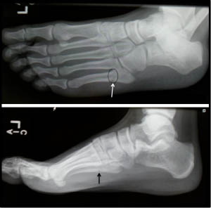
Figure Jones metatarsal fracture of the 5th metatarsal.
Metatarsal base fractures and Lisfranc-type injuries
Fractures at the base of the metatarsal bone are often associated with injuries to the tarsal and metatarsal joints - Lisfranc-type injuries. In order to detect such injuries, the clinician must evaluate the x-ray images very carefully. A Lisfranc injury can present with an enlarged gap between the first and second metatarsal bones, small fractures at the base of the first and second metatarsal bones, and an abnormal relationship between the sphenoid rim and the base of the second metatarsal bone. Computed tomography (CT) is the best way to rule out this injury.
If there is a suspicion of a Lisfranc injury, even if the x-rays show nothing, an MRI may also be indicated.
costs of the services
Initial consultation with the doctor treating you
- Getting to know the patient's complaints and clarifying the cause of the complaints
- Investigation
- assessment of symptoms
- Analysis of data from blood tests, X-rays, CT and MRI scans
- diagnosis
- Identification of treatment options
- Clarification and detailed review of the findings of the first visit to the treating specialist
- establishing the diagnosis
- Defining the treatment
' Blockade with glucocorticoids (price of the drug not included)
- Consultation with the attending physician
- taking blood
- Preparation of the platelet-rich plasma
- Injection of the obtained plasma into the patient area
- Stay in the medical facility
- Preparation of epidural anesthesia
- Arthroscopic surgical intervention
- Consumables (surgical material)
Postoperative visit to the specialist
- Post-operative examination
- Review of CT, MRI and X-ray findings
- Recommendations for recovery
- Injection of a preparation based on hyaluronic acid
- removal of the threads
opinions of doctors
These are the opinions of real people, taken from the doctor's page on prodoctorov.ru
My name is Helen Vorobyeva. In August 2021, Denis Sergeevich operated on my hip. In February 2022 I came for another check-up. The doctor determined that I had absolutely everything.
I went to Denis Sergeyevich for advice on knee replacement. Osteoarthritis of the knee had previously been diagnosed in the same clinic and surgery had been recommended. Doctor.
My grandmother (91 years old) had a complicated dislocated shoulder fracture. No one wanted to help her, and it was scary sending my grandmother to the hospital in shackles. Several doctors said they couldn't help her. from.
I, Mitropolevsky Tatiana Valentinovna, born in 1949, underwent surgery for a cemented total left hip arthroplasty. The first similar operation was performed on the right joint.
I live in the Rostov region. For the past two years, I've been plagued by hip pain and since nothing helped, I decided to have an endoprosthesis fitted. On the advice of a surgeon in Rostov, I went to .
For more than a year I experienced minute by minute wandering pains in the pelvic area that got worse after sitting or lying on my side. I went through all the doctors of gynaecology, urology, proctology, neurology, etc.
I went to the doctor with severe pain in my hip joint, I could hardly walk. Denis Sergeevich carefully examined the patient, made a diagnosis, and then began manipulation.
dr Yakushev Denis Sergeevich is an orthopedist and traumatologist, but he is also a doctor and a man of stature. dr Yakushev operated on my mother's right hip joint in 2016. The operation was an emergency.
I really liked everything about it! The doctor thoroughly explained everything and explained the situation. Clear and understandable! He was very accommodating and professional! It was a pleasure speaking with him. I recommend him to 100%!!!
I want to thank Denis Yakushev very much! I went to him for a ruptured biceps tendon. Diagnosis was made very quickly and surgery was scheduled immediately. The operation was performed by .
Cookie Settings
IMAIOS and some third-party providers use cookies or similar technologies, in particular to measure visitor numbers. Cookies allow us to analyze and store information such as your device's characteristics and some personal data (e.g. IP addresses, navigation, usage and location data, unique identifiers). This data is processed for the following purposes: to analyze and improve your experience and/or the experience of our content, products and services, to measure and analyze audiences, to interact with social media, to display personalized content, to measure performance and Content attractiveness. For more information, see our privacy policy: Privacy Policy.
You can give, withdraw or deny your consent at any time by using our cookie settings tool. If you do not consent to the use of these technologies, this will be treated as a refusal to lawfully store cookies. To consent to the use of these technologies, please click the 'Accept All Cookies' button.
Cookie Settings
When you visit the IMAIOS website, cookies are stored in your browser.
Some of these cookies require your consent. Click on the type of cookies to enable or disable them. To ensure maximum functionality of the IMAIOS website, it is recommended that you accept different types of cookies.
These cookies ensure the proper functioning of the website and allow it to be optimized (detection of problems encountered when navigating the website, logging into your IMAIOS account, online payments, troubleshooting and website security). The website cannot function properly without these cookies, so their use does not require your consent.
These cookies are used to measure traffic: traffic statistics help improve the performance of the website.
Human tarsal anatomy information:
The hock, the tarsus (foot root)is formed by seven short spongy bones, the ossa tarsi, which are arranged in two rows like the carpal bones. The posterior or proximal row consists of two relatively large bones: the talus and underlying calcaneus. The anterior row or distal row consists of a medial and a lateral part. The medial part consists of the navicular bone and three sphenoid bones. In the lateral part there is only one elbow bone.

Due to the upright posture of humans, the foot bears the weight of the entire supramalleolar region, resulting in a peculiar structure of the tarsal bones in humans compared to animals. Thus, the calcaneus, which is one of the most important support points of the foot, has attained its greatest size, strength and elongated shape in humans, which is elongated in the anteroposterior direction and thickened at the posterior end in the form of the calcaneal tuberosity.

The tarsal bone has adapted to the tibia (top) and hock (front), which explains its size, shape, and articular surfaces. The other bones of the tarsal, which are also heavily used, have become relatively massive and have adapted to the arched shape of the foot.
- talus, Consists of a body, corpus tali, which in front merges into a tapering neck, collum tali, and ends in an oval, convex head, caput tali, with an articular surface, facies articularis naviculars, which serves to articulate with the navicular bone. The shaft of the talus has a so-called block, trochlea tali, on its upper side, which serves to connect it to the tibia. The superior articular surface of the block, facies superior, which articulates with the distal articular surface of the tibia, is convex anteroposterior and slightly concave anteriorly. The two lateral articular surfaces of the block, facies malleolares medialis et lateralis, which lie on either side of the block, articulate with the ankles. The articular surface of the lateral ankle, facies malleolaris lateralis, curves down onto the lateral process of the ankle (processus lateralis tali), which emerges from the shaft of the ankle. Behind the block, the posterior calcaneal process diverges from the calcaneal shaft, separated by a groove for the flexor hallucis longus tendon. There are two articular surfaces (anterior and posterior) on the underside of the femur that connect to the heel bone. Between them runs the deep, rough sulcus tali.
- Heel Bone (Calcaneus calcaneus). On the top of the bone are the articular surfaces that correspond to the inferior articular surfaces of the talus. On the medial side, an extension of the heel bone branches off, which is called the sustentaculum tali or heel support. The name derives from the appendage as it supports the head of the talus. The articular surfaces in the anterior part of the heel bone are separated from the posterior articular surface of this bone by the calcaneal sulcus, which together with the talus sulcus forms a bony canal, the sinus tarsi, which opens into the dorsum of the foot on the lateral side. On the lateral surface of the heel bone is the sulcus for the long tendon of the fibula. On the distal side of the calcaneus, opposite the second row of the tarsal bones, is a saddle-shaped articular surface for connection with the cuboid bone, the facies articularis cuboidea. The shaft of the heel bone ends backwards in a rough outgrowth, the tuber calcanei, which forms two outgrowths towards the sole, the processus lateralis and the processus medialis of the tuberis calcanei.
- The heel bone (Os naviculare)Located between the head of the talus and the three parietal bones. On the proximal side it has an oval, concave articular surface for the heel head. The distal surface is divided into three smooth articular surfaces that communicate with the three sphenoid bones. On the medial side and inferiorly, a rough tubercle, the tuberositas ossis navicularis, protrudes from the bone and can be easily palpated through the skin. There is often a small joint pad for the elbow bone on the lateral side.
- The three sphenoid bones, the ossa cuneiformiaare named after their external appearance and are referred to as the mediate, intermedium and lateral cuneiform bones. Of all the bones, the medial is the largest, the intermedium is the smallest, and the lateral is the middle one. On the respective surfaces of the sphenoid bones are articular surfaces for connection with adjacent bones.
- The cuboid bone, Os cuboideum, Located on the lateral edge of the foot between the heel bone and the base of the 4th and 5th metatarsal bones. The articular surfaces are present. On the plantar side of the bone is the oblique tuberositis ossis cuboidei, which is preceded by the sulcus tendinis m. peronei longi.
Which doctors should you see for a tarsal exam?
Is there anything that worries you? Would you like to learn more about the tarsus or have an examination carried out? You can Make an appointment with your doctor – Clinic Eurolaboratory is always there for you! The best doctors will examine you, advise you, provide the necessary care and diagnose the problem. You can also doctor at home. clinic Eurolaboratory is open for you around the clock.
How to contact the clinic:
The phone number of our clinic in Kiev is: (+38 044) 206-20-00 (multichannel). The clinic secretariat will find a suitable day and time for you to visit the doctor. Click here for our coordinates and directions. Further details on all of the clinic's services can be found on the clinic's homepage.
If you have been examined before Be sure to bring the results with you to your doctor's office. If you have not yet done any examinations, we will carry out the necessary work in our clinic or with our colleagues in other clinics.
It is important that you take a close look at your general health. There are many diseases that at first do not make themselves felt in the body, but in the end, unfortunately, it is too late to treat them. It is simply necessary to be examined several times a year to be examined by a doctor several times a yearnot only to prevent a serious illness, but also to keep the body and the entire organism healthy.
If you want to see a doctor, you can find and read answers to your questions on the Internet Tips for self care. If you are interested in reviews of clinics and doctors, you can get information on the forum. You can also go to the medical portal Eurocoolregister to be kept up to date with news and information about Stępień, which will then be automatically sent to your email address.
Instead of a summary
It should be noted that the supporting surface of the human body standing upright is limited to the outer edge of the feet. According to the laws of physics, the greater the distance between feet, the greater the bearing surface and the more stable the balance of the human body. Any movement in space (walking, running) begins with a loss of balance (body moving forward – falling) and the subsequent creation of a new support surface (change of foot position). The foot is support and motor at the same time, as it lifts the body off the ground.
The authors: Olga Gurova, Biologist, Principal Investigator, Assistant Professor in the Department of Human Anatomy, PFUR; Igor Ponomarev
treatment methods
The choice of treatment methods depends on the type, severity and age of the injury:
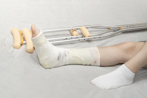
- With a fracture without displacement, a plaster cast is immediately applied.
- With a fracture of the foot with displacement, open or closed reduction of the fracture is performed under local anesthesia. A plaster cast is then applied.
- Skeletal Traction. Used for long-term injuries when closed repositioning is ineffective.
- Osteosynthesis is a surgical method of bone fusion for severe fractures with displacement.
- The Ilizarov apparatus is used for displaced fractures.
recovery period
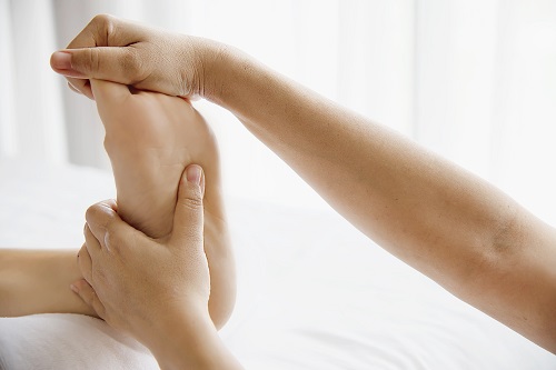
Rehabilitation therapists use the following techniques to restore function and anatomy to the injured limb:
- Injection of various drugs into biologically active points of the foot with the help of needles.
- Therapeutic massage. Promotes the restoration of blood circulation, reduces pain syndrome.
- Physiotherapy.
- Wearing a splint for at least a year after the injury.
- therapeutic exercises. A specialist will select a complex of exercises that will help you quickly return to normal life and avoid limping.
- use of ribbons. Muscles and ligaments are fixed with special ligaments.
- Pilates, yoga – exercises to relax, improve ligament flexibility and joint mobility.
- nutritional therapy. Foods are prescribed that accelerate bone formation.
definition
Sinus syndrome is a pathological condition that leads to compression of the neurovascular structures of the maxillary sinuses and pain syndrome.
The tarsal sockets are the area between the ankle and the heel bone. The joint in this area is called the subtalar joint.
The condition is often caused by dislocation of the ankle ligaments, but can also occur as a result of re-injury of the local ligaments. In most cases, it occurs when a person shifts their weight to the outside of the foot while walking or running.
In some cases, tarsal sinus syndrome can be caused by flat feet. This puts extra pressure on the subtalar joint and can increase pressure on the soft tissues around the ankle, leading to inflammation.
Tarsal Sinus Syndrome - Treatment
conservative therapies
Treatment of tarsal sinus syndrome often begins with nonsurgical or conservative methods. Non-surgical treatments often provide relief. Common non-surgical treatments include:
Non-steroidal anti-inflammatory drugs Nonsteroidal Anti-Inflammatory Drugs (NSAIDs): These may provide a combination of pain relief and anti-inflammation.
aids : Aids can immobilize the joint, provide support while walking and prevent further damage to the joint. Aids include braces, medical shoes, or shoe inserts that support the arch of the foot. Your doctor or physical therapist can recommend special shoes that provide better arch and ankle support.
steroids Steroids: Medications can help with inflammation by reducing pain and swelling in the joint. Steroids are often given by injection. This treatment can help when the disease does not respond to other treatments.
surgical treatment
For severe tarsal sinus syndrome or when nonsurgical measures have not resolved symptoms, your doctor may recommend surgical treatment. Surgical methods include:
Removal of scar tissue : In some cases, the surgeon may consider removing the scar tissue, usually using a minimally invasive technique called arthroscopy.
Surgical treatment of flat feet: The surgeon may use a combination of procedures to correct the ligaments, bones, and tendons that support the arch of the foot when flat feet are the cause.
ankle arthrodesis When symptoms are caused by inflammation of the subtalar joint, also known as hindfoot arthritis, surgical fusion of the joint may be necessary. Your doctor may use this option if the joint cannot be replaced.
Surgical options should be discussed with your surgeon or other doctor, who will explain the possible advantages and disadvantages of each procedure and make recommendations for your individual case. It's important to consider any additional medical conditions that may aggravate the condition, such as: B. obesity or diabetes.
Surgery for a metatarsal fracture
Fracture surgery is indicated when the metatarsal fracture is displaced more than half the bone width.
Percutaneous fixation with spokes
This method has been popular for many years and remains one of the most commonly used procedures in the world.
First, the clinician notes the displacement of the fracture, then spokes are drilled through the fracture in specific directions (dictated by the type of fracture).
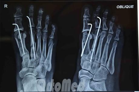
Advantages: Low trauma, quick, easy, cheap, no incision and consequently no postoperative scar.
Disadvantages: The spoke ends remain above the skin so that the spoke can be removed after the fracture has healed; risk of wound infection and penetration of infection into the fracture area; prolonged wearing of a plaster cast for a month; inconveniences in daily life.
Open reduction of a fracture
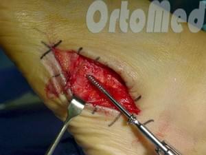
Open reduction of a metatarsal fracture, osteosynthesis of the bone with a plate and screws. The operation consists of a surgical incision, access to the fractured metatarsal by carefully pulling tendons, vessels and nerves, mobilizing the bone fragments, removing the dislocation and fixing it in the correct position.
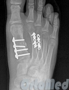
No cast immobilization is performed as the metal structure fixes the fragments.
Walking with heel support is allowed for one month.
Rehabilitation after a metatarsal fracture
Once the metatarsal fracture has healed and the pain has subsided, your doctor will allow you to put weight on your foot and gradually increase the load.
Do not self-medicate!
Only a doctor can diagnose and prescribe appropriate treatment. If you have any questions, you can call or Email your question.
| Treatment of foot fractures | Price, RUB |
| Manual repositioning | from 2,500 |
| plaster cast | from 1,500 |
| Osteosynthesis (without metal work) | from 38,000 |
| local anesthesia | from 700 |
| regional anesthesia | from 3000 |
| Bandages, suture removal | from 500 |
- Cracked metatarsal.
- The shaft of the heel bone.
- The tarsal and metatarsal bones.
- tarsus.
- Anatomy of the Lisfranc joint.
- metatarsal and metacarpal bones.
- metatarsal bones.
- Orthopedic insole junior.