The causes of erythema annulare are as follows

- Erythema annulare
- symptoms
- Causes of allergies
- What types of allergic eye diseases are there?
- How does it work?
- How to use it during pregnancy and breastfeeding?
- Symptoms of tendonitis in the ankle
- How ankle tendonitis is diagnosed
- What are small hemorrhages under the skin?
- What diseases do petechiae cause?
- Is it OK if the swelling goes away during pregnancy?
- We hope you found this article helpful. To receive such information, sign up for our newsletter. It will provide you with useful and up-to-date information, not only during pregnancy, but also in the first, difficult months of motherhood.
- Sources:
- pharmacokinetics
- Dosage and application
- Main types of pain
- Can we take painkillers?
- If you have a toothache
- What is Sweat Pox?
- Types of sweats
Erythema annulare
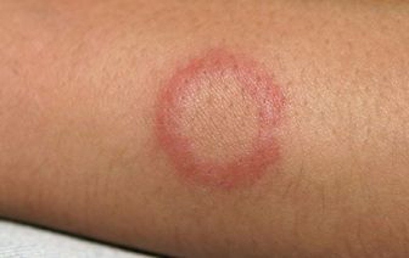
Erythema annulare (Erythema rheumatoidum) is a polyetiological skin disease with an erythematous-ulcerative character in which ring-shaped spots form on the skin. Children, adolescents and young men are most commonly affected. Erythema multiforme is a long-term, chronic skin disease. It is infectious, allergic and toxic.
- Erythema belongs to a group of skin defects with similar symptoms that only an experienced doctor can distinguish.
- It appears on the trunk in the form of ring-shaped spots.
- The spots gradually form a ring-shaped lesion (hence the name erythema annularis).
- There are dozens of types of erythema (without expertise it is impossible to distinguish between erythema centrifugalis and erythema rings).
- The course of the disease is protracted, recurrences are common, and some forms are difficult to treat.
- It lasts for several months (but can last up to ten years).
- Erythema annulare occurs equally frequently in children and adults.
- Some forms of erythema annularis, such as Darier's erythema, are more common in young men.
The causes of erythema annularis range from high air temperature to bathing in hot water. The redness can also be triggered by physical factors. Temporary redness is a normal (physiological) phenomenon. It usually disappears quickly and spontaneously as soon as the stimulus (temperature, medication, massage, stress, sun exposure) is eliminated.
Redness without an apparent cause should be cause for concern. It is a pathological sign and an appointment with a dermatologist should be made. The doctor will determine whether the patient has erythema annulare or another skin lesion. Pathological processes usually manifest themselves as permanent skin discoloration.
symptoms
Erythema annulare causes different symptoms depending on its clinical form. Some of these symptoms are described below.
- Erythema multiforme – In response to an allergic substance (food, medicine). The spots are red, there is no desquamation and the course is quite mild (the lesions disappear within a few days). In contrast to simple erythema, persistent erythema is long-lasting.
- Scaly form – A resulting red spot will soon begin to peel. Some pigmentation is visible in the center of the redness, which fades over time. The spots can grow up to 20 cm in size. The lesions hardly change in the middle and grow at the periphery. This form can take several months to develop. After the rash clears up, the skin is pigmented. Recurrences are common, and strange patterns (areas of hyperpigmentation) appear on the skin over the years. The scaly form is often caused by helminth infections and tumors.
- Erythema darius centrifugalis Erythema annulare – The etiology is unclear (viral, bacterial and fungal pathogens cannot, however, be excluded). It is characterized by the appearance of numerous small pink spots that rise slightly above the skin. The spots tend to enlarge and change shape. A characteristic feature of erythema darius is the circular shape of the lesions. It occurs most commonly on the trunk and less commonly on the face.
- Another form of centrifugal annular erythema is a rare variant of lupus, an autoimmune disease. The redness appears to spread from the center of the face to the edges and is not physically uncomfortable. It occurs as an independent disease (erythema annulare bietta) or as a symptom of another disease.
Main symptoms of erythema bietta
- The lesion is characterized by pink or red patches on the skin.
- The erythema in the center is almost normal in color or may be pale.
- Itching is not always present and can sometimes be burning.
- Belly, flanks, arms and sometimes the face (cheeks).
- The spots can grow quite quickly, up to 20 cm in diameter. If several spots are in close proximity to one another, coalescence occurs.
- They then lift easily from the skin.
- Over time, a 'pattern' of old and new lesions forms on the body.
- In the daisy type, the spots appear only for a short time, in the vesicular type (vesicles are small bubbles) they appear immediately and disappear just as quickly, in the exfoliative type the spots peel outwards.
Causes of allergies
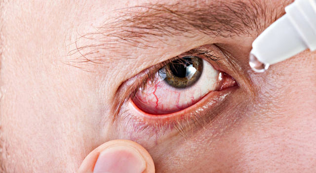
In fact, there are as many allergens as there are substances and compounds known to humans. In other words, an allergic reaction to the eyeball (conjunctiva) and the skin around the eyes can be triggered by anything: pollen, any food, household chemicals, animal hair, down and bird feathers, house dust (infested with microscopic mites), insect bites, Hygiene products and cosmetics, medicines, sweat, saliva or hair of other people, etc. At the same time, the etiopathogenetic mechanism that leads to the fact that an individual reacts violently (in some cases catastrophically) to a substance, while the organism of another person is completely indifferent to it Substance remains, has not yet been fully clarified. However, the role of an innate predisposition has been reliably proven, as has the cumulative (combined) nature of the action of allergens.
The eyes are one of the 'preferred targets' and areas where allergic reactions develop. The tissues and structures of the eyeball are thin, highly sensitive and have little protection from harmful environmental influences - and are therefore particularly vulnerable. The cornea, eyelids, eyelashes, sclera and conjunctiva come into direct contact with the air, which contains particles of countless substances, as well as with rain and tap water, cosmetics, shampoos, creams, eye drops and gels and contact lenses. In addition, many other, equally sensitive mucous membranes (nose, mouth, etc.) are in close proximity and in direct communication with the eye socket. The eyes are also susceptible to allergic reactions to things ingested by the body, such as: B. individually allergenic foods and medications.
What types of allergic eye diseases are there?
The symptoms of allergic eye reactions are varied. This can include more or less mild redness, itching, a vesicular rash, a burning sensation in the eyelid skin (allergic dermatitis), inflammation of the transparent mucous membrane (allergic conjunctivitis) with tearing, itching, etc. However, in some cases, the reaction is much more serious and leads to the rapid development of keratitis (toxic or infectious-allergic keratitis), uveitis (allergic inflammation of the ocular circulation) and even damage to the retina and optic nerve head.
The most common form of eye allergy, conjunctivitis, can be acute or chronic. In both cases, the symptoms are similar: persistent tearing, itching or itching of the eye, mucous sputum and the characteristic swelling of the vitreous humor of the conjunctiva (chemosis).
Pollinosis, seasonal hay fever, is a typical and common example of an allergic reaction to pollen, including an eye reaction. Such allergies usually peak in August and are caused by one of the strongest allergens, ragweed (ragweed is considered a quarantine weed and must be deliberately eradicated by a relevant authority, which does not seem to be done successfully everywhere). Pollen or fluff from dandelions, grasses, flowers and other plants can also trigger an allergy. Allergic conjunctivitis almost never occurs in isolation and is usually accompanied by a runny nose (catarrh), uncontrollable, repeated sneezing, rashes, and itching; Attacks of allergic bronchial asthma can also occur.
A so-called allergy is slightly different from pollenosis. Spring rhinitis is an allergic (kerato) conjunctivitis, which is also a seasonal disease that occurs at the beginning of the warm season. The cause is not clear, but there is a reasonable hypothesis that spring fatigue is caused by an individual sensitivity (hypersensitivity) to the ultraviolet part of the solar spectrum, which is dangerous for the eyes in many ways. The role of allergenic pollen cannot be ruled out either. A distinctive feature of this seasonal allergic inflammation is its occurrence only in childhood (mainly in boys), its chronic course, the presence of photophobia and increased lacrimal and mucous membrane discharge in the clinical picture; A specific symptom is also the accumulation of papillary eruptions on the inner conjunctiva of the eyelids ('paving stones'), less often on the limbus - the periphery of the cornea.
Using a special micronization process, the particles of the active ingredient Detralex® are reduced to their smallest size, which contributes to better absorption and direct delivery of the substance into the veins.
*Barbel R; Amiel M./Phlebology 1992; 7 (Suppl 2):p. 41-44 (Barbe R; Amiel M. Phlebologie 1992, 7 (Suppl 2), pp. 41-44).
Detralex® contains a micronized, purified flavonoid fraction of five flavonoids, substances of plant origin
Paysant J., Sansilvestri-Morel P., Bouskela E., Verbeuren TJ / International Angiology, 2008, 27, pp. 81-85
Instructions for the medical use of Detralex®. RU LP-#(000880)-(RG-RU).
Paysant J., Sansilvestri-Morel P., Bouskela E., Verbeuren TJ / International Angiology, 2008, 27, pp. 81-85
Instructions for the medical use of Detralex®. RU LP-#(000880)-(RG-RU).
Paysant J., Sansilvestri-Morel P., Bouskela E., Verbeuren TJ / International Angiology, 2008, 27, pp. 81-85
Instructions for use of the drug Detralex®. RU LP-NR(000880)-(RG-RU).
Paysant J., Sansilvestri-Morel P., Bouskela E., Verbeuren TJ / International Angiology, 2008, 27, pp. 81-85
Instructions for the medical use of Detralex®. RU LP-#(000880)-(RG-RU).
Paysant J., Sansilvestri-Morel P., Bouskela E., Verbeuren TJ / International Angiology, 2008, 27, pp.81-85
Instructions for the medical use of Detralex®. RU LP-#(000880)-(RG-RU)
How does it work?
Improves blood outflow and reduces venous congestion by increasing venous tone and reducing capillary permeability.
How to use it during pregnancy and breastfeeding?
There are no or limited data on the use of the purified micronized flavonoid fraction in pregnant women.
Animal studies have shown no reproductive toxicity.
As a precautionary measure, it is recommended not to use Detralex® during pregnancy.
It is not known whether the purified micronized flavonoid fraction (metabolites) is excreted in breast milk.
A risk for newborns and infants cannot be ruled out. The decision to stop breastfeeding or discontinue Detralex® therapy should be made taking into account the benefit of breastfeeding for the child and the benefit of therapy for the woman.
Symptoms of tendonitis in the ankle
The main symptoms of tendonitis in the ankle are:
Ankle tendonitis is often caused by overuse of the tendon. This can be caused by playing a particular sport. In other cases, ankle tendonitis may occur as a result of aging.
You can prevent ankle tendonitis by taking appropriate measures during exercise, such as: B. warm up before training and take breaks between exercises.
How ankle tendonitis is diagnosed
Ankle tendonitis can be diagnosed by an orthopedic examination. The orthopedist may recommend additional examinations, e.g. B. X-rays of the ankle or an MRI scan of the ankle to determine if the pain is caused by another condition.
In most cases, ankle tendonitis will heal within a few weeks with proper care and rest. The condition can usually be treated at home. In the first few days after the injury, try to rest as much as possible, use ice, and elevate the ankle. Take pain relievers and anti-inflammatory medications to reduce pain and swelling.
If your symptoms don't improve after a few weeks, your doctor may recommend surgery:
In severe cases, surgery is ordered to repair the tendon.
What are small hemorrhages under the skin?
Bleeding under the skin is caused by rupture of the capillaries. Due to the small amount of blood in the thinnest vessels, the spots are only a few millimeters in size. As a rule, there is only one spot with such spots. The person does not feel worse and may not even be aware of the presence of petechiae on the skin.
To understand that the rash is capillary damage, and not an allergy, just press the spot - the spots do not change color or disappear. The shapes become brighter after a while and then disappear.
What diseases do petechiae cause?
Blood diseases such as leukemia and aplastic anemia change the composition of the blood, particularly by reducing the production of platelets. When there is little blood clotting and weakened blood vessels, bleeding occurs and wounds heal more slowly. Petechiae can also occur against the background of other blood diseases and organs involved in blood formation.
Another cause of vascular diseases can be autoimmune diseases. For unknown reasons, the immune system begins to wage an imaginary battle against the body's own cells. A sustained immune attack leads to inflammation, which destroys blood vessels.
As a result, petechiae (red spots) may appear:
Bleeding can also occur temporarily, including:
Vitamin C and K deficiency leads to petechiae under the skin. Bruising then occurs in small amounts. Over time, as the body recovers from the disease and the nutritional content of the food consumed increases, the appearance of petechiae decreases.
A dangerous condition occurs when the patient notices a rash on the body in combination with other symptoms:
- Increased body temperature.
- Pain when turning the head.
- Headache.
- Difficulty with motor coordination.
- Muscle soreness and pain in the muscles.
- Blurred consciousness.
- Pain in small or large joints.
- Digestive system disorders.
- Bleeding wounds, gums.
- Bruises on the body.
Bleeding under the skin should not scare a person unless it occurs frequently and in small amounts and there are no other signs of a serious condition. If this is not the case, you should be examined at a medical facility.
Is it OK if the swelling goes away during pregnancy?
If you have recently changed your diet, spent more time outdoors or taken other measures, that is very good. It means your treatment is working and you are on the right track.
The situation is different if the swelling disappears on its own during pregnancy without your life changing. This is normal a few days before the due date: the progesterone has done its work, its levels drop and release additional water. However, if your due date is still a long way off, a spontaneous decrease in swelling is at least a suspicious sign. See your doctor to find out the cause of the sudden swelling and decide what you can do about it.
We hope you found this article helpful. To receive such information, sign up for our newsletter. It will provide you with useful and up-to-date information, not only during pregnancy, but also in the first, difficult months of motherhood.
Sources:
pharmacokinetics
After applying the cream/gel to the skin, ibuprofen penetrates into the deeper tissues (subcutaneous tissue, muscles, joints, synovial fluid) and reaches therapeutic concentrations there. The therapeutic effect at the site of application is achieved through direct distribution through the skin to the target tissues. It is detected in small amounts in blood plasma. With the recommended route of administration, the concentration in the synovial fluid is approximately 2 µg/ml.
inflammatory and degenerative diseases of the musculoskeletal system: joint syndrome with exacerbation of gout, arthritis (rheumatoid arthritis, psoriasis, gout), periarthritis of the shoulder, ankylosing spondylitis (Behterev's disease), deforming osteoarthritis, osteochondrosis with root syndrome, radiculitis, tendinitis, bursitis, synovitis, lumbago , sciatica;
Muscle pain (myalgia) of rheumatic and non-rheumatic origin;
Injuries (sports, industrial, household) without damage to the integrity of the skin (sprains, strains or tears of muscles and ligaments, bruises, post-traumatic swelling of soft tissues).
Dosage and application
Cream. Apply to the skin at the site of pain 3-4 times a day and rub gently until the medicine is absorbed. Depending on the size of the affected area, a 4-10 cm wide strip of the cream is applied.
The duration of treatment depends on the severity of the disease and the nature of the damage and is on average 2-3 weeks.
Gel. A 5-10 cm long gel strip is applied to the affected area and rubbed gently until completely absorbed, 3-4 times a day.
The duration of treatment depends on the severity of the disease and the type of lesion and is on average 2-3 weeks.
Main types of pain
During pregnancy, women suffer from headaches, toothaches and back pain. Cephalalgia occurs in the early stages. These can be pressing, cramping or pulling. The patient feels as if a tire is being pulled around her head. The symptoms worsen in the first half of the day and usually subside again in the evening.
Toothache can occur for the following reasons:
- Increased acidity of saliva (early pregnancy toxicity is a trigger);
- hormonal changes;
- changes in metabolic processes;
- Nutrient deficiency.
Back pain is caused by overuse of the muscles as a result of weight gain. The center of gravity is shifted by the growing belly.
Can we take painkillers?
Such medications are contraindicated at the very beginning of pregnancy. Later, non-steroidal anti-inflammatory drugs (NSAIDs) are allowed if necessary. These are relatively safe and are dispensed without a prescription.
Permitted painkillers for pregnant women:

- Paracetamol. Painkiller and antipyretic agent. Effect: pain-relieving, fever-reducing. Prescribed for mild pain syndromes. Tolerable for cephalalgia (especially migraines), trauma, myalgia and neuralgia. Suitable if the expectant mother has a fever. Crosses the placenta but does not harm the baby.
- Nurofen. Is a derivative of phenylpropionic acid. Effect: fever-reducing, pain-relieving, anti-inflammatory. It is taken for inflammatory and degenerative joint diseases. Can be used for soft tissue injuries, radiculopathy, synovitis, bursitis, tendinitis, arthritis, osteoarthritis, spondylosis. It helps reduce the amount of amniotic fluid. For this reason it is forbidden in late pregnancy.
- No-shpa. Is an isoquinoline derivative with strong antispasmodic effects. Acceptable for tension headaches, colitis (including spastic colitis with constipation), inflammatory bowel disease and spasms of the smooth muscles of the gastrointestinal tract.
- Riabal. Antispasmodic with M-choline blocking effect. Recommended for pain associated with spasms of the smooth muscles of the gastrointestinal tract, urinary tract and biliary tract.
If you have a toothache
Drugs based on mepivacaine and articaine can be used. Medications containing adrenaline should be avoided.
What is Sweat Pox??
Sweat pox is a disease that belongs to the group of simple dermatitis. Infants and small children are most commonly affected. However, under certain conditions, adults can also be affected.
It is characterized by the appearance of blisters or nodules on the surface of the skin. That depends on the type of sweating. However, a common symptom is redness and irritation of the skin. Next, the type must be determined, and there are some characteristic signs.
Types of sweats
Occurs when the superficial ducts of the sweat glands are blocked, resulting in accumulation of sweat in the surface layer. It is called crystalline because it appears as bubbles of clear liquid. They open harmlessly and there is nothing bad about them. They resemble pimples, but the fluid is clear and the blisters are painless. There is no evidence of inflammation in this type of sweating.
It is the most common form of this disease. And the most common cause of their occurrence – is overheating, high temperatures and, therefore, increased sweating. In this case, the blockage of the pores occurs in the epidermal part of the tubules. And that already leads to inflammatory processes. In this case, small red spots form on the skin.
This type of disease is quite rare as it is basically a complication of red sweat. Typically, the person treats the redness and does not progress to this stage. It is also known as yellow sweat stains. It is characterized by more severe and advanced inflammation. Accordingly, the spots visible on the skin are more intense.
Read more:- Description of the human foot bone.
- foot pattern.
- Ankle strap at heel.
- Front lower leg area.
- Ligament damage in the right ankle.
- Photo of the right leg.
- Pain in the periosteum of the tibia.
- Ankle of the right foot in Latin.