People with Marfan syndrome have distinctive facial features that give them the bird-like expression that all patients in this group have. The eyes are deep-set, the skull is elongated, and the teeth are close together due to the small size of the jaw and the 'Gothic palate'. The nose appears large, the ears are rather large and set low. It is rare for one person to experience all of these symptoms; The most common external characteristics are a tall stature and elongated fingers.
- Marfan disease (syndrome)
- What causes / causes of Marfan syndrome:
- Symptoms of Marfan syndrome in children
- Factors that increase the risk of developing Marfan syndrome in children
- Clinical manifestations
- Diagnosis
- How to recognize the lesions characteristic of this disease?
- Can weak connective tissue be dangerous?
- How can doctors tell if a person has Marfan syndrome?
- How do doctors treat Marfan syndrome?
- Causes of Marfan Syndrome
- Is Marfan syndrome hereditary?
- Diagnosis of Marfan Syndrome
- How does Marfan syndrome manifest itself?
- How is the disease treated?
- Classic symptoms of Marfan disease
- Correction and treatment methods
- Diagnosis of Marfan Syndrome in Israel
- Day 1: Consultation
- Day 2: Diagnosis
- Day 3: Completion of the consultation
- What is the cost of treating Marfan Syndrome in Israel?
- Benefits of Marfan Syndrome Treatment in Israel
Marfan disease (syndrome)
Marfan disease (Marfan syndrome) – is a congenital collagenopathy or systemic connective tissue defect with a specific type of inheritance and various clinical manifestations: skeletal, cardiovascular and ocular diseases. Patients with Marfan syndrome have aortic aneurysms, gigantism, myopia, dolichosthenomelia and arachnodactyly, thoracic deformity, hip joint protrusion, lens ectasia, flat feet, dura mater ectasia, and kyphoscoliosis.
The disease is named after French pediatrician Antoine Marfan, who in 1886 first described a five-year-old girl with long, thin legs and 'woven' toes.
The frequency of this congenital condition is around 1:10,000 and varies little by gender. However, in children and adolescents the percentage is up to 6.8 %, with most cases occurring in boys.
What causes / causes of Marfan syndrome:
Marfan syndrome is a congenital anomaly that is inherited in an autosomal dominant manner. The syndrome is caused by mutations in the FBN1 gene, which is responsible for the synthesis of fibrillin, a structural protein of the intercellular matrix that provides elasticity and contractility to connective tissue. Abnormalities and deficiency of fibrillin lead to abnormal formation of fibrous structures and loss of strength and elasticity of connective tissue, which cannot withstand physiological stress. Histological changes occur in elastic vascular walls. In 75 percent of cases, Marfan syndrome is inherited in families, and in 25 percent there is a primary mutation. If the father is over 35 years old and has a history of Marfan syndrome, there is a very high chance that he will pass the disease on to his child.
Scientists differentiate between different forms of Marfan syndromewhich depend on the number of organs and systems affected:
- Sterile – with mild lesions in 1 or 2 systems;
- severe – with mild lesions in 3 systems; or severe lesions in at least 1 system; or severe lesions in 2-3 or more systems.
In Marfan syndrome, the severity of the lesions is divided into mild, moderate and severe. A distinction is made between progressive and stable Marfan syndrome.
Symptoms of Marfan syndrome in children
Marfan syndrome affects the body's skeletal, cardiovascular and ocular systems. However, according to several studies, the pulmonary system and skin are also affected.
The following are symptoms that affect one or more of the above systems:
- elongated face (dolichocephalic facies);
- deep-set and slanted eyes;
- flattened ears;
- poor appetite;
- tall and slim body;
- long arms, legs and toes;
- sagging and excessive skin with stretch marks;
- poor wound healing on the skin;
- crooked spine;
- flat feet;
- bulging of the chest known as 'pigeon chest';
- large labia or increased joint mobility;
- reduced proportions of the upper body in relation to the lower body.
The combination of these symptoms may indicate Marfan syndrome.
Factors that increase the risk of developing Marfan syndrome in children
Marfan syndrome is inherited in an autosomal dominant manner, meaning an abnormal gene in one parent can cause Marfan syndrome in a child. The risk of having a child with Marfan syndrome is 50 % in each pregnancy, even if one parent has Marfan syndrome.
Three out of four cases of Marfan syndrome are caused by genes inherited from parents. In some cases, the disease is caused by a mutation of a (new) gene in the child.
Clinical manifestations
Marfan syndrome is characterized by damage to the cardiovascular system, the musculoskeletal system and the eyes. The pathology is characterized by a number of symptoms that vary from patient to patient. The syndrome has a chronic course, but the speed of progression of symptoms is individual.
Patients with this disease are distinguished by their height. The limbs are longer than the trunk (dolichostenmelia). Long webbed fingers (arachnodactyly) are a characteristic symptom. The physique is asthenic with little subcutaneous fat and underdeveloped skeletal muscles. The face is narrow and high. There may be abnormalities in the teeth and a bulging palate. Marfan syndrome can already be suspected in newborns. The body length at birth is usually greater than 53 cm for boys and 52.5 cm for girls.
If Marfan syndrome is suspected, a geneticist should be consulted. The specialist will examine the child externally and, if necessary, order further tests.
Symptoms of Marfan syndrome include hypermobility of the joints. This is a condition in which a person can move their joints to a greater extent than a healthy person. Connective tissue changes lead to thoracic deformities, spinal curvatures, flat feet and other orthopedic disorders.
The clinical course and outcome depend on the severity of the cardiovascular lesions. The patients have malformations of the elastic vessels (aorta, main arteries), valvular defects and defects in the cardiac septum. The aorta shows dilatation in the first segments, disrupting the aortic valve in the left ventricle, as well as multiple aneurysms. The congenital heart defects are diverse: pulmonary artery stenosis, ventricular and atrial septal defects. Children with this disease have rhythm changes of varying degrees.
Read more:Cardiovascular diseases are an important factor affecting life expectancy. Congenital malformations may need to be corrected surgically.
Diagnosis
Diagnosis of the syndrome is based on family history, physical examination, ECG, EchoCG, x-rays, laboratory tests and molecular genetic studies. The diagnostic criteria are:
- Changes to the ascending aorta in the form of dilation or dissection;
- Abnormal position of the lens;
- deformity of the sternum, which is an indication for surgical treatment;
- The upper body segment is longer than the lower body segment;
- Severe scoliosis;
- limited mobility of knees and elbows.
The ECG shows cardiac arrhythmias and hypertrophic changes in the heart muscle. EchoCG (cardiac ultrasound) shows valvular insufficiency, backflow of blood during heart contractions and other structural abnormalities. Cardiac hypertrophy and aortic lesions are diagnosed by chest X-ray, CT or MRI.
All patients with Marfan syndrome must be examined by an ophthalmologist. The specialist will perform biomicroscopy and ophthalmoscopy to assess the position of the lens. Other methods are also recommended: X-rays of large joints, MRI of the brain and spinal cord, etc.
The differential diagnosis includes the exclusion of other genetic and chromosomal diseases. These include homocystinuria, hereditary arthro-ophthalmopathy, Ehlers-Danlos syndrome, etc.
How to recognize the lesions characteristic of this disease?
Marfan syndrome has a number of symptoms that can be attributed to connective tissue defects.
- Strong growth from childhood, caused by greater susceptibility of cartilage to tensile processes against the background of weak connective tissue. Intensive growth processes predispose to inadequate bone mineralization with early development of osteoporosis after the age of 30.
- Asthenic physique with disproportionately long arms in relation to the torso.
- Typical fingers – very thin, webbed fingers on the hands.
- Chest deformity – concave chest.
- High palate.
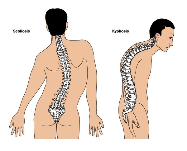
- Abnormal posture up to kyphoscoliosis, caused by a weakening of the ligaments in the spine.
- Excessive mobility is one of the most noticeable symptoms of Marfan syndrome. Weak ligaments around the joints also result in hypermobility of the joints, which can lead to high mobility of the individual joints and cause various subluxations.
- Connective tissue changes in the heart are associated with prolapse of the heart valves, which leads to impaired valve function and incomplete closure during heart contraction. The most dangerous heart defect is associated with mitral or aortic valve regurgitation. These lesions lead to aortic dilatation with incomplete valve closure and subsequently form an aneurysm.
- The visual changes are manifested by high myopia, which is accompanied by lens subluxation, which develops as a result of weakness of the zygomatic ligament.
- The respiratory changes are associated with congenital lung pathology with pulmonary hypoplasia, congenital emphysema or bronchiectasis.
Can weak connective tissue be dangerous?
There is an increased risk of cardiovascular disease due to impaired valve function. During pregnancy, women with these lesions are at increased risk of aortic dissection due to hypersecretion of estrogen, which slows the influx of collagen and elastin into the aorta.
Weakness of the ligamentous apparatus of the eye manifests itself in adults in the form of glaucoma or the development of retinal degeneration with subsequent retinal detachment. Severe scoliosis and hip joint protrusion occur during active, rapid growth. Radiologically, these symptoms are clearly visible on x-rays.
It is recommended that people with external signs of Marfan syndrome be examined by an ophthalmologist and a cardiologist and that a genetic test be performed to identify the altered gene.
How can doctors tell if a person has Marfan syndrome?
When doctors suspect Marfan syndrome, they ask about the symptoms and family history of Marfan syndrome. They will perform a physical exam and:
If the person has Marfan syndrome, the doctor will examine the heart, bones and eyes with other tests, such as: B:
ECG Electrocardiography Electrocardiography is a test that measures the electrical activity of the heart. It is quick, painless and harmless. The results of the examination are recorded. Read more (A painless test in which the heart's electrical currents are measured and recorded on a piece of paper)
How do doctors treat Marfan syndrome?
Doctors cannot cure Marfan syndrome. But you can treat some of the symptoms:
prescribe medications that help blood flow more efficiently, thereby preventing aortic problems;
recommend that children wear orthotics to treat abnormal curvature of the spine (or sometimes undergo surgery to repair the spine);
heart surgery to correct problems with the aorta or heart valves;
Your doctor will examine your bones, heart, and eyes annually to treat complications of Marfan syndrome.
Causes of Marfan Syndrome
Marfan syndrome is a genetic disorder with autosomal dominant inheritance and is caused by mutations in the FBN1 gene. This gene is responsible for the production of fibrillin-1, an important structural protein that forms ligaments and elastic vessels. When this protein is abnormal, connective tissue structures become more elastic and lose their resistance to deformation. The most serious effects of such changes affect the eyes and the cardiovascular system. Fibrillin-1 is essential for the proper function of the cinnamic band that connects the lens to the ciliary body. Therefore, when its synthesis is impaired, which is the case in patients with Marfan syndrome, there is a weakening of the cinnamic band, which in patients initially manifests itself as myopia, a subluxation of the lens, but later leads to secondary glaucoma, a partial one or complete loss of vision.
Other functions of fibrillin in the body include the formation of the extracellular matrix, which ensures the integrity of connective tissue and the proper functioning of cell growth factors.
The aorta is an elastic vessel and contains many elastic ligaments on which its resistance to stress depends. If its function is disrupted, the aorta expands and its walls dissolve. It is a life-threatening situation that can lead to sudden death if the aorta ruptures under intense stress. Aortic dilatation is dangerous for women during pregnancy, especially in the third trimester, but also during childbirth and for a few months afterward.
Other common cardiac abnormalities associated with Marfan syndrome include mitral valve lesions, which require surgical treatment.
Is Marfan syndrome hereditary?
Marfan syndrome is a genetic disorder that is inherited in families in 75 percent of cases, with the remaining 25 percent due to a primary gene mutation. In families where the father is older than 35, the risk of having a child with Marfan syndrome is increased.
Diagnosis of Marfan Syndrome
The diagnosis of Marfan syndrome is made on the basis of a physical examination, family history, examination by a cardiologist and an ophthalmologist, and a chest x-ray.
The following criteria are also important for diagnosis:
- Presence of thoracic malformations requiring surgical intervention;
- Presence of abnormal tissue and organ changes in the visual, cardiovascular system, characteristic of Marfan syndrome;
- ectopia of the lens;
- The upper trunk is significantly shorter than the lower trunk, with a ratio greater than 0.86;
- Shoulder width is greater than height (ratio greater than 1.05);
The renal excretion of glucosamine is increased many times over in Marfan syndrome, which is also used in the diagnosis.
EKG and echocardiography can reveal cardiac abnormalities. Respiratory abnormalities can be detected by examining the external respiratory system. Patients with Marfan syndrome often suffer from pulmonary edema, hypercapnia, irregular air distribution in the lungs, and various abnormalities in respiratory mechanics.
DNA testing (direct automated sequencing) can detect and identify mutations in the FBN1 gene.
How does Marfan syndrome manifest itself?
Typically, patients with Marfan syndrome are taller than the age and family average, and their arm span is greater than their height. People with this syndrome also have arachnodactyly (long, abnormally elongated fingers). Patients may also have a keel-shaped chest (displaced outwards) or a funnel-shaped chest (displaced inwards). Joint hypermobility, flat feet, kyphoscoliosis (abnormal curvature of the spine), diaphragmatic and inguinal hernias may also be present.
Symptoms may include vision problems: ectopic lens (subluxation or upward displacement of the lens) and iridodontia (fluttering of the iris). Patients with Marfan syndrome may develop cystic lung disease and recurrent spontaneous pneumothorax. Cardiovascular problems may include aortic aneurysm (enlargement or bulging of the aortic wall) and mitral valve prolapse (disease involving valve dysfunction between the left atrium and the ventricle).
How is the disease treated?
Untreated people with Marfan syndrome live a maximum of 30-40 years and die from either an aortic dissection, an aneurysm or heart failure.
Treatment is mainly symptomatic and aims to relieve some of the symptoms of the disease. Patients must undergo a comprehensive physical examination annually, which must also include an ophthalmologist, a cardiologist and an orthopedist. With regular symptomatic treatment, patients can live to old age, but life expectancy is still lower due to cardiac and vascular complications.
For the symptomatic treatment of Marfan syndrome, most clinical studies recommend the prophylactic use of beta-blockers from an early age. This is intended to prevent the formation of an aortic dissection aneurysm. In cases of severe aortic root dilatation, surgical correction is performed.
Classic symptoms of Marfan disease
The classic symptoms are short stature, slim build, curvature or scoliosis, long arms and legs that are out of proportion to the torso, long, slender 'webbed' fingers and underdeveloped muscles. Their skin is fragile and stretches easily, while they have an increased tendency to bleed. Examination of the cardiovascular system reveals expansion of the aortic arch and various variants of heart valve defects.
Myopia is quite common and occurs in more than half of all patients with Marfan syndrome. It is due to the spherical shape of the lens, changes in the refractive power of the cornea and the deformation of the eyeball itself.
There are also changes in the iris that are due to increased tissue stretch. Iris defects called colobomas then form, and the angle of the anterior chamber can be closed by the stretched iris tissue, leading to an increase in intraocular pressure, that is, the development of glaucoma.
The stretching and weakening of the ligaments that hold the lens of the eye together, called cinna ligaments, results in a partial or complete rupture. Either the lens subluxates when the ligaments are partially torn but the lens is still supported by the remaining ligaments. Or the ligaments come off completely and the lens sinks into the eye socket and changes position at will - the lens is dislocated. In addition, lens opacities or cataracts develop earlier and more frequently than in healthy people.
Glaucoma occurs when the drainage of eye fluid through the anterior chamber angle is impaired due to an altered iris or a displaced lens that blocks the drainage path for the eye fluid.
The retina also becomes overstretched, increasing the risk of peripheral chorioretinal dystrophy—local thinning of the retina that can lead to retinal detachment.
Correction and treatment methods
Myopia can be corrected with glasses or contact lenses.
If there is a cataract or there is a significant displacement of the lens that limits vision or poses a risk of glaucoma, the lens is surgically removed and an artificial intraocular lens is inserted.
For retinal dystrophy with a high risk of retinal detachment, prophylactic laser treatment is performed - small laser burns are used to strengthen the retina in areas of retinal thinning. If a detachment occurs, surgical treatment is essential.
Diagnosis of Marfan Syndrome in Israel
The correct diagnosis is based on the clinical symptoms of the disease and requires a multidisciplinary approach, which, unfortunately, is not yet available in many post-Soviet hospitals. If you have doubts about the accuracy of your diagnosis or want to find out about all possible treatments, you should seek a second opinion from independent Israeli experts.
You can now consult doctors from Israeli clinics via a secure internet connection. With the video consultation, you not only save the cost of flights and accommodation abroad, but you are also entitled to a free personal consultation with a doctor if you need one.
Day 1: Consultation
The doctors at Assuta Clinic begin the diagnostic process with a thorough physical examination and a detailed medical history of your family. Taking a medical history is also important to determine which manifestations of the disease are prone to progression.
Unfortunately, there is no single reliable test that can confirm or refute the diagnosis of Marfan disease. Therefore, it is very important that the clinical symptoms of the disease be assessed by an experienced specialist. In addition, an accurate diagnosis can reduce the cost of treating Marfan disease in Israel and save the patient from unnecessary treatments. Doctors at Assuta Clinic deal with dozens of patients suffering from the syndrome every year and have been able to develop effective diagnostic protocols.
Day 2: Diagnosis
Based on the patient's symptoms, any family history and the results of the physical examination, the treating doctor will draw up a plan for further diagnostics. In most cases these include:
Day 3: Completion of the consultation
Experts from the fields of cardiology, heart surgery, pulmonology, orthopedics, ophthalmology and, if necessary, other medical disciplines examine the examination results together and determine a treatment plan that best suits the needs of the individual patient.
What is the cost of treating Marfan Syndrome in Israel?
The final cost depends on the specific treatment the patient receives. Some patients only need careful monitoring or medication, while others require more serious procedures or surgery. However, if you compare the pricing policy of Israeli, American and Western European clinics, medical services in Israel are 30-50 % cheaper.
Benefits of Marfan Syndrome Treatment in Israel
- Specialized doctors with experience in diagnosing and treating this genetic disorder.
- A wide range of new, advanced techniques to slow disease progression
- Use of innovative surgical techniques to treat cardiac complications
- Comfortable hospital environment, qualified and compassionate nursing staff
- Provision of a personal case manager for the duration of the stay.
- Marfan syndrome at a glance.
- Marfan syndrome.
- Marfan Syndrome - Summary.
- Ehlers-Danlo Syndrome.
- Treatment of short leg syndrome.
- Syndrome of the tibial nerve.
- Price of the insole of Dr. Khoroshev.
- tarsal and metatarsal joints.


