The anatomy of the human lower limb is a complex system of interconnected muscles, bones and ligaments. The musculature of the lower limbs determines their structure, as do the musculature of the hips or pelvis - all these areas are responsible for upright walking, and this type of movement is associated with a high level of stress. The entire muscle complex of the lower leg, the intermuscular septa, and the tibial fascia (FG) are responsible for the proper functioning of the knee, ankle, and foot.

- Tibialis longus muscle anatomy information:
- Which doctors should you see for a lower leg muscle exam?
- tibialis segment
- Extensor (long) toe
- Front thigh muscle group
- Quadriceps femoris (thigh muscle)
- Large lateral thigh muscle
- Medial vastus muscle
- The medial broad thigh muscle
- Straight thigh muscle
- Hamstrings
- Biceps muscle of the arm (bicipitalis)
- semitendinosus muscle
- semimembranosus muscle.
- Human Lower Limb Muscles Anatomy Information:
- Which doctors should you see for a shin muscle exam?
- Human Tibialis Muscle Anatomy Information:
- Which doctors should you see for a lower leg muscle exam?
- muscles and tendons
- Arteries and veins of the lower limbs
- muscles of the pelvic girdle
- movements of the lower limbs
- exercises in the gym
- Raises are the best exercise for your calves
- Jump rope and other leg exercises
Tibialis longus muscle anatomy information:
The M. peroneus (fibularis) longus (fibula muscle)The long peroneus muscle (fibula muscle) is superficial and originates in the head and proximal third of the lateral surface of the fibula, as well as the anterior and posterior intermuscular septum and tibial fascia. The tendon bypasses the lateral ankle from behind and inferiorly and lies in the synovial sheath under the retinaculum mm. peroneorum superior. The tendon then merges into a groove on the lateral surface of the calcaneus and is passed through the retinaculum mm. peroneorum inferius held on the bone. The tendon then wraps around the lateral edge of the foot, lies beneath it in a groove on the elbow bone where it is surrounded by a synovial sheath, and crosses the plantar bone obliquely, attaching at its medial edge to the medial vertebrae and the 1st ( Inn. L5-S1. Superficial peroneal nerve.) Attachment to the medial talus bone is unique to man (not found in monkeys) and reflects the tendency of the tibia and foot muscles to migrate to the tibial side and to the transverse arch of the foot receive.

M. peroneus (fibularis) brevis, short fibulaM. peroneus (fibularis brevis), short fibula, lies below the short fibula. Its tendon runs behind the lateral malleolus in a common sheath with the preceding muscle and inserts at the fifth metatarsal tuberosity. Sometimes it forms a thin bundle with the V finger extensor tendon. (Gosp. L5-S1. Superficial peroneal nerve). functions. Both peroneus muscles flex, pronate the foot by lowering the medial edge and elevating the lateral edge, and retract the foot.
Which doctors should you see for a lower leg muscle exam?
Are you worried about something? Would you like to learn more about the lower leg muscles or would you like to be examined? You can. Make an appointment with your doctor. – Clinic Eurolaboratory is always there for you! The best doctors will examine you, advise you, provide the necessary care and diagnose the problem. You can also doctor at home. clinic Eurolaboratory is open for you around the clock.
How to contact the clinic:
Telephone number of our Kiev clinic: (+38 044) 206-20-00 (multichannel). The clinic secretariat will find a suitable day and time for you to visit the doctor. Click here for our coordinates and directions. For more information about all the services of the clinic, please visit our personal page.
If you have been examined before Be sure to bring the results with you to your doctor for a consultation. If you have not yet done any examinations, we will carry out the necessary work in our clinic or with our colleagues in other clinics.
It is important that you take a very close look at your general health. There are many diseases that do not initially make themselves felt in our body, but unfortunately are treated too late. This simply means that you have to be examined several times a year to be examined by a doctor several times a yearnot only to prevent a serious illness, but also to keep the body and the organism healthy as a whole.
If you want to consult a doctor, you can find and read answers to your questions on the Internet Self Care Tips. If you are interested in opinions about clinics and doctors, you will find the necessary information in the forum. You can also go to the medical portal EurocoolSign up to keep up to date with the latest tibialis segmentus news and information on the site, which will then be automatically sent to your inbox.
tibialis segment
Belongs to the group of anterior muscles of the lower limbs. This system controls part of the ankle apparatus of a specific part of the limb. The tibialis anterior (TMA) muscle begins to grow on the outer plane of the bone of the same name. It then proceeds to the inferior and superior retained flexor fibers, which are elongated processes of the glabial fascia and foot and develop at the tibia. The PBM then inserts at the base of the growth of the first metatarsal and the medial side of the sphenoid.
The muscle is easy to feel through the skin and is particularly prominent at the beginning of the foot because the connective tissue fibers protrude outwards. It serves as the extensor muscle of the lower leg and also acts as a supinator.
Extensor (long) toe
The DRP is located above the aforementioned element at the origin. Growth begins at the tip of the tibia and the anterior border of the fibula, at the FH and at the intercondylar membrane. At foot level, the fiber divides into 5 tendons (TJ):
The long toe tendon fulfills an understandable function, which is also mentioned for the foot. Because the tendon attaches to the outside of the foot, this component can also cause pronation.
Front thigh muscle group
The extensor muscles (extensors) are located at the front of the thigh. Their main function is limb extension.
Quadriceps femoris (thigh muscle)
This muscle is synonymous with the quadriceps muscle. It is located on the front lateral surface and is a fibrous structure consisting of four muscles:
In the anterior group, all tissues have separate heads that unite into a single tendon that runs downwards. It approaches the femur and attaches to the kneecap. Below the knee it merges into the patellar ligament, extends to the shinbone and begins at the iliac crest.
The job of the quadriceps muscle is to straighten the thigh and lower limbs in the knee joint.
Large lateral thigh muscle
It covers the outer, lateral part of the thigh (extends from the hip joint to the knee joint) and is part of the quadriceps. It provides the ability to straighten the leg and squat.
Medial vastus muscle
Comes from the rough line of the thigh. It is a thick and flat muscle fiber that stretches along the back of the thigh. Its lower end runs forward to the knee joint.
Also read: How to identify hyperpronation of the foot?
The medial hamstring muscle group allows for jumps, squats, and lunges with the foot in any direction.
The medial broad thigh muscle
A thin plate that separates and overlaps the lateral and medial muscles at the bottom. Above that is the rectus muscle.
It has a similar function to the previous muscles.
Straight thigh muscle
The longest muscle in the group, it includes all other muscles. It connects to the large pelvic bone at the top and to the patellar ligament at the bottom. It stands out well from the limb and forms the boundary of the limb.
Hamstrings
They emanate from the tibial tubercle, run under the gluteus medius muscle and connect to the adductors below. After that, they are further separated from each other.
Biceps muscle of the arm (bicipitalis)
It begins at the ischial tuberosity. The crest runs in a spindle shape over the entire length of the affected area. It consists of two heads:
The biceps muscle (biceps) allows the limb to flex at the knee joint and helps maintain balance.
semitendinosus muscle
It extends down towards the knee, tapers at the end and is shifted towards the middle. He contributes to the extension of the bent part and pulls the leg back at the hip.
semimembranosus muscle.
Long and flat, runs along the posterior side of the inner thigh, the first end connects to the pelvic bones, ending on the fascia of the various muscle tissues of the lower leg. It performs the same functions as the previous one.
Also Read: Structure, Function, and Pathologies of the Coccyx Muscle
Human Lower Limb Muscles Anatomy Information:
muscles of the lower limbs The muscles of the lower limbs move the distal part of the limb - the foot - and, like the muscles of the thigh, have the task of keeping the body upright and moving it on the ground. So there is no fine specialization of the individual muscles, as can be observed in the forearm in connection with the function of the hand as a working organ. Rather, large muscle masses combine and receive a common tendon that focuses their efforts to produce the strong and large movements necessary to maintain an upright posture while walking upright. Most of the muscles responsible for the movements about the anterior axis of the ankle and toe joint lie on the anterior and posterior surfaces of the tibia, between the two tibia bones in front (anterior muscles) and back (posterior muscles). In response to movement of the foot about the fibula axis, the muscles also lie laterally, along the fibula (lateral muscles).


Due to their origin, the first and third groups belong to the dorsal muscles of the lower limbs and the second group to the gastrocnemius muscles. The posterior group is more developed than the others and consists of two layers: superficial (calf muscles) and deep. All of the tibial muscles are elongated and attached to the foot, some attaching to the tarsal bones and bases of the metatarsal bones and others to the phalanx of the foot. Since the fleshy portions of the muscles are proximal to the tibia while the muscles merge into tendons distal to the foot, the tibia is therefore tapered. Functionally, the anterior muscles lengthen the foot, and the muscles leading to the toes lengthen the toes. The posterior and lateral muscles, the tendons of which attach to the foot from the back or soleus side, are responsible for flexing the foot. In addition, some of the posterior muscles flex the toes. Pronation and supination of the foot are primarily produced by the muscles of the lower leg that attach to the medial or lateral edge of the foot.
Which doctors should you see for a shin muscle exam?
Are you worried about something? Interested in learning more about the tibialis muscle or need an exam? You can make an appointment with dr. – Clinic EuroThe laboratory is always there for you! The best doctors will examine you, advise you, provide the necessary care and diagnose the problem. You can also doctor at home. clinic Eurolaboratory is open for you around the clock.
How to contact the clinic:
The phone number of our clinic in Kiev is: (+38 044) 206-20-00 (multichannel). The clinic secretariat will find a suitable day and time for you to visit the doctor. Click here for our coordinates and directions. Further information on all of the clinic's services can be found on the clinic's website.
If you have already been examined, Bring the results of these tests with you to the doctor's office. If examinations have not yet been carried out, we will carry out the necessary work in our clinic or with our colleagues in other clinics.
You must be very careful about your general health. There are many diseases that do not initially make themselves felt in the body, but then unfortunately it is too late to treat them. It is enough a couple of times a year visit a doctor several times a yearnot only to prevent a bad illness, but also to keep the body and the entire organism healthy.
If you want to see a doctor, you can find and read answers to your questions on the Internet Self Care Tips. If you are interested in reviews of clinics and doctors, you can get information on the forum. You can also go to the medical portal EurocoolSign up to keep up to date with the latest lower leg muscle news and information on the site, which will be automatically sent to your inbox.
Human Tibialis Muscle Anatomy Information:
M. tibialis anterior, tibialis anterior muscleThe tibialis anterior is the most medial muscle of the group. It begins at the lateral condyle and lateral surface of the tibia in its two proximal thirds, as well as at the intercondylar membrane and crural fascia. Descending along the tibia, it passes into a strong tendon passing through the most medial fibular canal under the retinaculum mm. extensorum superius et inferius to the medial border of the hindfoot, where it attaches to the medial cuneiform bone and the base of the first metatarsal. Function. Straightens the foot and raises its medial edge (supination); together with the M. tibialis posterior it drives the foot. When the foot is strengthened, this muscle tilts the shinbone forward, bringing it closer to the back of the foot. (Inn. L4-S1. Deep peroneal nerve).

M. extensor digitorum longus, extensor of the long toe.This muscle originates on the lateral condyle of the tibia, on the head and anterior surface of the fibula, on the interosseous membrane and the fascia of the tibia, downwards the muscle divides into a tendon, which divides into four parts, which pass through the lateral Channel running to the hindfoot where the tendons fan out and attach to tendon insertions on the dorsal of toes II-V. A small muscle bundle separates from the distal part of the extensor digitorum longus on the lateral side, giving rise to the fifth tendon, which passes under the retinaculum of the inferior extensor muscle and inserts at the base of the V metatarsal. This bundle is called the peroneus (fibularis) tertius. It is thought to be the first stage of isolation of a muscle new to humans (not found in apes), the pronator of the foot, essential for upright gait. functions. Along with the M. The peroneus tertius lengthens the foot, raising the lateral edge of the foot (pronation) and extending the foot laterally. When the foot is straightened, its action is similar to that of the anterior tibialis muscle. He also stretches the four toes (II-V). (Inn. L4-S1. Deep peroneal nerve).
Which doctors should you see for a lower leg muscle exam?
Are you worried about something? Would you like to learn more about the lower leg muscles or do you need an examination? You can. make an appointment with dr. – Clinic Eurolaboratory is always there for you! The best doctors will examine you, advise you, provide the necessary care and diagnose the problem. You can also doctor at home. clinic Eurolaboratory is open for you around the clock.
How to contact the clinic:
The phone number of our clinic in Kiev is: (+38 044) 206-20-00 (multichannel). The clinic secretariat will find a suitable day and time for you to visit the doctor. Click here for our coordinates and directions. You can find more information about all of the clinic's services on the clinic's website.
If you have already done research, remember to take the results to your doctor for consultation. If examinations have not yet been carried out, we will carry out the necessary work in our clinic or with our colleagues in other clinics.
It is important that you take a very close look at your general health. There are many diseases that do not initially make themselves felt in our body, but unfortunately are treated too late. This simply means that you have to be examined several times a year to be examined by a doctor several times a yearnot only to prevent a bad illness, but also to keep the body and the organism as a whole healthy.
If you want to see a doctor, you can find and read answers to your questions on the Internet Self Care Tips. If you are interested in clinic and doctor reviews, you can get information in the forum. You can also go to the medical portal EurolaboratorySign up to keep up to date with the latest tibialis muscle news and updates on the site, which will then be automatically emailed to you.
muscles and tendons
The entire muscular system of the lower shoulder girdle is divided into different sections:
The tendons are the immobile part that connects the muscles together, making sure they work properly and are firmly attached to the bones.
leg muscles and tendons
- bend the knee
- strengthen the position of the foot and its hold
- bend the ankle.
foot muscles
The main function of the foot muscles is to control the bones as a kind of lever and to set them in motion. The leg muscles are among the strongest muscles in the body because they are what make people walk.
Arteries and veins of the lower limbs
The lower limbs are subjected to heavy loads, which is why a continuous supply of muscles and a high blood flow of nutrients are required.
The venous system of the lower limbs is characterized by branching, there are two types:
- The deep veins. They ensure the outflow of blood from the lower limbs and carry away the blood that has already been filtered.
- Superficial Veins. They ensure blood flow to the joints and muscle tissue and supply them with vital substances.
veins of the lower limbs
The arterial network is less diverse than the venous network, but its function is extremely important. Blood flows under high pressure in the arteries and all nutrients are transported via the venous system.
There are four types of arteries in the lower limbs
- iliac artery (iliac artery);
- Thigh;
- carotid artery;
- tibial arteries.
Arteries of the Lower Limbs
The main source is the aorta, which arises directly from the heart muscle area. When blood does not circulate properly in the lower limbs, there is a burning sensation in the joints and muscles.
muscles of the pelvic girdle
The pelvic muscles are divided into two groups: internal and external. The internal muscle group includes the iliac muscle, internal obturator, and glenoid muscle. The external pelvic muscle group includes the gluteus maximus, gluteus minor, broad fascia, quadriceps, and external obturator.
The hamstrings are divided into 3 groups: anterior (hip flexors), posterior (hip extensors) and medial (hip drive).
Because of their large mass and high extensibility, these muscles are able to develop a great deal of force, acting on both the hip and knee joints. The hip muscles have static and dynamic functions in standing and walking. Like the pelvic muscles, the thigh muscles in humans have reached their maximum development due to upright posture.
The muscles of the lower limbs, like the other muscles of the lower limbs, are well developed, which is determined by the function they perform in relation to the upright posture, statics and dynamics of the human body. The muscles of the lower limbs, which have extensive origins in the bones, intermuscular septa, and fascia, act on the knee, ankle, and ankle joints.
A distinction is made between the anterior, posterior and lateral tibial muscle groups. The anterior group includes the tibialis anterior muscle, the long toe extensor, and the long thumb extensor. The posterior group includes the triceps (consisting of the calf muscle and the camel muscle), the chin and adductor muscles, the long flexors of the toes, and the posterior tibialis. The muscle group of the tibialis lateralis includes the short and the long calf muscle.
In addition to the tendons of the lower leg muscles, which attach to the foot bones and belong to the anterior, posterior and lateral groups, the foot has its own (short) muscles. These muscles are attached to the skeleton of the foot and have a complex anatomical-topographical and functional relationship to the tendons of these lower leg muscles, whose attachment points are located on the foot bones. The muscles of the foot are located in the hindfoot and plantar muscle.
movements of the lower limbs
The hip moves around three axes in the hip joint (triaxial – multiaxial joint). Flexion and extension (around the front axis) are possible up to 80° with the limb straight and up to 120° with the tibia flexed at the knee. Abduction and adduction (around the sagittal axis) can be performed up to 70-75° and rotation around the longitudinal axis up to 55°.
Thigh flexion: M. iliopsoas, M. rectus thigh, M. tailor, M. broad fascia, M. pectoralis.
Thigh extension: gluteus maximus, biceps femoris, semimembranosus, semitendinosus.
Advancing the thigh: large adductor, long adductor, short adductor, pectoralis muscle, small muscle.
Thigh retraction: gluteus medius and small muscles.
Inward rotation of the hips: gluteus medius (anterior bundles), gluteus minimus, tensor fasciae broad.
External rotation of hips: gluteus maximus, gluteus medius, portacarpus, iliopsoas, quadriceps, external, and internal retinaculum.
exercises in the gym
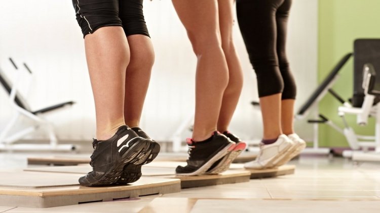
In gyms there are special calf trainers for performing toe raises. The principle is the same as the exercises described above: after raising your toes as high as possible, lower your heels over the edge of the platform until the calf and cambialis muscles are fully stretched. If the exercise is performed while standing, the arms are placed under special pads and standing on tiptoe is combined with lifting weights, which increases the load on the target muscles. During the movement, the knees should not be bent - only the ankles are used. In the seated position, the resistance caused by the load is also overcome, but here the knees are brought under the rollers. In order to work the entire triceps muscle evenly, you should train both standing and sitting. Remember that the psoas are well trained when seated and the calf muscles when standing.
A leg press can be used to train the calves. Stand in a 45 degree reclined position with your toes on the bottom edge of the platform. The distance between your feet is equal to or slightly less than the width of your shoulders. As soon as your body weight is no longer on the supports, you can begin the exercise. This involves bending and straightening the foot and giving the appearance of a toe press with the knees stationary. First the toes push the platform up, then the foot flexes and the platform lowers. There is a 1-2 second pause at the highest point. After completing all of the planned exercises for the lower leg muscles, stretch the tight calves with stretching exercises.
Raises are the best exercise for your calves
Standing and seated deadlifts are important exercises for training your calves. There are even special machines for deadlifts, but you can do without them at home.
- Sit on the edge of a chair with your weight resting on your knees.
- Raise your heels as high as possible. Don't twist your body backwards, keep your back straight.
- Lower your heels.
- Repeat the exercise 10 times.
- Do four sets.
Use a barbell, heavy dumbbells, or plastic bottles filled with water as weights. You can place your front foot on the edge of a low platform and drop your heel under your toes—this stretches the lower leg muscles more, making them more efficient.
An alternative to squats are single-leg raises:
- Sit on the edge of a chair and place your left foot on your right knee.
- Place your body weight on the heel of your left foot.
- Raise yourself on tiptoe - 10 reps.
- Support yourself with your left foot (place your right foot on it).
- Put extra strain on yourself.
- Perform a series of raises, 10 reps.
- Do two sets.
- Lean back in a chair with your hands on.
- Raise yourself on tiptoe. Pause for 2 seconds.
- Then lower your heels an inch off the floor.
- Repeat this 10 times.
- Do three sets.
Once you've mastered this exercise, you can try doing the raises while standing and unsupported. It's even better if you pick up a weight - dumbbells or water bottles. You can do single-leg exercises: two sets of ten repetitions on each leg are sufficient.
Jump rope and other leg exercises
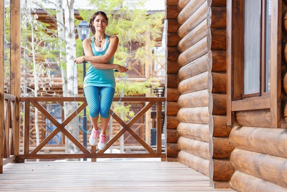
Jump rope exercises are not only good for the calves, but also for the glutes and hamstrings (especially the hamstrings). Various types of jumps can be used when exercising at home, with or without a skipping rope. One possibility is tuck jumps:
When performing squats, you can lean on your whole foot or just your toes. To make the exercise harder and speed up calf pumping, hold the weight in your hands. You don't have to do a vertical squat: you can hop back and forth, or try doing the exercise on one leg.
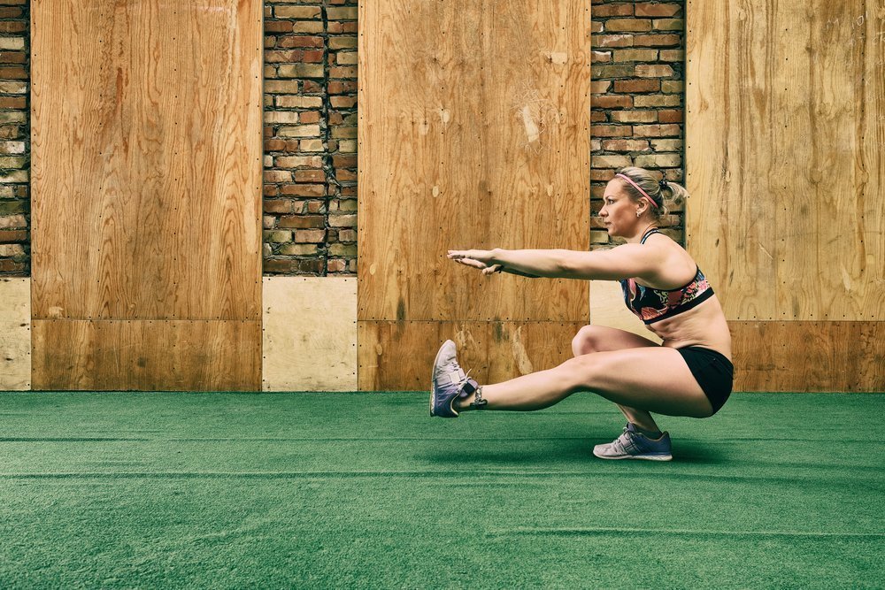
Another good exercise to strengthen your calves is the heel squat. Method:
- Stand up, put your feet shoulder-width apart. Don't turn your toes outward - keep your feet parallel to each other.
- Begin the squat the usual way — pull your pelvis back and keep your back straight.
- Lift your heels off the floor.
- As you rise from the squat, keep your heels in the air.
- Repeat the exercise 10-15 times.
- Do two sentences.
Strengthen your lower leg muscles by walking on tiptoe. Take small steps and try not to bend your knees. Do the exercises for at least 15 minutes at a time to let the muscles take full advantage of this unconventional gait.
Four leg exercises are sufficient in one workout. After the main load, at the very end of the exercise, a stretch should always be performed. The following exercise can be used:
- Take a big step backwards. Don't put your heel on the ground.
- Bend your body forward.
- Hold the stretched pose for 15-20 seconds.
- Repeat the exercise with the other foot.
- Innervation of the lower leg muscles.
- Tibialis posterior muscle.
- The flexor muscles of the foot.
- Long fibula muscle.
- Long primer.
- Lamb Muscle Soreness.
- tendons of the foot.
- Tendon of the tibialis anterior muscle.