If sitting is difficult, the exercises can also be done lying on a mat:
- Rehabilitation program after ACL reconstruction
- Full extension of the knee joint should be achieved through the following exercises:
- Movements to flex the knee joint:
- Postoperative Rehabilitation (Day 1 – 14)
- Hamstrings in a lying position
- Shin Curls vs Straight Leg Press [ edit edit source ]
- Stretch with a towel and a pillow
- yoga
- muscular structures
- front group.
- frequently asked Questions
- Recommendations for implementation
- How to strengthen the knee joints
- pain above the knee
- exercises
- Anatomy of the quadriceps muscle of the thigh.
- General movements and functions of the quadriceps femoris muscle.
- Adjacent muscles and landmarks (muscle topography).
- Superficial anatomy of the quadriceps femoris.
- Internal topography of the quadriceps muscle.
- Diagnostic tests for the quadriceps muscle.
- Errors in leg curl exercises
- Leg curls on the trainer - what to replace them with?
- Leg curls on a bench with dumbbells.
- Leg curl with weights
- Standing leg curls with elastic/elastic band.
Rehabilitation program after ACL reconstruction
In the time before the operation, the damaged knee joint should be protected. For this purpose, orthopedic aids (ortheses, bandages) are used. Persistent use of orthotics must be limited to avoid quadriceps femoris atrophy.
It is also important to reduce swelling of the damaged knee joint and restore range of motion in the joint whenever possible. By the time of the operation, the patient should have developed a normal gait.
Shifting body weight to the injured leg is acceptable as long as it does not cause pain. The administration of non-steroidal anti-inflammatory drugs (Nurofen, Ibuprofen, Nais) is recommended. Duration of use: 7-10 days after injury.
Full extension of the knee joint should be achieved through the following exercises:
1) Passive extension in the knee joint.
- Sit in a chair and place your foot on the edge of a stool or chair. Relax your hip muscles. Allow the knee joint to straighten under its own weight.
- Place your foot on a rolled up towel.
- Allow the leg to relax into a straight position.
- 3-4 times a day for 10-15 minutes. See Figure 1.
Figure 1: Support the heel with a rolled up towel.
Figure 2: Passive leg extension. The knee should go beyond the edge of the table.
Movements to flex the knee joint:
1) Passive flexion of the knee joint:
2) Increasing the degree of diffraction by wall slipping.
- Lie on your back, place the injured leg against the wall and, bending the knee, slide the leg along the wall while applying downward pressure with the other leg.

Figure 3: Sliding on the wall
Postoperative Rehabilitation (Day 1 – 14)
1) Watch out for swelling. Place the limb in an elevated position. It is possible to apply cold to the knee joint. Get up and walk, but stay in bed the rest of the time.
2) Do not sit with your leg down for a long time, it will cause significant swelling of the knee joint and the entire limb. If you have to sit for a long time, the operated leg should be elevated (on a chair in front of you).
3) It is necessary to take anti-inflammatory and analgesic drugs to control the pain.
4) Once the pain and swelling have gone down, you can begin walking with crutches.
It is not advisable to put weight on the injured leg. This can cause swelling.
The orthosis should be worn for up to 6 weeks after the operation. Flexion angle in the orthosis during this period: 0-10°
Early development of movement and extension in the joint
1) Passive knee extension with a rolled up towel. The towel should be large enough to lift your shin and thigh off the table. See Figure 1.
- Take off the knee brace every 2-3 hours to do the exercise.
- Allow the knee joint to fully passively extend for 10 to 15 minutes. During this time, the muscles of the upper and lower leg should be completely relaxed.
This exercise can also be done while sitting. Hold the heel with the sound leg and try to fully extend the knee joint.
2) Active stretching can be performed with the healthy leg. Avoid overextending the joint. See Figure 7:
Figure 7: Extension of the knee joint with the sound leg
Exercises that affect the quadriceps femoris muscle
1) Isometric contractions of the thigh muscles should be started as early as possible.
This exercise prevents atrophy and contracture of the quadriceps muscle and reduces swelling and fluid in the knee joint.
Hamstrings in a lying position
For advanced and professionals
Source: 'Body building. A book for coaches.
Publisher: Oksana Usoltseva Published.: Eksmo 2013.
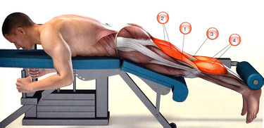
Muscles that work on the exerciser when bending the leg: 1 – semitendinosus; 2 – biceps; 3 - semitendinosus; 4 - calf muscle
level of trainingBeginner to advanced.
The main load is on the back of the thighs.
- step 1. Lie face down on a trainer with the rollers just above your heels.
- Step 2 .. Exhale and slowly bend your knees at maximum amplitude.
- Step 3 .. On an inhale, slowly return to the starting position.
Make sure your body doesn't move away from the surface of the machine during the movement, as this takes stress off the hamstrings and shifts the stress to the glutes. The one-leg curl variation is great for isolated training and non-harmonic hip development.
Shin Curls vs Straight Leg Press [ edit edit source ]
It is generally accepted that training with free weights is more effective than training on machines. However, there are exceptions to all rules. Training hamstrings while lying down results in greater muscle gain than lifting weights on straight legs. The American researcher Brad Schoenfeld published these results in the Journal of Strength and Conditioning [1].
Ten men who had previously trained with weights took part in the experiment. Athletes performed straight leg deadlifts and supine shin curls. Muscle activity and conductivity were measured during exercise using electrodes attached to the muscles. The greater the electrical activity in the muscle, the more tightly the muscle was tensed and the greater the growth stimulus it received.
Flexing the leg elicited greater electrical activity in the lower leg than straight leg extension. Flexion also mobilized more muscle fibers in the outer and inner parts of the biceps femoris.
The upper posterior surface of the thigh responded equally to straight leg flexion and straight leg extension. The more repetitions performed in lower limb flexion, the greater the degree of muscle tension, indicating greater isolation of the biceps femoris in this exercise. However, that doesn't mean the straight leg press is an ineffective exercise.
'It is enough to slightly increase the duration of the approach, for example by slowing down the tempo, and the deficits identified are compensated by the appearance of more growth factors in the muscle cells, as well as the additional recruitment of new muscle fibers.
Comparison of electrical activity in hindlimb muscles
| flexion of the lower leg | Push straight leg | |
|---|---|---|
| Lower outer part | 110 | 40 |
| Lower inner part | 79 | 42 |
| Upper outer part | 82 | 75 |
| upper body | 120 | 122 |
Stretch with a towel and a pillow
How do you stretch hamstrings with a towel? Roll it up and lie on an exercise mat. Place one leg bent at the knee on the floor and loop the towel around the other leg and pull up 90° to stretch the muscles. Try to straighten the leg as much as possible and keep going until the ligaments burn.
The second method is to kick a pillow or punching bag with your toes straight to loosen and stretch the tendons.
Also read: Shin Splint Exercises
yoga
Performed at home, asanas can help safely stretch ligaments and leg muscles.
- Supta Padangushthasana is performed as follows:
- Lie on your back;
- Place the corset over the foot;
- Stretch outstretched leg up;
- Extend your arms without lifting your shoulders off the floor;
- The other leg points to the floor;
- raise your head;
- Repeat the exercise as often as possible.
- Uttanasana technique with a chair:
- Place the roll on a chair;
- stand 0.5 m from the chair, feet hip-width apart;
- spread your elbows and place your hands and head on the roll;
- The feet are perpendicular to the ground;
- Lift for a breath while straightening the spine.
- How Prasarita Padottanasana is performed:
- Feet in limb width.
- arms at the sides.
- Gently bend forward while twisting the hip joints.
- As you inhale, remain still.
- As you exhale, bend down until your head is in line with your feet.
- As you inhale, gradually return to the starting position.
- Perform what is known as the downward facing dog pose as follows:
- Sit on your knees and breathe in.
- As you exhale, bend forward.
- Extend your palms forward and press them against the mat.
- Hold this pose for 15 seconds to a minute.
- Methods for performing variations of the pigeon pose:
- Sit on your mat and pull your left leg back.
- Lift it up and bend it slightly.
- Bend your right leg and place it on your side, like in the lotus position.
- Tilt your head back with the back of your head resting against your back leg.
- Stay like this for about 1 minute.
muscular structures
The knee is the bulging part that connects the thigh to the lower leg. The muscles are divided into 3 classes:
They also work in the hip joint. They fulfill movement and resting functions, such as the gluteus maximus muscle.
front group.
Allows bending of the leg at the knee joint. It consists of:
The first muscle extends from the anterior crest of the pelvis to the tibia and descends to the broad tibial fascia. It allows flexion of the thigh.
The last complex is a strong extensor muscle of the leg. Types of Quadriceps Muscle Group:
- The rectus femoris muscle of the thigh is the most stretchable. It runs from the buttocks and extends to the surface of the thigh. It is characterized by the presence of two joints. When the pelvis is stationary, pull the thigh towards the breastbone and straighten the knee.
- Medial - Located on the inner area of the front surface of the hip. It moves down and merges into the thick tendons of the thigh.
- Lateral - is the largest of all the thigh muscles described. It arises from the tendon ligament and connects to the rectus.
- Median hamstrings - is the weakest and most vulnerable of all quadriceps parts. He is between the previous types. It starts at the origin of the kneecap.
The medial or medial complex includes two variants that form the inner layer of the thigh. These are:
The posterior group of muscles of the upper limbs includes the following:
- The biceps femoris (thigh muscle) is spiral and elongated. It originates at the top layer of the medial ischial tuberosity, then lengthens with the second head and merges into the flat tendon. Allows limb flexion, balancing and knee joint extension.
- The semimembranosus tendon runs parallel to the previous tendon. Approximately in the middle of the thigh it merges into the long tendon and connects to the medial layer of the tibia at the top.
- The semitendinosus muscle begins at the ischial tuberosity and attaches to the abdominis muscle in the middle of the thigh.
frequently asked Questions
Osteoarthritis has many limitations when it comes to physical activity. All types of physical activity are categorically contraindicated, especially competitive sports and other sports with a high risk of joint injuries. Light jogging, walking, fitness with limited loads on sore joints is allowed. Exercise bikes make sense.
You can and should support the joints with medication and physical therapy.
You can and should combine different techniques, especially gymnastics and swimming. In any case, your doctor will make the final decision on how to administer physical therapy.
Read this article about other treatment options for osteoarthritis.
- Ulashchik, WS Therapeutic physical training//Physiotherapy: a universal medical encyclopedia. -Knizhny Dom, 2008, pp. 308-315, 640 pages.
- Therapeutic physical training/ Ed. N. Popov. Fizkura i Sport, 1988, 271 p.
- Dubrovsky VI Therapeutic physical education: a text-book for students. – Moscow: VLADOS, 1998-608 p.
- Epifanov VA Therapeutic physical education.-M.: Medicine, 1987, – 528s.
Recommendations for implementation

The exercises must be useful and not aggravate the patient's condition. Once you know what exercises you can use to work on a painful knee joint, you need to look at the general recommendations.
The most important rule is that you should train regularly because a single exercise doesn't make much of a difference. The aim should be to do the exercises regularly, even if they are difficult at first.
It is important to gradually increase the load and not try to immediately overload the knees. Too much exercise can lead to torn ligaments and strained muscles. The exercises should be done judiciously, and one should not try to reach one's full potential right away.
The average workout takes about 30 minutes a day. That is enough for positive effects to set in and the pain to go away. Do not train too long so as not to overload the problem area.
It is important that the exercises are done under the supervision of a trainer or doctor. Your doctor will tell you which exercises you can and cannot do.
During the exercises, there should not be any serious discomfort, and if there is severe pain, you should stop doing the exercises. It is worth discussing the problem with a specialist. He or she will tell you how to exercise so that you are not in severe pain.
If only one joint is affected, both should be loaded. This ensures even blood flow and facilitates movement of the limbs.
Stick to the rules so that training does not harm your health, but benefits it. If problems arise during training, immediately contact a specialist and adjust your training program.
How to strengthen the knee joints

Many people wonder how to strengthen their knee joints and ligaments. Exercise can help here and prevent diseases. The main goal of the exercises is to increase the flexibility of the ligaments and improve blood circulation.
- Knee stretches in the supine position. The person should lie on their back with both legs bent at the knees. The heel is pressed to the floor and one leg is lifted and straightened. Hold this position for 2 minutes, then slowly lower. Repeat the exercise 5 times, alternately straightening your legs.
- knee extension. The patient sits on the table and puts his feet on the floor. He slowly straightens his knee and holds it in this position for a minute. Then he returns to the starting position. This is repeated 15 times.
- Asymmetrical movements. Lie on the floor with your right leg bent at the knee and your left leg straight. Alternate the position of the legs and hold them in this mass for up to 2 minutes.
Simple exercises will help relieve pain and prevent disease.
pain above the knee
Topical ointments and oral non-steroidal anti-inflammatory drugs are very helpful in this case.
Muscle pain below the knee is a sign of many diseases:
- mechanical injuries of muscle fibers and tendons;
- varicose veins on the lower limbs;
- atherosclerosis and arteritis;
- Inflammation of muscles and nerves.
The most common cause of muscle pain in the legs below the knee is heavy physical exertion. Minor tendon injuries do not cause any serious clinical symptoms, apart from occasional pain when walking. Persistent mechanical injury to the muscles over time increases inflammation and causes swelling below the knee.
The second most common cause of knee muscle pain is a spinal disorder that can pinch nerve endings:
They increase with palpation of the posterior shin area and with sudden flexion of the body. The cause of pain in the leg muscles below the knee is bursitis (inflammation of the muscle covering in the tendons). This is caused by inflammation of the knee joint or damage to the meniscus. If the joint space in the knee joint is severely narrowed, the cartilaginous structures are damaged and inflammatory fluid forms. This accumulates in the tendon sheaths, causing swelling in the back of the knee.
Pain in the muscles below the knee can occur due to
- Nerve damage (polyneuropathy) occurs
- bone inflammation (osteomyelitis);
- taking medicines (statins);
- decreased levels of calcium, potassium and magnesium in the blood;
- Bone formation disorder in children and adolescents (Osgood-Schlatter disease).
The pain in the muscle below the knee is not localized. They extend 10-15 cm, suggesting that the anatomical structures of the lower limbs are expanded. The pain syndrome increases with exertion and decreases when the exertion is stopped. Muscle pain below the knee bothers smokers. Severe pain is caused by deep vein thrombosis in the lower leg. They are located below the knee and increase with dorsiflexion of the foot and pressure on the front of the tibia.
exercises
To relieve pain in the muscles above the knee, physiotherapists recommend doing gymnastic exercises every day. Lie on your back and bend your knees, keeping your feet hip-width apart and pressing down on the floor. Lift your pelvis and support your weight on your ankles, tighten your abs, bring your knees perpendicular to your ankles (your body should form a straight line from your shoulders to your knees), hold this position 3 to For 5 seconds, then slowly lower your pelvis to the floor.
To strengthen your hamstrings, lie on your back and stretch your arms out to your sides. Wrap your legs around a large exercise ball and raise it perpendicular to the floor. Squeeze the ball with your inner thigh muscles 10 times. Do this exercise in 2 or 3 sets.
To strengthen the outer thighs, lie on your side, which doesn't hurt. You can lie down on an exercise mat so as not to lie on a hard floor. Lift the sore leg 6 inches off the floor. Hold it in the air for 2-3 seconds, then lower it back onto the other leg.
- Lie on your back and stretch your arms out to the sides;
- Bend the leg you are stretching by placing your foot on the floor;
- Keep the other leg straight on the floor with the toe pointing up;
- Lower bent leg to side as far as possible;
- Lower the bent leg for five seconds and return to the starting position.
Squeeze the glutes. To do this, roll a towel into a tight cylinder, lie on your back and bend both legs, planting your feet on the floor. Place the towel between your knees and squeeze to engage your inner thighs and glutes. Squeeze for 3-5 seconds, then relax your buttocks.
To reduce muscle pain in the legs below the knee, do an exercise that improves venous outflow through the blood vessels of the lower limbs. Have a fitness mat or pillow ready. While lying down, place a pillow under your lower back and straighten your legs against a wall. Press your glutes firmly into the floor. Begin to climb your legs down and back up the wall for a minute, then pause for a minute and repeat the exercise. Do 5 repetitions.
Anatomy of the quadriceps muscle of the thigh.
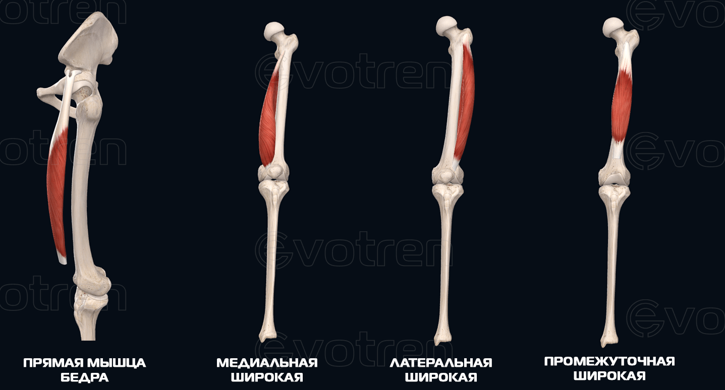
Rectus muscle of thigh (m.rectus femoris).
Origin:
– Lower iliac spine (Anterior inferior iliac spine);
– Upper part of the acetabulum.
Features and functionality:
– Spindle shape on both sides;
– takes a medial position;
– straightens the tibia in the knee joint;
– Flexion of the hip joint
Medial broad muscle (m. vastus medialis).
origin:
– Lower part of the intervertebral line;
– The medial lip of the cruciate ligament (Labium mediale lineae asperae femoris).
features and function.:
– is a thick, flat muscle;
– occupies the anterior and medial surfaces of the femur;
– Stretches the tibia in the knee joint.
Broad lateral muscle (m. vastus lateralis)
origin:
– greater trochanter;
– intervertebral line;
– lateral lip of thigh raw line (Labium laterale lineae asperae femoris).
Features and Functions:
– The broadest of all heads;
– located on the lateral and partially posterior surface of the thigh;
– Stretches the tibia in the knee joint.
Medium broad muscle (m. vastus intermedius)
origin:
Front surface of the thigh (below the intertrochanteric line, Intertrochanteric line).
characteristics and function:
– Lies under the rectus femoris muscle in the form of a tendon plate;
– Stretches the tibia in the knee joint.
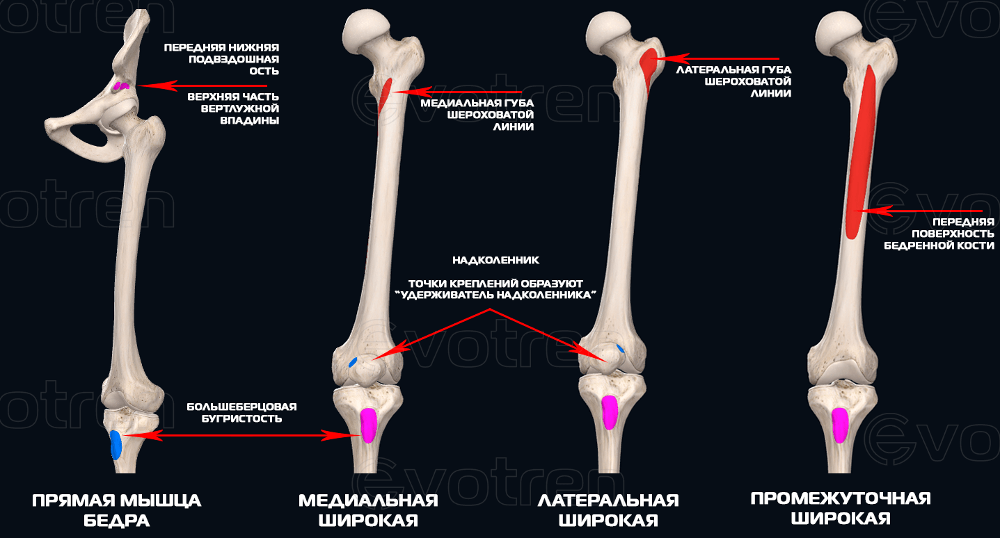
Insertion of the musculus tetriceps femoris:
- All four heads form a powerful tendon that attaches to the kneecap and extends into the medial ligament of the kneecap (Ligament patellae);
- The patellar ligament attaches to the tibial tubercle (Tibial tuberosity);
- Part of the tendon fibers of the lateral and medial broad muscles of the thigh on the sides of the kneecap pass laterally and form the patellar retinaculum (Retinaculum patellae).
General movements and functions of the quadriceps femoris muscle.
- hip flexion (with the pelvis at rest);
- Forward rotation of the pelvis (pelvic tilt, with leg at rest) -. not to be confused with the examination of the pelvic position in statics;
- stabilization and centering of the kneecap;
- Extension of the lower extremity in the knee joint.
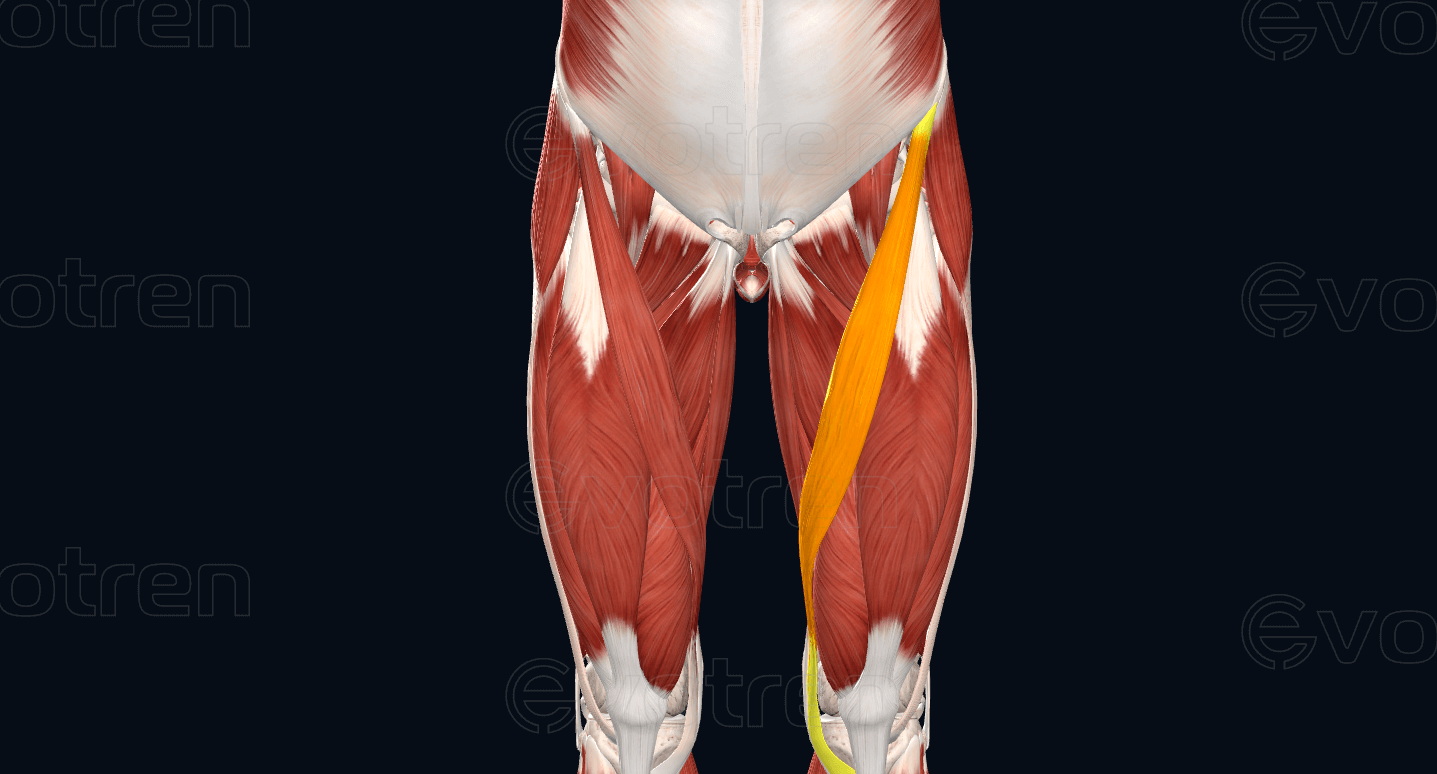
Adjacent muscles and landmarks (muscle topography).
Superficial anatomy of the quadriceps femoris.
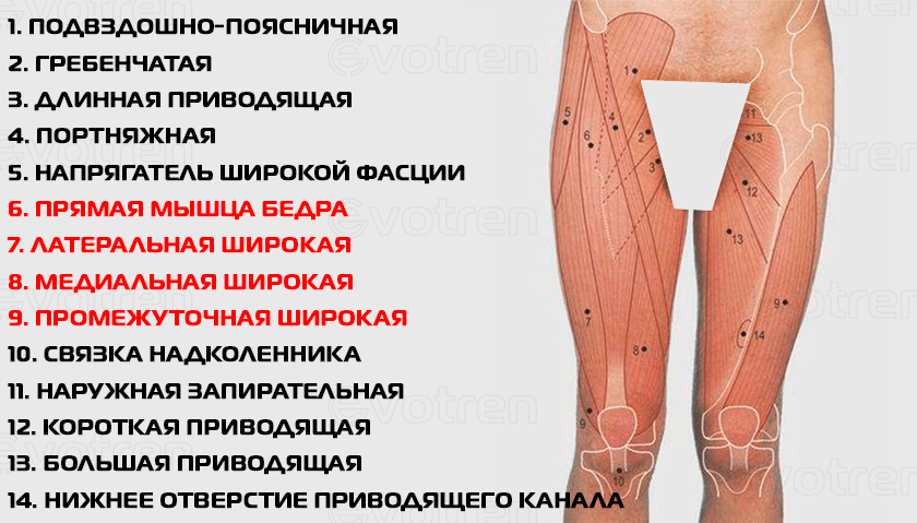
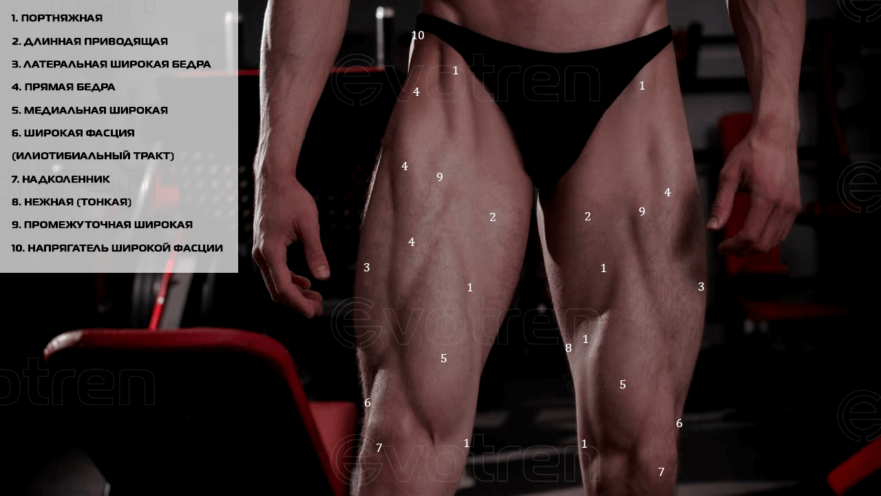
- upper – groin fold, pelvic bones;
- laterally – wide fascia (oriotibial tract);
- medial – Crown fascia, adductor longus, tender (thin) spot and goosebumps;
- Below: – From below: patella ('patella'), patellar ligament and tibial tuberosity.
Note that the sartorius begins above the rectus femoris, runs along its medial border, and then descends behind the medial border of the medialis muscle and inserts at the goose foot area of the tibia
Internal topography of the quadriceps muscle.
Since the quadriceps is a superficial muscle, from the front it is only overlapped obliquely (downward and medially) by the biceps muscle.
From the side, the broad fascia of the thigh (tractus iotibialis) partially overlaps the lateral broad muscle.
Diagnostic tests for the quadriceps muscle.
The following video shows the test options for analyzing the quadriceps muscle.
Chiropractic Muscle Test for the Quadriceps Muscle:
Errors in leg curl exercises
It might seem difficult to make a mistake with this exercise, but we'll tell you the most common mistakes.
- Too deep a grip. If you place the pad on the heel or Achilles tendon, forget about quality work on the back of the thighs: part of the load will be absorbed by the calves, and you will feel fatigue much faster.
- Elevate your pelvis as you bend your knees. Be sure to keep your pelvis firmly pressed against the mat or you risk injuring your lower back. If your pelvis is loose, it's a sure sign that you need to reduce your working weight.
- Lift your legs by the power of inertia. You'll always be tempted to swing your legs slightly due to inertia, especially when you're doing leg curls after a core exercise and you lack the energy. Just control each movement and remember that otherwise all the weight will be on your knees.
- Raise your legs with a jerk. This is especially important if the weight is too heavy, because then you can't bend your legs evenly and you're straining your back.
- Support yourself with your knees on a bench. Yes, just go easy on them.
Leg curls on the trainer - what to replace them with?
If you don't have such a machine at your gym or you train at home, try doing the leg curls with a a similar exercise.. Of course, you won't get full consistency, but with the right technique, you'll work the hamstrings just as effectively. Try the following variations:
Leg curls on a bench with dumbbells.
You perform this exercise the same way you do a bench leg curl, but with the following elements A dumbbell clamped between your legs. We know the main problem here is finding someone to hand you the dumbbells, but you can still get creative. This has an important benefit: As with any free weight work, you increase the stress on your stabilizers.
Leg curl with weights
Grab a vertical support with your hands and keep your body straight. Alternately Leg curls with weights. Bend the heel of your buttocks as much as your muscles will allow, do not throw your foot on the floor.
The most important thing is that your knee doesn't wobble'. In this case, of course, you are limited by the weight, but you can fully control your technique and do more repetitions. Trust me, the main goal is achieved.
Standing leg curls with elastic/elastic band.
By performing some kind of loopYou attach the elastic to a vertical object, pull the bottom of the loop over your foot, and perform the same leg curl. We really like this variant because the tension of the band allows the exercise to be performed evenly and perfectly engages the stabilizing muscles.
If you don't have any equipment at all, do lunges. But this is another story.
Read more:- The flexor muscles of the foot.
- Which muscle extends the lower leg and flexes the thigh.
- Triceps femoris muscle.
- Lamb Muscle Soreness.
- abdominal muscle.
- muscle work while running.
- Knee splint for knee arthrosis.
- lower leg muscles.