Patients most commonly report pain 'in the middle' of the heel and in some cases the arch of the foot. The inflammation is strongest in these areas.

- ALMOST
- ontogenesis
- ICD-10
- Causes of enthesopathy
- Diagnosis of plantar fibromatosis
- Treatment of longitudinal fibromatosis
- Dissection of the plantar fascia, where to do it, where to go in St. Petersburg?
- Treatment of heel spurs with UHT
- Heel spurs: causes, symptoms
- The advantages of our treatment:
- diagnosis
- Why do heel spurs form?
- Internal causes of fasciitis
- external causes
- How can fasciitis be distinguished from other foot problems?
- clinical picture
- Additional investigation methods
- Soft tissue diseases in the foot area
- Surgery for plantar fasciitis of the foot
- Advice and recommendations
ALMOST
FASCIA [fascia (PNA, JNA, BNA); Latin fascia bandage] are sheaths of dense fibrous connective tissue that cover muscles and their tendons, some organs and neurovascular bundles. Fascia is part of the soft skeleton (see) and has supporting and trophic functions.
The structure of the fascia and its functional significance are discussed in the works of NI Pirogov (1838), PF Lesgaft (1905), AP Samarin (1912), VN Shevkunenko (1938),
В. V. Kovanov and TI Anikin (1961, 1967), AP Sorokin (1964), E. Singer (1935), J. Obersteg (1948), R. Last (1956), J. Lang (1960) and others. Most researchers assume that the formation and development of fascial sheaths around muscles, organs and vessels is related to movement. The formation of fascia is considered to be the response of connective tissue to the pressure it is exposed to due to changes in the volume of individual organs as they function. There are different opinions in research about the definition of the term 'fascia'. The term 'aponeurosis' is often used instead of 'fascia'. AP Samarin considers all types of connective tissue membranes - from thin-cell to aponeurotic - to be fascia (see aponeurosis). Last considers only those connective tissue lamellae that have a fibrous structure and aponeurotic character to be fascicles. В. V. Kovanov and TI Anikina define fascicles as connective tissue sheaths that cover muscles, tendons, nerves and organs; In their opinion, there is little difference between fibers, fascicles and aponeuroses (see connective tissue).
ontogenesis
The formation of fascicles from mesenchyme begins from the 2nd to 4th month of intrauterine development. In the 5th month, the connective tissue fibers of the fetus are bound and converted into collagen fibers (see Collagen). Elastic fibers (see elastin) develop later than collagen fibers. Where the fetal connective tissue comes into contact with other tissues, its boundary layer thickens, particularly around the muscles. The fascial sheaths of the nerves and vessels develop in cellular, cell-fibrous, fibrillar, and fibrous stages; the transition from one stage to another is gradual. At the end of intrauterine development, the fascia consists of fibroblasts and individual collagen fibers, which then come together to form thin bundles. In newborns, the bundles of collagen fibers are scattered throughout the fascia. As the fibers grow, the bundles adhere tightly to each other. In adults there are only individual compression cells in the fascia. The curvature of collagen fibers with normal curvature does not become visible until the third year of life. At the age of 7-10 years, the curvature of the collagen fibers in the fascia reaches a maximum. With age, the curvature of collagen fibers is straightened, and in some places they disappear and gaps form between the fiber bundles. At the age of three, thick elastic fibers begin to appear in the fascia, and by the age of 10-11 they reach the same size and shape as adults.
The age-related changes in the fascia are primarily due to the condition of the vessels and nerves that ensure their blood circulation and innervation. As we age, the fascia becomes thin and interspersed with fatty tissue.
ICD-10
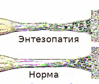
Enthesopathies (Latin: enthesis – connection between tendon and bone) are a common group of diseases of the musculoskeletal system. The name is derived from the word 'enthesis' or 'enthesis', which refers to the place where connective tissue formations attach to bony structures, and has been used in medical literature since the 1960s.
Some experts interpret the term more broadly to include not only enthesopathies, but also inflammation of adjacent tendon locations and inflammatory processes in the area of the tendon sheath. Statistically, enthesopathies are diagnosed in 35-85 % of patients with joint diseases. They often occur as part of autoimmune diseases in athletes and people in certain professions. They tend to have a long-term course with gradual progression, degenerative-dystrophic tissue changes, functional deterioration and increased likelihood of injury to the altered anatomical structure.
Causes of enthesopathy
In traumatology and orthopedics, two types of enthesopathies are distinguished, which are based on the etiological factors: primarily inflammatory and primarily degenerative. Primary inflammatory pathology occurs when the inflammation in arthritis spreads from neighboring joints. The primary degenerative process occurs as a result of repeated minor injuries with constant overload or is the result of a one-time major injury (wound, tear in the area of the enthesis). The cause of overload can be both high physical activity and impaired biomechanics of movement due to diseases of the musculoskeletal system. Factors that increase the likelihood of overuse injuries include:
- Consistent physical activity. This pathology often occurs in athletes (tennis players, runners, footballers, weightlifters, etc.) and in certain professions (construction workers, loaders, painters, circus and ballet dancers), due to repetitive movements that cause excessive stress and repeated microtraumatization of the joints. Repetitive microtrauma is often exacerbated by more severe injuries with the formation of scar tissue.
- joint diseases. Experts consider enthesopathies to be a fairly specific feature of seronegative spondyloarthropathies, including Bechterew's disease, psoriatic arthritis, Reiter's disease and other reactive joint injuries of urogenital origin, as well as reactive arthritis secondary to infectious enteritis, nonspecific ulcerative colitis and Crohn's disease. This pathology can occur with osteoarthritis and is especially common in osteoarthritic and dystrophic lesions of the knee and hip joints.
- Trophic Disorders. Deterioration of tissue exchange in the area of enthesis can be caused by neural dysregulations in radicular syndromes, insufficient local blood supply in cardiovascular diseases, and changes in the hormonal background during menopause in women.
- Connective tissue dysplasia. Congenital incompleteness of the connective tissue structures is associated with a high probability of microinjuries to the tendon and ligament apparatus, followed by the development of inflammation even with minor loads. Hereditary collagenopathies are a major cause of enthesitis lesions in young adults.
Diagnosis of plantar fibromatosis
Confirmation of the diagnosis of plantar fasciitis is strong pressure of the thumb on the heel bone during dorsiflexion of the foot, causing pain. Pain along the medial edge of the plantar fascia is also possible. If the clinical findings are inconclusive, the presence of a heel spur on the radiograph may confirm the diagnosis; however, the absence of lesions on the x-ray does not exclude the diagnosis, and visible spurs are not the cause of plantar fasciitis symptoms. Additionally, in some cases, heel spurs are difficult to detect on x-ray and appear as brittle foci of newly formed bone, suggestive of spondyloarthropathies Overview of Seronegative Spondyloarthropathies Read more (e.g., ankylosing spondylitis or reactive arthritis Read more ). An MRI scan is indicated if an acute fascial tear is suspected.
Other conditions that cause heel pain can mimic plantar fasciitis
Acute severe heel pain with redness and warmth may indicate gout Podagra Podagra is a condition caused by hyperuricemia (blood uric acid levels >6.8 mg/dL [>0.4 mmol/L]), which leads to the precipitation of sodium mononitrate crystals in and around the joints. Read more .
Treatment of longitudinal fibromatosis
To reduce stress and pain in the fascia, you should take shorter steps and avoid walking barefoot. Activities that place impact on the foot, such as walking. B. running should be avoided. The most effective treatments for calf fasciitis include calf and plantar fascia stretching exercises and the use of orthotics to stretch the calf and plantar fascia muscles while you sleep. Orthotics can also reduce fascial tension and pain. In addition, exercise correction, weight loss in obese patients, nonsteroidal anti-inflammatory drugs (NSAIDs), cooling massage and sometimes glucocorticoid injections are used. However, the administration of corticosteroids can exacerbate degenerative changes in the plantar fascia, so many clinicians limit their use (see 'Use of corticosteroids in obese patients'). Recommendations for the use of corticosteroid injections Use of glucocorticoid injections ).
In refractory cases, physical therapy, oral glucocorticoids, and immobilization may be recommended before surgical treatment is considered. The newest form of treatment for unresponsive plantar fasciitis is extracorporeal shock wave therapy (ECWT), which uses a hand-held applicator to deliver low-frequency pulsed waves to the painful area. Pulsed shockwave therapy is a safe, non-invasive method that stimulates metabolism and increases blood flow as well as repairs damaged tissue and accelerates healing. EHT is used in large medical centers (1 Treatment Note Plantar fasciitis is characterized by pain at the attachment of the longitudinal fascia to the heel bone (calcaneal enthesopathy), which may radiate along the medial edge of the longitudinal bone. Read more).
Dissection of the plantar fascia, where to do it, where to go in St. Petersburg?
At 'AndroMeda' Clinic, surgical manipulation of aponeurosis dissection is initiated with local anesthesia to reduce pain in the surgical area. The surgeon makes 2-3 longitudinal punctures in the skin in the metatarsal area. The surgeon then turns the scalpel transversely and cuts the PA from the depth of the soft tissue outward. After surgery, it is important to follow the doctor's instructions to ensure rapid healing of the limb.
To find out the prices, how much an aponeurosis dissection costs in St. Petersburg, how much an aponeurosis dissection costs – call the clinic or make an appointment with a specialist in our clinic.
| Dissection of the subscapular aponeurosis, price | 8,000-15,000 rubles. |
| Specialist consultation for dissection of the plantar aponeurosis, price | 600-1,600 rubles. |
Treatment of heel spurs with UHT
Heel spurs: causes, symptoms
The human foot is a perfect mechanism given to us by nature. The thin bones of the foot support the weight of the human body, similar to arched bridges made of thin elements that support the weight of a car driving over them.
| On the plantar side, the sole of the foot is supported by the plantar aponeurosis, a strong ligament that connects the front and back of the foot. It prevents the foot from flattening under the weight of the body. The rear end of the plantar aponeurosis is attached to the heel bone, the front ends are attached to the toes. |
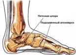
| This is where the spur is most likely to form. The plantar aponeurosis and the tissue surrounding it are put under a lot of stress in an upright position. The ligament can usually withstand this stress, and rare micro-tears heal spontaneously and without much pain. |
The advantages of our treatment:
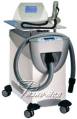
- Treatment with the new generation Zimmer UHT device from Germany.
- Magnetotherapy treatment when paying for UHT treatment for free.

Heel Spur Pictures of different spurs HERE.
Below are various photos of heel spurs. This is what a heel spur looks like on an x-ray. As you can see in the photo, spurs can be large and small, but the pain does not depend on the size of the spur. There are spurs that are larger than 1 cm and do not hurt (heel spur photo 1), while very small spurs hurt a lot (heel spur photo 2). There are also cases where the spur is not present at all, but pain still occurs. This can be explained by the fact that the spur itself does not cause pain, but the pain comes from the torn ligaments. If the Achilles tendon is torn, the spur grows upward on the ligament (heel spur photo 3).
diagnosis
A simple analysis of the patient's complaints, a visual inspection, palpation of the sole of the foot and an X-ray are sufficient in most cases to correctly diagnose heel spurs. X-rays and palpation of the heel spur help differentiate plantar fasciitis from possible bone fractures. The absence of bony articulation with pain requires an additional differential diagnosis, which refers to the possible presence of systemic inflammatory diseases such as ankylosing spondylitis, rheumatoid arthritis, Reiter's syndrome and some others. Such diseases can also cause heel pain. The presence of a bony growth is not always a reliable symptom, as 70% of adults have such a growth but do not experience pain. Therefore, the diagnosis should be made by a specialist who will be able to correctly determine the cause of heel pain and also rule out other diseases with similar symptoms.
Effect of geroprotectors - short peptides: - Stabilization of metabolic processes in bones and connective tissue - Acceleration of the regeneration of musculoskeletal tissue in the event of inflammation - Pain relief when walking - Increasing plasticity.
Composition:
– AC4 peptide complex – Alanine – Glutamic acid – Aspartic acid
Method of use: 1 capsule 2 times a day before meals. Course of 20 capsules.
Why do heel spurs form?
Plantar fasciitis – is a micro-injury to the fascia with subsequent inflammation. At a young age, the constant impact on the heel is compensated for by the normal work of the skeletal muscles responsible for cushioning.

Internal causes of fasciitis
An internal cause impairs neuromuscular damping and causes uncoordinated muscle work:
- Neuromuscular attenuation is impaired;
- unbalanced load on the foot and spine.
- Infections (chlamydia, gonorrhea);
- arthritis (inflammation of the joints);
- Connective tissue diseases (lupus erythematosus).
Excessive muscle strain during sporting activities, especially running and jumping, is also harmful. Another cause is an imbalance of blood and lymph flow in the legs due to arteriosclerosis or diabetes.

A heel spur does not form overnight. It is a long process that is preceded by inflammation in the fascial tissue. A big mistake is not seeking medical help for heel pain.
Unconsciously, you begin to protect the painful area and step on your toes or wrap your foot, which increases the destructive stress.
Attempts to endure the pain or self-medicate with home remedies only make the situation worse.
external causes
External causes of plantar fasciitis are more likely to affect women over 40 and men over 50. The main cause is:
How can fasciitis be distinguished from other foot problems?
Fascia inflammation – is an inflammatory process that occurs in response to the progressive death of fascial cells as a result of repeated microtrauma. The body is unable to break down and remove the dead cells and swelling and inflammation occur. Characteristic symptoms are:
- 'Initial' pain in the heel, which occurs not as a result of exertion, but beforehand, when the feet have been resting for a long time - in the morning or after sitting for a long time;

The foot hurts much more in the evening as a result of all-day exertion.
The symptoms of plantar fasciitis can be similar to other foot conditions, so it is important to make the correct diagnosis.

Plantar fasciitis is treated by a Podiatrist or Podiatrist or surgeon.
clinical picture
Patients with heel spurs complain of pain when taking the first steps in the morning or after sitting at a desk for a long time or while driving a car - the so-called start-up pain. The pain occurs in the heel and can be very severe (Figure 1).
Improvement often occurs when you take the first steps or stretch the shin muscles and the fascia of the foot. However, the pain usually returns during the day, especially if the patient walks or stands a lot. A burning pain is not typical of plantar fasciitis and can occur when there is nerve irritation (e.g. Baxter's neuritis).
The main causes of plantar fasciitis are
- Old
- Recent increase in physical activity (e.g. new running program)
- Work that requires standing on your feet for long periods of time
- Weight gain
- stiff (rigid) calf muscles
On clinical examination, the pain usually appears on the inside of the heel from the sole of the foot. The pain also occurs with direct pressure (palpation) on this area.
Stiffness in the lower leg muscles is also a common symptom. Symptoms may be exacerbated when the toes are contracted, stretching the plantar fascia (see Figure 3). There is a link between flat feet and the development of plantar fasciitis, but the condition can occur in any type of foot.
Plantar fasciitis is the most common cause of heel pain, but there are other, less common causes:
- Heel overuse pain syndrome
- Soft tissue atrophy in the foot
- Compression of the first branch of the lateral median nerve (Baxter nerve)
- Tarsal Tunnel Syndrome
- Stress fracture of the heel bone
- Periostitis
- Inflammation due to seronegative arthritis
Additional investigation methods
The diagnosis of plantar fasciitis is usually made based on the patient's complaints and clinical examination. X-rays of the feet are not necessary to make the diagnosis. However, when x-rays are taken, heel spurs are visible in the lateral projection.
It is important to know that overuse of the plantar fascia can cause excessive bone formation in the form of a heel spur. However, the presence of a heel spur does not directly correlate with symptoms.
Many patients have x-rays of the foot that show a spur but no symptoms, and conversely, patients with plantar fasciitis do not have a heel spur on x-rays.
Initially, patients are not prescribed an MRI scan of the foot. However, if symptoms persist after treatment, an MRI scan may be ordered to rule out other causes of heel pain - such as a stress fracture of the heel bone.
Soft tissue diseases in the foot area

MMuscles, tendons, tendon sheaths, ligaments, fascia, aponeuroses and the joint capsule play an important role in joint stability. Periarticular soft tissue pathologies can be considered both as concomitant diseases of arthritis and as an independent pathology. The following terms are commonly used to describe soft tissue pathologies:
- Tendinitis – inflammation of the tendon tissue;
- Tenosynovitis/Tendovaginitis – inflammation of the tendon tissue and tendon sheath;
- Enthesitis/Enthesopathy – Inflammation of the tendon tissue where the tendon attaches to the bone;
- Bursitis – Inflammation of the synovial capsules, thin-walled cavities lined with synovial membrane that facilitate the movement of tendons and muscles over bony prominences.
pAnkle and foot pathologies as well as periarticular soft tissue lesions in this area are a common reason for a visit to the doctor and, according to national and international literature, account for between 6 and 21 % of all musculoskeletal pathologies.
pThe causes of soft tissue abnormalities in the ankle and foot area can be caused by both external and internal factors. Extrinsic factors include overload (altered movement patterns), trauma (single or repeated microtrauma) and local injection of corticosteroids (ICS) into the tendon, which can cause degeneration of tendon tissue, intrinsic – congenital anomalies of the joint structures, leading to abnormal Biomechanics lead, imbalance of the muscles surrounding the joint, hypodynamia (immobilization), impaired blood supply to certain tendon zones, age-related regression of the musculoskeletal system. There is often a combination of several factors.
Surgery for plantar fasciitis of the foot
Surgical intervention is necessary in patients whose symptoms persist despite appropriate therapy. Removal of the bony growth produces positive results. An undesirable consequence of surgical treatment of plantar fasciitis can be weakening of the arch of the foot.
Plantar fasciitis can be cured if a complex of therapeutic measures is initiated in a timely manner. Patients are recommended to see a specialist at regular intervals in order to prevent a possible recurrence of the disease or to minimize residual consequences. After the operation, rehabilitation is required, the duration of which varies from case to case.
Advice and recommendations
Stretching is one of the most important ways to treat plantar fasciitis. This technique focuses on stretching the muscles of the foot and the entire limb. Targeted exercises should be performed several times a day. Muscles can be strengthened by rolling a tennis ball or roller.
This article is intended for information and awareness purposes only. Please remember: self-medication can harm your health.
Specialist in orthopedics and traumatology, pediatric orthopedist and traumatologist
Head of the Department of Traumatology and Orthopedics, Category II orthopedic traumatologist, trainee doctor.
Read more:- tibial fasciitis.
- Treatment of plantar fasciitis.
- Tear of the foot fascia.
- Treatment of plantar fasciitis at home.
- How to cure plantar fasciitis forum.
- Structure of the human heel.
- fasciitis.
- Treatment of plantar fasciitis on the sole of the foot.