The pectoralis major muscle (Latin: pectoralis major muscle) is a large, fan-shaped superficial muscle located at the front of the rib cage. Below it is the triangular pectoralis minor muscle.

- The hip flexors become weak from sitting for too long.
- What happens when the hip flexors become weak or stiff?
- beginning
- Clinical Significance
- Stretching-guru.ru
- Long toe flexor
- Structure and function of muscles
- DELTA MUSCLES AND ABS
- references in the literature
- Related terms (continued)
- Causes of muscle dysfunction
- diagnosis
The hip flexors become weak from sitting for too long.
Everyone knows that you should get up from your desk or use a standing workstation as often as possible. This activates and stretches the hip flexor muscles in particular. But what are the hip flexors and why are they so important—and what happens when we allow them to get weak and stiff (rigid)?
The hip flexors – are powerful muscles located at the front of the thigh. They include:
The hip flexors activate when you draw your knees toward your chest. They are important when walking and running. They're also important in sports because they flex the hip and work with the quadriceps to extend the knee when running or kicking. An athlete with a damaged hip flexor will have great difficulty running or kicking. The hip flexors also work with the glutes and other core muscles to stabilize the spine and are therefore important for posture.
What happens when the hip flexors become weak or stiff?
Weak hip flexors can make climbing stairs, running, or even walking on a level surface difficult or painful. This can also cause other hip muscles to have to work harder to compensate. This changes your gait pattern. The hip flexors weaken when we sit too much, but simple stretching and strengthening exercises can make the muscles less stiff.
A tight hip flexor can make walking and standing difficult because it pulls down the spine. This causes you to lean forward, which strains your lower back muscles (which work in the opposite direction to keep you upright). An imbalance between the hip flexors and the opposing muscles that pull the trunk in the opposite direction can lead to lower back pain.
Stiffness in the hip flexors can reduce the range of motion in the knee joint. This can lead to a stiff gait where the knee doesn't bend as much as it should. Over time, this can lead to pain in the knee joint. In general, weak or tight hip flexors can cause joint or muscle dysfunction, which can lead to injury.
beginning
It begins in the middle of the anterior surface of the radial bone and on the adjacent part of the intercondylar membrane. The SSFPC also arises from the medial epicondyle of the humerus, the coronary process of the ulna, and the superficial flexors of the fingers [3].
A study by Ballesteros et al. [4] showed that the DSBPC accessory head (Ganzer muscle) [5] originated from the superficial flexors of the fingers in 47.1 % of the cases, from the epicondyle of the humerus in 29.4 %, and from the epicondyle of the humerus in 23.5 % of the cases Processus coronarius proceeds from the ulna. Hemady et al. [6] showed that the head of the DPSSF process originates from the medial epicondyle of the humerus in 55.5 % of the cases and from the coronary process of the ulna in 16.6 % of the cases.
Clinical Significance
Trigeminal finger – inability of the DSBPK tendon to move smoothly in the pulley system due to stenosis secondary to tenosynovitis; A1 block is most commonly affected.
The presence of an accessory DSBPC head has several clinical implications. It can compress the median nerve and the anterior intercostal nerve, the latter resulting in paresis or paralysis of the HSPC, DSBPC, and pronator quadratus, while compression of the median nerve results in paralysis of some tenor muscles and sensory deficits.[8 ][9]
The tendon attachment of the DSBPC and HSPC of the second finger, called the Lindburgh-Comstock anomaly, can result in the inability to flex the interphalangeal joint of the thumb and the distal interphalangeal joint of the index finger separately.[10]
Folkman's contracture, a secondary complication of compartment syndrome.,. causing muscle and nerve ischaemia and/or eventual necrosis; once the necrotic tissue is repaired, tightening of the scar tissue results in shortening of the affected tissue. The fingers of the deep flexor and DSBPC are usually involved, and their apparent shortening due to necrosis explains the classic volar deformity of Volkmann's ischemic contracture.[11]
Stretching-guru.ru

Twine is accessible to everyone, regardless of age and level of mobility! However, without preparatory exercises, getting started with the splits is not only difficult, but also risky: you can overstretch your muscles and injure yourself.
Here are some of the best exercises for learning the longitudinal and lateral splits to gently and painlessly stretch your muscles and joints.
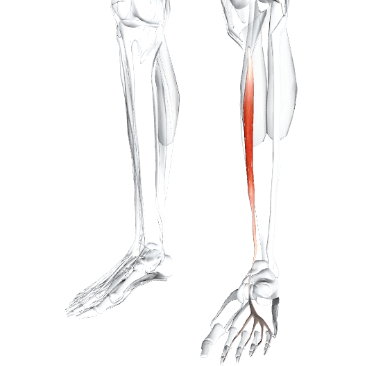
Long toe flexor

Latin name lex, to bend; digit, fingers; longus, long.
The insertion of the tendons of this muscle on the four toes is similar to the insertion of the tendons of the deep flexors of the fingers of the hand.
Place of origin: Medial part of the posterior surface of the tibia below the camboid line.
attachment point: The bases of the distal phalanges of the second through fifth toes.
Function: Flexes all joints of the four toes (allows the foot to be stable on a surface when walking). Participates in sole flexion of the ankle and extension of the foot.
entertaining: Tibial nerve L5, S1, (2).
vascular supply: Posterior tibial artery (from the popliteal artery).
Main functional movement: Examples: walking (especially barefoot on uneven ground). Stand on tiptoe.
This article was written with the help of wikipedia.
Do you have a question?
If you still have any questions or misunderstandings after reading this article, please ask us in the comments below or contact us in our official Facebook group. You will receive an answer as soon as possible.
Structure and function of muscles
Muscle Structure and Function All protrusions of the vertebral arch are covered by strong, elastic ligaments that connect the vertebrae and intervertebral discs from the base of the skull to the sacrum. These ligaments allow us to keep our body upright. Another arrangement
Deltoids and Abdominals - Narrow-grip chest raises and bar lowers; - Bench press with barbells behind head; - Dumbbell press sit-up; - Side dumbbell presses seated, standing and squat.
DELTA MUSCLES AND ABS
Dumbbell pull-ups (barbell behind head) Front and back presses (variations) Dumbbell raises (seated, standing, bent over) Twisting and pivoting movements of the trunk
2.1 Joints: structure, function and biomechanics Joints are flexible connections between two bones. The structure of the joints is responsible for the movements, their direction and amplitude. Figure 2.1: Schematic representation of a joint: 1 – joint head; 2 - cartilage; 3 – joint capsule; 4 -…
references in the literature
(c) the strength of the foot flexors (triceps, subscapularis, posterior tibialis, long toe flexors, long fibula and short fibula) (Lysov PK, Nikityuk DB, Sapin MR, 2003).
1 – process of the radius bone; 2 – ligament of the triceps brachii muscle; 3 – partition between muscles; 4 – pectoralis major muscle; 5 – clavicle; 6 – part of the sternocleidomastoid; 7 - sphincter muscle; 8 – Latissimus Dorsi; 9—musculus seroma anterior; 10 – External oblique muscle; 11 – rectus abdominis muscle; 12 - aponeurosis muscle; 13 – latissimus hamstring; 14 – rectus thigh muscle; 15 – transverse thigh muscle; 16 – medial thigh muscle; 17 – long fibula muscle; 18 – calf muscle; 19 – anterior tibial muscle; 20 – long toe extensor muscle; 21 - kidney muscle; 22 – tendon insertion of extensor muscle; 23 – tendon insertion of extensor muscle; 24 – tendon insertion of extensor muscle. forefoot bones; 23 - lateral ankle; 24 - middle ankle; 25 – Achilles tendon; 26 – long toe flexor; 27—tibia; 28—semimembranosus muscle; 29—striatum; 30 – patella; 31 – patellar ligament; 32 - tender muscle; 33 – long adductor muscle of the thigh; 34 - tailor's muscle; 35 – scapular muscle; 36 – Iliac Muscle! ligamentous muscle 37—inguinal ligament; 38 - anterior iliac spine; 39 - white line of abdomen; 40 - rib border; 41 - sternal line; 42 – large pectoral muscle; 43 - long head; 44—triceps brachii muscle; 45 - epiphyseal muscle; 46—pronator roundis; 47—ulnar spur; 48—flexor radius; 49—flexor ulnaris; 50—extensor longus; 51 - short head
Related terms (continued)
The gluteus maximus muscle (Latin gluteus maximus) is the largest of the three gluteal muscles and is closest to the surface. It largely determines the shape and appearance of the buttocks.
The chewing muscles (Latin: Musculi masticatorii) are the muscles of the head that enable the chewing process.
The cervical plexus (Latin: plexus cervicalis) is a nerve plexus consisting of the anterior branches of the four superior cervical nerves, which are connected by three arcuate loops. It is located on the anterolateral surface of the deep muscles of the neck (scapular, medial ladder, and cervical ligaments) at the level of the four upper cervical vertebrae. It is covered anteriorly and laterally by the sternocleidomastoid muscle.
Skeletal traction is a comprehensive method of treating traumatic limb injuries. The aim of the method is to gradually reduce fractures with weights and keep them in the correct position until the original callus layer has formed.
The musculus temporalis (Latin: musculus temporalis) fills the fossa temporalis. It begins on the temporal surface of the frontal bone, the greater wing of the sphenoid bone, and the scala part of the temporal bone. The muscle bundles converge to form a strong tendon that runs inward from the zygomatic arch and attaches to the coronoid process of the mandible. The temporalis muscle has a fan-shaped structure. Its anterior fibers run vertically upwards, the middle fibers run obliquely backwards, and the posterior fibers run almost horizontally backwards.
The supraspinatus muscle (lat. m. epicranius) is one of the facial muscles of the head, covering almost the entire skullcap and connected to the tendon helmet (lat. galea aponeurotica).
Synostosis (from Greek σίν- 'of' and oστός 'bone') is a type of continuous connection of bones by bone tissue. The term belongs to the Basel Nomina Anatomica. Bone tissue at synostosis is formed from mesenchyme in desmal osteogenesis and from cartilage in cartilaginous osteogenesis. In the second case, there is a synostosis with ossification of the synchondrosis.
Causes of muscle dysfunction
The tendons of the foot can lose strength or suffer other damage for a variety of reasons:
- age-related atrophy due to impaired tissue nutrition,
- abnormalities of the endocrine system,
- connective tissue diseases,
- enzymopathies,
- polyneuritis,
- complications after trauma,
- excessive physical exertion.
The main cause of damage is tendinitis. This is an inflammatory disease of the tendon that can also affect the surrounding muscle tissue. Dystrophic damage can develop into a chronic condition that is very dangerous and almost incurable.
Foot pain can also be caused by salt deposits and overgrowth in bone tissue. They can be caused by taking certain medications, etc.
diagnosis

The long extensor of the big toe or the entire metatarsal can be injured. On examination, the doctor will notice a 'thumping' sensation when walking or pulling. The doctor will feel the muscle and do a series of tests to assess the nature of the injury. When the muscle is damaged, you may feel weakness and pain when moving with or without resistance. If weakness is found throughout the metatarsal, including the little toe, the nerve may be compressed.
Read more:- The flexor muscles of the foot.
- flexor muscle of the big toe.
- Long fibula muscle.
- How muscles and bones are related.
- Anterior tilting of the pelvis.
- Tibialis posterior muscle.
- Short flexor of the big toe Latin.
- Pronator - what does that mean?.
