For this reason, traumatologists often immediately perform an endoprosthesis. This is a difficult and time-consuming procedure, but the most effective. Prostheses are used:

- Unstable ankle
- Symptoms of CHNS
- Anatomy of a Dislocation
- severity and healing time
- Treatment of minor to moderate injuries
- Treatment of grade III injuries
- Preparation
- surgery
- Grade II deformity
- Deformity grade III
- Need to strengthen the ankle
- choice of technique
- Right nutrition
- Principles of Postoperative Recovery.
- Arthroscopy of the ankle
- Main causes of ankle ligament inflammation
- Clinical signs of ankle ligamentitis
- Symptoms of ankle ligament injuries.
- What should be examined?
- Ligament sprains: treatment
- How else can dislocations be treated?
Unstable ankle
Many people sprain their foot without realizing that even a single ankle ligament injury can lead to chronic ankle instability (ACL) if not treated properly. This condition impairs foot support, gait instability, nerve conduction, and the nervous system's control of movement.
Sprains are the most common cause of ankle ligament injuries. People who play sports often suffer from this type of ligament injury.
A major danger with CKHS is that if not recognized and treated early, it can develop into an uncomfortable complication: osteoarthritis. And treating osteoarthritis requires surgery to replace part of the joint (arthroplasty).
The highly qualified and experienced doctors of ON CLINIC help to restore the health of our patients' joints, even in complex cases!
Symptoms of CHNS
The occurrence of ankle instability is accompanied by the following symptoms
- pain that increases with prolonged stress on the ankle,
- restriction of mobility of the joint,
- involuntary painful flexion of the sole of the foot when walking and other activities, especially in hilly terrain,
- swelling of the injured area,
- Bruise,
- local increase in skin temperature,
- Pain at the site of injury on palpation.
- I: Selective fibular ligament injury, ankle function is fully intact, slight pain or discomfort on exertion, slight swelling, rarely instability,
- II: Fragmentary rupture of ligaments, regular instability and moderate swelling in the ankle, restricted mobility of the leg, pain can also occur without weight bearing,
- III: Complete rupture of ligaments, pronounced pain syndrome with every movement and significant swelling of the painful area.
A doctor should be consulted if there are injuries and signs of ankle instability. Self-treatment can lead to irreversible or very difficult to correct consequences! The doctors at the ON CLINIC will help you eliminate pain, swelling and instability, rebuild the ligaments, and maintain full mobility and quality of life.
Free consultation with the head of the Center for Orthopedics and Traumatology, orthopedist-traumatologist, doctor of the highest category, candidate of medical sciences Samoilov VV for joint surgery.
joint endoprosthesis. Treatment of arthrosis in the ON CLINIC
Anatomy of a Dislocation
Ligaments are made of strong connective tissue. They usually stretch to a certain extent and then return to their normal state. With a severe sprain, the ligament tears and you may feel or hear a pop or pop.
A sprained ankle ligament is a common injury. Twisting collateral ligament injuries account for about 85 % of all cases. According to statistics, the frequency is highest in early 2020 between the ages of 15 and 19. Half of all ankle ligament sprains occur during sports activities such as basketball (41.1 %) and soccer (9.3 %). It occurs in 77-83 % of all sports injuries.
The most common risk factor is a sprain in the past. It can compromise the strength and integrity of the stabilizers by disrupting sensory nerve fibers. Gender, height, weight, limb dominance, postural variation, and foot anatomy are internal risk factors that may also affect the possibility of injury. External causes include the attachment features of the protection system, the type of footwear, the duration of the competition and the intensity of the activity.
In addition to sports injuries, ankle injuries can also result from simply stepping on the foot incorrectly. For example, stepping on the side of the foot instead of the sole of the foot. This can happen when climbing stairs or running.
severity and healing time
Ankle sprains are categorized by severity:
- 1st degree - slight sprain and microscopic damage;
- Grade 2 - partial tear;
- Grade 3 - the ligament is completely torn.
Most sprains take between 4 and 6 weeks to heal. It can take several months to recover from a serious sprain.
When the ligament apparatus is damaged, the following symptoms appear:
- increase in body temperature to subfebrile - 37.5°C;
- chills and fever;
- weakness and dizziness;
- Local swelling around the injury;
- Paralysis - the ankle is hot when palpated;
- localized hyperthermia;
- Pain on palpation and movements in one or more directions, and when walking.
With a complete rupture of a ligament, in addition to the above symptoms, there are the following:
- Excessive mobility in the joint projection;
- inability to stand on the affected leg;
- loss of consciousness;
- pain shock;
- Circulatory disorders, which manifest themselves as bruising in the fingers, numbness and tingling in the projection of the injury.
Even with a mild clinical picture, a trauma surgeon should be consulted. He will help you choose treatment tactics and restore joint and ligament function.
Treatment of minor to moderate injuries
First degree ligament injuries are asymptomatic. In the first 24 hours, the patient does not feel any pain even with intensive physical activity. Swelling occurs later, the hematoma area increases and post-traumatic inflammation develops. The injured person noticeably limp, trying to get rid of the pain when walking.
With a grade 2 ligament sprain, acute pain occurs immediately, and an hour later there is swelling and hematoma from multiple bruises. To rule out an ankle fracture, the victim should see a trauma surgeon. Treatment of grade 1-2 injuries does not require hospitalization. Cold compresses and limb immobilization are indicated for the first 2-3 days of treatment:
- An elastic bandage or medium stretch open toe bandage should be worn for about 10 days;
- A cast should not be worn for more than a week, otherwise it will take a long time for muscle tone to recover.
Nonsteroidal anti-inflammatory drugs are the first choice in treating ligament injuries. Topical medications are applied to the ankle in the form of ointments, creams, or gels:
Topical anti-inflammatories have anti-inflammatory and pain-relieving effects. If ointments with NSAIDs do not reduce the severity of the hematoma, the traumatologist prescribes drugs that improve blood supply to tissues. Direct-acting anticoagulants are most commonly used:
After its application, capillary permeability is restored and microcirculation is improved. Nutrients and biologically active substances begin to reach the injured ligaments, which accelerates their regeneration.
Treatment of grade III injuries
Even 'neglected' joint problems can be treated at home! Just remember to take such a swab once a day.
A serious injury to the ankle results in a complete rupture of one or more ligaments. The severity of the symptoms resembles and sometimes even exceeds the clinical picture of a bone fracture. The patient is unable to put weight on or move the foot. This is caused by severe pain and a complete loss of function in the foot. Supporting the injured foot is not possible because the anatomical conditions of the joints have changed. Swelling and hematoma quickly spread to the entire surface of the ankle.
When treating grade 3 ankle ligaments, conservative methods are rarely used for the following reasons
- torn fibers heal slowly;
- the functionality of the foot may not be fully restored;
- If surgery is not performed, injuries often occur due to the instability of the ankle.
When the ligament is completely detached from the bone, elective surgery is performed. Its continuity is restored after tendon and bone sutures are applied. After the operation, the patient is placed in a plaster cast for 3-4 weeks. Throughout the postoperative period, the patient is prescribed drugs that improve blood circulation in the ankle. Treatment with drugs that expand the lumen of blood vessels is carried out:
Taking these drugs improves blood flow to the tissues and causes the swelling and hematoma to subside quickly. Venotonic drugs (Troxevasin, Troxerutin, Lyoton) accelerate regeneration processes.
Read more:Chondroprotectors are rarely prescribed for mild to moderate ligament injuries. It is advisable to take them after an operation. Chondroprotectors accelerate the healing of the ligament-tendon system. And when they accumulate in the ankle, they have an anti-inflammatory and pain-relieving effect.
Preparation
Before the endoprosthesis, the doctor assesses the patient's condition and selects a suitable prosthesis model. The patient is examined by a traumatologist and an orthopedic surgeon, and if necessary, specialists from other specialties, such as endocrinology, neurology or rheumatology, are also consulted. They adjust the dosing regimen of drugs that affect the biochemical processes in bones, cartilage and soft tissues. Consultation of an allergist is indicated if the patient is hypersensitive to certain groups of pharmacological agents. The anesthesiologist selects the drugs for anesthesia according to his opinion. The required size and configuration of the endoprosthesis is determined after an instrumental examination:
- contrast medium angiography of the ankle;
- X-rays in multiple projections;
- Ultrasound examination of all joints of the lower limbs;
- computed tomography and positron emission tomography;
- Diagnostic arthroscopic examination.
With instrumental diagnostics, the condition of the joints, the ligament-tendon apparatus, nerves, muscles, blood and lymphatic vessels is assessed. Based on the results of biochemical studies, it is possible to determine whether or not there is an inflammatory process in the structures of the joints, even if it is of an infectious nature.
When choosing an endoprosthesis, the patient's posture and gait resulting from the joint deformity are taken into account. Swelling and pain sensations are eliminated as much as possible and inflammation is controlled.
The patient is directly involved in the preparation of the procedure. Within 1-2 months, the patient must give up alcohol and smoking, which can affect blood flow to the ankle. The patient is referred to a physical therapist who selects the crutches and rigid orthotics needed during the rehabilitation period.
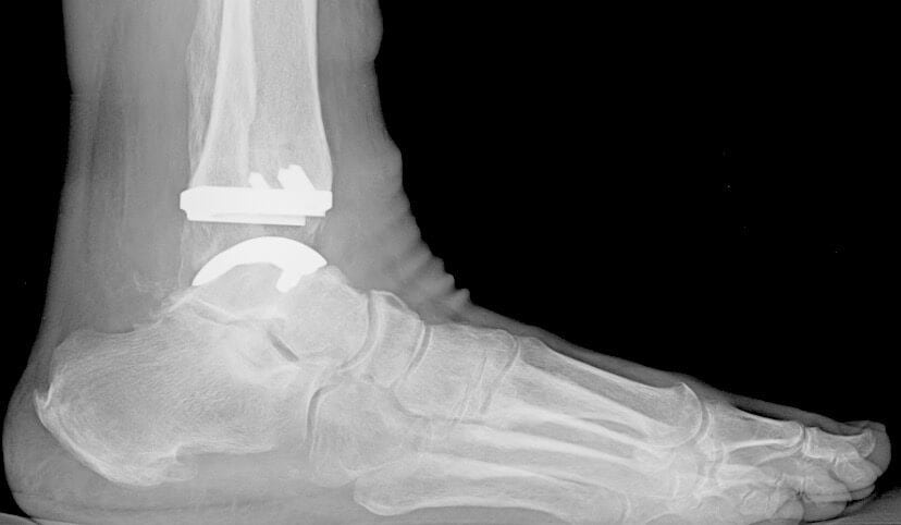
surgery
In an ankle replacement, spinal anesthesia, or epidural anesthesia, is performed, in which medication is injected into the epidural space of the spine through a catheter. The aim of the operation is to amputate part of the talus and tibia. Due to the complexity of the ankle, a total joint replacement is performed, although some clinics also perform a partial joint replacement. How orthopedic surgeons perform the operation:
- An incision is made along the ankle;
- Opening the joint by retracting nerves, tendons, blood and lymph vessels;
- uncovering of Calvary;
- Removal of part of the joint, the cartilaginous surfaces of the joint elements that make it up.
This type of surgery is considered benign because much of the joint structure is preserved. In the past, long staffs could be used in surgery. Today, the plates are fixed with pins, which ensures a close anatomical fit between the bone surfaces and the implant shell. A special porous material is used that promotes the ingrowth of vessels and tissues. This ensures a natural and firm fixation of the plate. The short pin is used to attach the bottom cover plate while the core is not attached at all. It is designed to ensure smooth sliding between the top and bottom metal plates. The top has a stop that prevents the core from sliding.
In the last phase of the procedure, the removed tissue is fixed in an anatomical position and sutured.
The artificial prosthesis used has the same functional properties as the ankle. During implantation, surgeons use software to control the balance and fixation of the implant. This contributes to the longevity of the prosthesis and freedom from discomfort during use. The range of motion is fully restored and friction is reduced thanks to the polymeric components of the design, compensating for the stress on the patient's own bone tissue in contact with the implant.
Grade II deformity
It is a tear of several fibers of the ligament apparatus.
The severity of the injury is defined by:
- bleeding and swelling 'spreading' over the outer surface of the foot;
- tenderness when palpating the ligament attachment area;
- Limitation of mobility of toes and ankle with pain;
- Walking can be difficult and the injured person should try to put as little stress on the foot as possible.
Deformity grade III
This deformity is characterized by a complete tear or rupture of the ligament fibers. Parts of the bone at the site of the injury are also often torn off (this can only be determined by x-rays).
A violation of this severity can be recognized by the following symptoms
- Severe pain in ankle when trying to put foot down;
- Extremely heavy bleeding and swelling affecting not just the joint but the entire foot, including the sole of the foot;
- Severe and acute pain in the area of the ligaments;
- The mobility of the joint is severely restricted.
Need to strengthen the ankle
The articular surfaces of the fibula and tibia as well as the talus block are located in the area of the ankle joint. The joint, ligaments and muscles connect the lower limb to the foot. In this area, the following types of injuries are distinguished:
Anomalies arise even without excessive physical exertion. For example, a person may trip and fall, resulting in a fracture of the fibula head. There are also other causes of injuries:
- Hard physical work;
- wearing high heels;
- incorrect positioning of the feet when walking;
To eliminate the risk of permanent injury and to strengthen the muscles and ligaments, a number of ankle strengthening products are recommended. The tissue becomes more active and stronger. Orthopedists recommend the use of complex musculoskeletal rehabilitation techniques to increase bone strength.
choice of technique
Many patients believe that active training is enough to strengthen the ankle musculoskeletal system. However, doctors recommend a number of techniques to strengthen the entire body. The table below lists the main techniques recommended by therapists, orthopedists and osteopaths.
method of strengthening characteristics Result vitamin therapy Consumption of high quality foods, oral or injected multivitamins Improving the composition of bones, muscles and ligaments Physical movement of the lower limbs Gymnastics, stretching, complementary exercises Strengthening of the ankle Comprehensive physical activity swimming, running Strengthening of the musculoskeletal system physical therapy UHF, electrophoresis, magnetic therapy, laser Normalization of blood flow with improved supply of nutrients and oxygen to the lesion massage General ankle tension and point massage. Blood flow is normalized. Muscle relaxation in hypertonia A combination of methods is recommended. For example, vitamin therapy and exercise can be used at the same time. Massage can be done from time to time to improve circulation. Physical therapy is recommended for patients who frequently injure themselves.
Right nutrition
If tissue weakness is the cause of frequent musculoskeletal injuries, proper nutrition will strengthen the body. Food with a high content of vitamins, trace elements and minerals should be included. The following substances are good for muscles, bones and ligaments:
Principles of Postoperative Recovery.
Because the arthroscopic reconstruction technique is considered minimally invasive, recovery is significantly easier than with open surgery. The patient can get up and move the next day, and the sutures are removed from the limb after an average of two weeks.
The rehabilitation program is individually tailored to each patient. The doctor takes into account the patient's age, physical condition, any medical conditions that may interfere with the rehabilitation process, and other personal factors. The following measures are recommended:
- A set of therapeutic exercises under medical supervision to restore joint mobility;
- use of orthotics and other orthopedic fixation devices throughout the recovery period to keep the joint in the correct position during the healing process;
- the subsequent wearing of special orthopedic shoes (insole) to avoid flat feet and protect the joint from further injury.
Ankle instability is an uncomfortable complication of ligament injuries in this area. This pathology can reduce the quality of life of patients and deprive them of mobility.
Only timely and thorough treatment by an experienced doctor can prevent future ankle problems.
If you turn to the doctor, a Petrosyan orthopedic traumatologist, the function of the joint will be restored, and the equipment of the clinic makes it possible to treat even complicated cases of the disease!
Arthroscopy of the ankle
Main causes of ankle ligament inflammation
Post-traumatic ankle ligament inflammation is common in young and old alike and can be caused by:
- fractures of the tibia and fibula;
- fractures of the talus and calcaneus;
- sprains and tears of the ligamentous and tendon apparatus;
- penetrating wounds of the soft tissues surrounding the joint;
- Post-traumatic scar deformities;
- Ankylosis and contractures caused by prolonged immobilization of the limbs.
In middle age, degenerative dystrophic changes in the joint tissue come to the fore. They may be due to the negative effects of the following causes
- Vascular pathologies such as lower limb varicose veins, atherosclerosis, diabetic angiopathy, obliterating endarteritis, etc;
- Disruption of tissue innervation due to tunnel syndrome, lumbosacral osteochondrosis with radiculitis and radiculitis syndrome, changes in the dural covering of the spinal cord, etc;
- Impairment of the microcirculation of blood and lymph in the affected area;
- slowing down the diffuse nutrition of cartilage, ligament and tendon tissues;
- Rheumatic diseases such as articular form of Bechterew's disease, systemic lupus erythematosus, rheumatoid arthritis, etc;
- Gout and gouty arthritis in the ankle.
In addition, pregnancy is a common trigger for the development of chronic ankle ligamentitis. During this time, numerous changes take place in the body. Among other things, the hormonal background changes, which can prepare the birth canal by softening the cartilage. Consequently, all cartilaginous synovial membranes in the joints are also negatively affected by the softening. For this reason, doctors advise pregnant women to wear only comfortable, low-heeled shoes. This helps at least a little to protect cartilage, ligaments and tendons from damage. If a woman does not follow the doctor's advice, she puts herself at risk of serious diseases of all major joints of the lower limbs.
Clinical signs of ankle ligamentitis
The ligament inflammation of the ankle is clinically noticeable only in acute phases. In most cases it is a chronic, recurring and slowly progressing form. The first signs of the symptoms appear suddenly. The ankle area is swollen. The skin is red. Palpation is difficult and painful. Stepping on the foot is painful. All these signs indicate an acute inflammation of the soft tissues in the ankle area. In some cases, general well-being may suffer and body temperature may rise.
These symptoms gradually subside, even without treatment. The disease goes into a subacute phase and then into a chronic phase. Gradually, the ligament and tendon fibers scar and become post-inflammatory deformed. This leads to the characteristic clinical signs of chronic ankle ligamentitis:
- Dull or aching pain that gets markedly worse after any physical activity;
- noises when walking, such as grinding, cracking, rubbing, crepitations, etc.;
- deformity of the foot (in an attempt to relieve the affected ligaments and tendons, the patient walks on the inside or outside edge of the foot, resulting in flat feet or club feet);
- Limitation of the range of motion of the joint and the gradual development of ankylosis and contractures;
- changes in gait, slight limp;
- fatigue of the calf muscles;
- Stiffness of the affected joint after a long period of rest (sleeping at night, working in a static position, etc.).
A visit to an orthopedist is necessary to diagnose ankle ligament inflammation. The doctor can make an initial diagnosis during the initial examination. By palpating, he can determine which ligaments and tendons have become permanently inflamed and will scar as a result. In order to be able to make an accurate diagnosis, various laboratory tests must be carried out:
Symptoms of ankle ligament injuries.
Patients suffer from acute pain in the ankle, which severely limits the function of the joint.
The joint and the rear part of the outer surface of the foot are swollen. Extensive bruising occurs on the 2nd or 3rd day after the injury. Pain is noted on the anterolateral surface of the ankle and foot on palpation, and the pain is more intense when palpation is performed with slight plantar flexion and foot adduction. Active and passive movements in the joint are severely restricted by the pain. After anesthesia, excessive inward and plantar pressure is noted. Upward pressure on the heel bone (axial loading) does not cause pain.
Patients limp, rotate the limb outward when walking, and support themselves only on the heel. Tight bandaging of the joint reduces pain and makes the limb easier to use.
Laboratory and instrumental studies
X-rays may show a tear in the cortical layer where the ligament attaches.
[1], [2], [3], [4], [5], [6], [7], [8], [9], [10]
What should be examined?
Conservative treatment of ankle ligament injuries
Conservative treatment of ankle ligament injuries is used in more recent cases. After procaine blockade of the injury site (1%ige solution, 10-15 ml), a circular plaster cast is applied from the upper third of the tibia to the ends of the toes. The foot is positioned at a 90° angle and rotated outwards (hypercorrection, valgus). The duration of the immobilization is 6 weeks. After that, reconstructive treatment is indicated. The joint is immobilized with an 8 mm thick gauze bandage for 1-2 months.
Surgical treatment of ankle ligament injuries
For chronic tears, ligament repairs are most commonly performed using the Watson-Jones method. The short fibula tendon is used as the material. The duration of the immobilization is 2 months. Postoperative treatment of ankle ligament injuries is the same as with the conservative method.
Ligament sprains: treatment
The symptoms of a dislocation dictate the treatment of the injury: eliminating pain, reducing swelling, and improving mobility through dislocation therapy.
In addition to their anti-inflammatory effects, nonsteroidal drugs (NSAIDs) have good analgesic effects in the treatment of sprains. Depending on the severity of the pain, they can be used externally (gels, ointments, creams) or internally (tablets, suppositories, powders, solutions for intramuscular injection) to treat sprains. The treatment duration for these sprains is usually 6-8 days. Such treatment is usually sufficient to reduce inflammation, but prolonged treatment can lead to undesirable side effects (ulceration and erosion of the gastric mucosa, liver and kidney dysfunction).
Swelling can be reduced by applying ice wrapped in a towel for 10-15 minutes every 3-4 hours. An elevated position of the injured limb is also recommended to reduce swelling in the first few days after injury.
If a large hematoma has formed, treatment for dislocations may also include prescribing creams and ointments containing direct anticoagulants (drugs that reduce blood clotting). They help the hematoma subside faster, normalize microcirculation and prevent the formation of venous clots in the injured area. However, they should not be used if there is a tendency to bleed, the integrity of the skin is compromised, or there is a skin condition in the area of application.
When treating a sprain, it's important to immobilize the joint where the ligament is torn. This is achieved by wearing a special device that fixes the joint in an anatomically correct position - an orthosis.
How else can dislocations be treated?
In recent years, sprains have been treated in the following ways Kinesiology taping – Attaching special adhesive fabric tapes to the injured area using a special technique. The positive effect of taping during stretching is to improve microcirculation and lymphatic flow in the injured area, correct the direction of movement in the joint, and reduce muscle hypertonicity.
The undeniable benefit of taping is that it stabilizes the joint while maintaining mobility. At the same time, however, the procedure also has disadvantages: taping is quite expensive material and can only be carried out by a specially trained specialist; Taping treatment cannot be performed if there are skin diseases, deep vein thrombosis of the lower limbs, severe kidney and circulatory diseases or complicated forms of diabetes at the application site.
Reconstructive treatments for dislocations are started after the acute symptoms and inflammation have subsided. They consist of special exercises therapeutic exercises.These exercises help bring muscles weakened during immobilization back into the joint's original range of motion. It is important to remember that physical therapy should be supervised by an instructor as improper exercise can not only be harmful but can also lead to ligament stretching.
In addition, to speed up the restoration of motor activity after the inflammation subsides, it is advisable to use a therapeutic massage A course consisting of 8-10 sessions. In order to avoid undesirable consequences, the procedure should be performed by a specialist doctor with medical education and special training.
Separately, they want about the use of physical therapy speak for sprained ligaments. Physiotherapy is the perfect answer to the question of how to heal a sprain quickly'. They help reduce swelling and pain in sprained ankles, resolve the hematoma faster, accelerate healing of damaged tissues, reduce the duration of immobilization, and restore skeletal muscle tone, which speeds healing of sprained ankles.


- dislocation of the ankle.
- Rupture of the ligaments of the ankle.
- Injury to the ligaments of the ankle.
- Damaged ligaments of the ankle.
- Structure of the human ankle.
- contusion of the ankle.
- Injury to the ankle.
- Treated subluxation of the ankle.