To date, it has been suggested to work in conjunction with SMAS to restore facial 'youth' [Okuda et al. [Okuda et al. 2019]..
- Myofascial manipulations
- What is osteopathy?
- What is fascia?
- The fascia tissue has many different functions, the most important of which are:
- Why is it important to monitor the condition of this tissue?
- Fascia and facial skin
- See also.
- Anatomical tour
- SMAS definitions
- Nuances when performing musculo-fascial massage
- Technology
- Age-related changes in the face
- The importance of facial spaces in facial surgery
- What are fascicles and their special importance for surgery?
- The backwardness of German surgery and the criticism of our European colleagues
- After the operation
- How does connective tissue affect our health?
- Musculo-fascial back massage
Myofascial manipulations
The development of human society occurs in cycles. The field of medicine is no exception. Modern man, with tens of thousands of medications at his disposal, has recently sought a drug-free cure, just as his ancestors did. They chased away disease with their hands, a healthy diet, and herbs. Today, various alternative medicine practices are becoming increasingly popular, based on the science that the human body is a self-regulating system that just needs a little help in dealing with illness. This also includes osteopathy, which has only been officially recognized in Russia since 2003.
What is osteopathy?
The word itself is made up of two Latin words – bone and disease. However, this branch of medicine deals not only with diseases of the musculoskeletal system, but of the entire body. The osteopath – is a general practitioner with in-depth knowledge of the physiology, structure and function of the human body. He carries out diagnosis and treatment with a single tool - his own hands. With them he 'hears', 'sees' and feels all pathological changes. He corrects her with his hands. And he corrects them gently and painlessly, through stroking, light rubbing and tapping.
Osteopathy is similar to manual therapy and massage. In fact, it is a holistic system of understanding human nature that is based on the unity of mind, body and soul. In this it is similar to the Eastern teachings of self-improvement.
- Musculo-skeletal-fascial (muscles, bones, connective tissue);
- Visceral (internal organs, blood and lymphatic systems, nervous system);
- Cranio-sacral (skull field, spine, sacrum, cerebrospinal fluid, spinal cord and brain).
The basic principle of this medical science is the slogan: 'Movement is life'. It's not so much about the movement of a person in space, but rather about the movement in his body (blood, lymph, cerebrospinal fluid and other things). It is believed that the cessation of movement in one area is the cause of various diseases. For this reason, one of the most important methods is osteopathy, among others musculo-fascial manipulation.
What is fascia?
fascia – Connective tissue that surrounds the nerves, vessels, ligaments and muscles and forms the skeleton around these systems. This tissue consists mainly of protein fibers, collagen and elastin, which form dense networks.
The fascia tissue has many different functions, the most important of which are:
– Transmission of nerve impulses from muscle to bone.
Interesting fact: It has been proven that the fascia, not the muscles, mainly shapes the body. The muscles provide the volume and the fascia tissue provides the shape of a particular body part. Thomas Myers, an American chiropractor, was one of the first to point out this fact. His opinion was later shared by other scientists.
Why is it important to monitor the condition of this tissue?
1. to ensure a normal condition of the skin
The shape of the face and body as well as the condition of the skin are mainly influenced by the fascia. If the connective fibers are very thin and weak, the skin becomes saggy. By paying proper attention to the fascia tissue, we take care of the condition of our skin.
How quickly a person recovers from injuries depends on the mobility of the fascia. Not all injuries are sports-related. There are enough situations in everyday life that can lead to injuries to our ligaments and tissues. We don't notice this until we reach a certain age because young people have well-developed fascia tissue. But after age 35, when the fascia becomes less flexible, injuries at home take a greater toll. But if you train your connective tissue, similar problems will not be a problem even at age 50.
Fascia and facial skin
When we talk about facial fascia, it is appropriate to introduce a new term – SMAS. This is the entirety of the subcutaneous muscles and ligaments that make up the facial skeleton. The SMAS consists of the superficial layer of facial muscles, subcutaneous fat, superficial facial fascia and other tissues. As with the body, the fascia determines the shape of the face. The skin loses elasticity because the connective tissue is weakened. And these changes are usually visible earlier than on the body. That's why you should take care of the facial fascia as early as possible.
Interesting fact: In many sources you can read that there is no fascia on the face. In fact, this is a misconception, because there are three fasciae in the facial head: the superficial, the internal and the visceral. The methods we propose are based on the assumption that they act on the superficial tissue.
See also.
- ↑ Moscow Law No. 1 of January 15, 1997 [www.businesspravo.ru/Docum/DocumShow_DocumID_61882.html 'On administrative responsibility for the production, distribution and demonstration of Nazi symbols in the territory of the city of Moscow']
- ↑ [www.fsin.su/fsin/simvolika/ Official symbolism of the FSIN].
– Which department are you? – shouted the adjutant, approaching.
– Eighteenth.
– What are you doing here, you should have been ahead long ago, you won't be able to get through before the evening.
– What a stupid order! "You don't know what you're doing," the officer said and left.
Then the general came and shouted something in anger, but not in Russian.
- Tafa lafa, but you can't understand what he's mumbling,' said the soldier, imitating the general. – I should have shot them, those bastards!
– At nine o'clock we were ordered to take up position, and we were not even halfway there. That is an order! – were repeated from all sides.
And the feeling of energy with which the troops went into action began to turn into irritation and anger at the useless orders and the Germans.
The reason for the confusion was that the Austrian cavalry was advancing on the left flank while the higher-ups decided that our center was too far from the right flank and all cavalry was ordered to the right. Several thousand cavalrymen advanced ahead of the infantry, and the infantry had to wait.
At the front there was a clash between an Austrian column leader and a Russian general. The Russian general shouted and demanded that the cavalry stop; the Austrian argued that it was not his fault, but that of a higher authority. Meanwhile, the troops stood around, bored and discouraged. An hour late, the troops finally moved and began to descend the mountain. The fog that spread over the mountain only thickened in the lowlands where the troops had dismounted. Up ahead, in the fog, one shot after another rang out, at first at irregular intervals: tratta tat, and then more and more frequently, and the matter was at Goldbach.
Not expecting to meet the enemy down by the river, and coming upon them in the fog, without hearing a word of encouragement from the higher commanders, with the realization spreading through the troops that it was too late, and especially in the dense fog with nothing to see in front of them or around them. The Russians sluggishly and slowly exchanged fire with the enemy, advancing and then stopping again without receiving orders from their commanders and auxiliary troops, who were in the fog wandering in unknown terrain and unable to find their troops. So began the fall of the first, second and third columns, which perished. The fourth column, with which Kutuzov himself was stationed, was on the Pratzen Hills.
There was still thick fog in the lower area where the action began, but above it had cleared, but you still couldn't see anything of what was happening in front of us. Whether the entire enemy force was ten vortexes away, as we assumed, or whether it was here in this line of fog, no one knew until nine o'clock.
It was nine o'clock in the morning. The fog spread over the ground like a solid sea, but at the village of Šlapanice, on the height where Napoleon stood surrounded by his marshals, it was completely clear. Above him was a clear blue sky, and on the surface of the milky sea of fog rippled a huge ball of sun that looked like a big empty purple car. Not only all the French troops, but also Napoleon himself and his staff were not on the other side of the streams and the Sokolnice and Schlapanice villages, behind which we wanted to take up positions and start the battle, but on this side, so close with our troops, that Napoleon could distinguish between horsemen and foot soldiers in our army with the naked eye. Napoleon stood slightly in front of his marshals on a small gray Arabian horse, dressed in a blue coat, the same coat in which he had made the Italian campaign. He stared silently at the hills that seemed to stand out from the sea of fog through which the Russian army moved in the distance, and listened to the sound of cannon fire in the hollow of the ground. During this time his immobile, gaunt face did not move a single muscle; his bright eyes were fixed on one point. His guess turned out to be correct. The Russian troops had already partially descended into the ravine to the ponds and lakes and had partially cleared the Pratzen Hills, which he intended to attack and regarded as the key to the position. He saw in the fog how, in the hollow formed by the two hills near the village of Prats, the Russian columns all moved in the same direction towards the hollow, flashed their bayonets and disappeared one by one into the sea of fog. From the information he had received since the evening, from the sounds of wheels and footsteps that he had heard during the night at the outposts, from the disorderly movement of the Russian columns, from all the guesses, he could clearly see that the Allies considered him too far forward, that the columns moving at Pratzen formed the center of the Russian army and that this center was already weak enough to be successfully attacked. Still, he hadn't started this case yet.
Anatomical tour
Using various examination techniques (including dissection, arteriography, macroscopic and microscopic examinations, ultra-radiography and histology), the researchers determined that SMAS is always present in the area of the cheek and parotid glands. 'Sometimes it's a thick layer, sometimes it's thin, but it's tight with the rest of the Superficial fascia of the face and neck… The SMAS divides the subcutaneous fat into two layers. Superficially, the small fatty stroma is enclosed in fibrous septa (septa), which are distributed from the SMAS towards the dermis. The deeper you penetrate into the tissue, the richer and more lush the fat lobules become; they lie between the deep facial muscles and are no longer separated by such fibrous septa' [Mitz, Peyronie 1976: 80]..
In addition, the SMAS is connected to the rear part of the Frontalis in the upper third of the face and with the Platysma – in the lower third of the face [Mitz, Peyronie 1976: 81]..
The authors further note that the SMAS is thick in the parotid region, but as it spreads into the cheek region, it becomes thinner and narrower.
In the parotid region, the SMAS appears on macroscopy and microscopy as a 1 to 2 cm thick net-like structure separate from the fascia. It can consist of 1-3 layers between the salivary gland fascia and the skin. Fibrous layers are sometimes also visible.
In the buccal area, however, the SMAS becomes thinner and is a connected network that sometimes extends into the dermis. This network particularly includes the facial muscles. Risorius, Frontalis, Orbicularis oculi, Platysma..
By the way, Vladimir Mitz still practices it today. In 2016, in private correspondence with Korean plastic surgeons, he explained why he assigned the acronym SMAS to this 'layer' - because he found muscle cells in the following areas, among others Risoriusand aponeurotic cells (fibrous and multilayered) On the same surgical shift. (Hwang K, Choi JH 2018: 3)..
SMAS definitions
It is not possible to find a single canonical definition that can describe SMAS. Different doctors and anatomists describe it differently.
- MeSH database.
«SMAS – The layer that lies between the superficial fat capsules and the superficial facial muscles of the head and neck'. - SMAS – an organized fibrous network consisting of the platysma, the parotid fascia and the fibrous layer that covers the cheek area. This system separates the deep and superficial facial fat tissue and has a specific morphology. Anatomically speaking, the SMAS lies below the zygomatic arch and above the platysma muscles. The fibrous layer of the SMAS is connected to the superficial temporal fascia, the frontalis muscle, and the platysma. SMAS is also often described as a fibrous degeneration of the platysma muscle itself.
- Whitney, Zito [2019] describe several physiological variants SMAS.. They believe the differences are due to the lack of consistent large-scale cadaveric studies with histological and macroscopic examinations. They cite Khawaja et al., who, after more than 800 facelifts, identified the SMAS as a superficial musculo-aponeurotic fatty system (SMAFS). They found that there are six different variants of SMAFS, including membranous, fatty, mixed (membranous fat, fleshy fat), insular (discontinuous), fleshy, and fibrous.
In addition, fat packets are distributed throughout the SMAS [Cotofana, S. and Lachman, N. 2019]..
It is believed that it is the distribution of fat that is responsible for the uniqueness and beauty of the face. According to Khawaja et al. The variants described are defined in the deeper layers of SMAS, and some variants may be associated with a congenital anomaly or trauma.
This classification is important for SMAS lifting surgery because correctly identifying the tissue type affects the surgical outcome.
Cadaver studies have shown that 80 % of subcutaneous fat is distributed on the face and only 20 % on the neck. On the face there are 57 % of fat above the SMAS and 43 % below the SMAS, while on the neck there are 65 % of fat above the platysma and 35 % below the platysma.
The zygomatic region has the highest concentration of fatty tissue [Raskin, LaTrenta 2007]..
Nuances when performing musculo-fascial massage
Musculo-fascial massage uses various techniques and even elements of manual therapy to support the interaction of muscles and fascia tissue. To achieve an optimal effect, the treatment should be carried out in a pleasant environment - this way the patient remains relaxed and calm.
Before treatment, it is advisable to remove make-up and other impurities from the skin, otherwise oxygen will not be able to reach the area and achieve the desired effect. Only when these rules have been followed can the treatment begin and the head, neck and face can be worked on.
The session will look like this and is worth preparing for so you feel confident:
- A professional massage therapist will begin the session with a relaxed conversation - this way he or she can build a relationship and create a relaxed atmosphere.
- It is important that the professional asks you how you feel and whether there are any contraindications or other problems that you should pay attention to.
- Then a diagnosis is made - the patient lies on his back and the masseur determines the problem areas by examining special points on the face and applying pressure to the skin.
- The massage plan is created individually for each patient, so the therapist must decide which techniques and methods to use based on the diagnosis.
- All movements are carried out in the rhythm of breathing.

As a rule, the first session lasts no longer than 30 minutes. Later the duration of the session will be increased.
Important: If the patient experiences discomfort, acute pain or discomfort during the massage, the treatment should be stopped immediately.
Technology
Myofascial facial massage is based on three basic principles that are applied flawlessly regardless of the type of problem or purpose. So the specialist will certainly apply the following during treatment:
- Stretching and relaxation. With this technique, the practitioner works on the deep layers of fascia - the movement enables the muscle fibers to be worked through and stimulates cellular regeneration. At the same time, the technique is carried out very slowly in order to find the right resistance of the fibers and not to injure the skin. An important nuance is that the master's hands should never rub the skin. The result of the technique should be to free the client's facial tissue from stagnation.
- Skin suspension. The phase that produces the lifting effect and involves work on the subcutaneous layers. This technique makes it possible to create a strong connection between tissues, tightening skin that has lost firmness. It looks like this: The masseur holds the problem area in suspension for a few seconds and relaxes it. No pinching movements - just tightening, from which the dermis should not pull away.
- Massaging muscle mass. Once the facial muscles are relaxed, the master should work on the problem areas by pulling them to the sides and back. This technique activates the fascia and stimulates a rejuvenating effect.
Useful tip: The most important thing is to always (regardless of the type of massage) follow certain rules, and every patient should familiarize themselves with these rules immediately before visiting a specialist.
With this knowledge, you can determine whether you really entrust your skin and face to a professional, and not an amateur who has no experience and only a diploma from a two-week massage course.
Here are just a few aspects that shouldn't be overlooked, otherwise you could end up with an irresponsible massage therapist and get exactly the opposite of the desired effect.
Age-related changes in the face
The age-related changes in the facial spaces are shown in Fig. 18.1, B.
Facial areas change in many ways as we age, but these changes are not stereotypical. The extent of the changes depends on the mobility of the soft tissues in the area corresponding to the visual space in question; The greatest mobility in the face is in the lower jaw area and the least in the temple area.
The age-related changes include a reduction in the tension of the spaces due to the stretching of the overlying soft tissues. In later life, the laxity of the 'roof' of the facial space manifests itself as external soft tissue protrusions, e.g. B. as malar sacs in the midface and 'eyebrows' on the jawline. This usually happens secondary to a weakening of the ligaments at the border of the visual space, e.g. B. the intraorbital fat bulges beyond the lower edge of the eyeball. Facial spaces that change with age are easier to pass because they are easier to open and the connective tissue in their cavity is easily shed.
The importance of facial spaces in facial surgery
The facial spaces are essentially 'safe' for the surgeon because they do not contain any important anatomical formations. All nerves, vessels, ligaments and muscles lie outside the facial spaces. Supporting straps reinforce the walls of the rooms and fix them in a precisely defined position. In some areas of the face, branches of the facial nerve attach to the walls of the facial spaces from the outside.
The detachment technique used in surgical procedures in the sub-SMAS layer has its own peculiarities depending on the area in which the procedure is performed. In the facial spaces, blunt dissection is sufficient to perform rapid detachment to the edges of the space. When the space is dissected, the gap between the soft tissue layers opens and there is no bleeding. This is a significant difference from conventional facial subcutaneous dissection, in which the plane of dissection extends only a few millimeters above the surface of the facial spaces, that is, beyond the surface of the muscle that forms the 'roof' of the space, be it the platysma or the orbit muscle. If the dissection is performed correctly, bruising is avoided and the risk of damage to the facial nerve branches is significantly reduced. During out-of-space surgical manipulation, the dissection technique is modified to precisely separate the branches of the facial nerve from the facial ligaments before severing the fiber bundles of the ligaments, if necessary.
The basic principle of surgical correction of age-related changes under the SMAS is to pull and fix the flaccid full-thickness flap (the 'roof' of the space) in the desired direction. Typically, the area of loose tissue tension that requires surgical correction is located medial to one of the facial spaces. The extent of surgical dissection of the medial border of the space depends on the condition of the supporting ligaments that reinforce the medial wall of the space.
What are fascicles and their special importance for surgery?
Pyrogov was one of the first surgeons to undertake a comprehensive analysis of the fascia, which forms the partitions between muscles and covers the blood vessels ('sheaths' of muscles and internal organs). The fascia is rich in elastic fibers and contains fatty tissue, nerves, arteries, veins and a network of blood vessels in its thickness. The reader has probably seen more than once the shiny grayish sheaths that cover the muscles on animal meat - these are the fascia.
The surgical value of fascia is extremely large and diverse. In order to correctly diagnose a disease and perform an operation, a surgeon must know the exact location of the fascia on the human body. 'fasciaPirogov,' Pirogov wrote. play an extremely important role in hernias, aneurysms, abscesses, etc.; A clear and complete picture of the development of these diseases can only be obtained by one who, with a scalpel (knife) in his hand, has carefully examined the relative position and mutual relationship of the fascial plates'. Nikolai Ivanovich Pirogov emphasized the special importance of fascia in finding the arteries suitable for the surgeon and ligature them.
The backwardness of German surgery and the criticism of our European colleagues
Pirogov's biographer, Dr. Volkov, writes: 'Pirogov's theory of fascia is the key to the entire anatomy - it is the entire genius of Pirogov, who clearly and clearly recognized the revolutionary value of his method'. In 'Surgical Anatomy' the fascia system is not understood as a fixed anatomical dogma, but rather as a surgical guideline. Nikolai Ivanovich dedicated his work to St. Petersburg doctors, to whom he gave a six-week course in surgical anatomy at the late Obukhovsky Hospital. In the preface to this work, Pirogov speaks of the scientific backwardness of the famous German professors of surgery.
'Before my stay in Germany, it had never occurred to me that an educated doctor, thoroughly engaged in his science, could doubt the usefulness of anatomy to a surgeon'. In 'Surgical Anatomy' Pirogov criticized the anatomical publications of a number of well-known authors, including such household names as Antonio Scarpa, Alfred Velpo and Pirogov's compatriot IV Buyalsky. For this work, Pirogov was awarded the Demidov Prize by the Academy of Sciences.
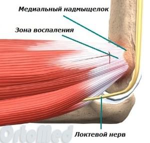
After the operation
Postoperative recovery for medial epicondylitis takes one to three months.
Immediately after the operation, the elbow joint is fixed in a movable orthosis (restricting movement), which holds the arm at a 90 degree angle.
A few days after the operation, the patient gradually begins to exercise the elbow joint and flexor muscles, with the range of motion increasing day by day. Active rehabilitation begins approximately two weeks after surgery. It will continue until the patient has fully recovered.
Surgical treatment of epicondylitis 34,000 p.
Do not self-medicate!
Only a doctor can make the diagnosis and prescribe appropriate treatment. If you have any questions, please contact us by phone or Please contact us by email.
How does connective tissue affect our health?
Over the years, with stress, sedentary work and trauma, the connective tissue deforms. It shrinks like a sweater that has 'shrinked' after washing. This not only affects our muscles and spine, but also the sanctuary of our health - the immune system. 'Contracted fascial connective tissue squeezes our cells like a boa constrictor. This reduces blood and lymph circulation, impairs cell nutrition and causes congestion in the deep tissues of the body. And when one considers that the lymphatic system is closely linked to the immune system, the mechanism by which shortening of connective tissue influences the onset of many diseases becomes clear. The age-related shortening of connective tissue not only poses the risk of muscle stiffness and limited mobility of the spine and joints, but also leads to immune-mediated pathologies. And the list is long, from chronic colds to cancer. Fortunately, there are ways to improve the condition of the connective tissue, one of which is myofascial massage. It is worth being clear about this beforehand so that age does not become a synonym for illness.
Myofascial massage has been successfully used to treat myofascial syndrome. However, there are a few things you need to know to make it as effective as possible.
What is myofascial syndrome?
All our movements are triggered by the contraction of muscles on command from the brain. When the nerve impulse becomes chaotic, the muscle fibers contract chaotically. This leads to muscle spasms and trigger points. Trigger points are small areas of tense muscle fibers. The presence of trigger points means that myofascial syndrome is present.
How does myofascial syndrome manifest itself?
The main symptom of myofascial syndrome is pain. It occurs due to overload, hypothermia, lack of tissue nutrition, emotional tension and many other factors. First, these factors activate a trigger point, which then automatically triggers a contraction and pain throughout the muscle.
Musculo-fascial back massage
The musculo-fascial back massage is effective against muscle pain. It is indicated for both acute and chronic musculofascial syndromes.
In our opinion, the importance of myofascial back massage is underestimated by many professionals. This is primarily due to a misunderstanding of their role and opportunities. But it is also short-sighted to overestimate the expectations of this method.
We have already said that it is unprofessional to use myofascial back massage as the sole treatment for myofascial syndrome. This is because it does not have sufficient therapeutic power for this purpose. However, it may have some short-term impact. However, the price of myofascial back massage should not be ignored. Will you not be satisfied with a short-term effect? Everyone wants to get a decent, complete result for their money. And such an outcome is possible. It just needs to be done in the right way.
As I said, the maximum effect of myofascial back massage can only be achieved in combination with other methods of gentle manual therapy. And then it will not be a temporary measure, but a normal, full-fledged result that will not let you down.
Read more:- Photo of the ligatures.
- tibial fasciitis.
- shin fascia.
- How muscles and bones are related.
- Anatomy of the syndesmosis.
- Tibialis muscle pain.
- Perseus shop.
- Longitudinal soleus muscle.