The LFC includes exercises for the large back muscles and the abdominal muscles. Stretching exercises (extension of the spine) in a lighter form are mandatory during this period. A reclined position in bed (head end raised by 40-60 cm) reduces the load.

- Rehabilitation after hip fracture (without surgery)
- Online protocol
- Consequences after a leg fracture
- How to properly stabilize the leg after a fracture
- Recovery in specialized centers
- recovery time.
- Useful tips
- rehabilitation
- Features of the problem during pregnancy
- Characteristics of fractures in children
- The opinion of the doctors
- gallery
- Evaluation of the results of your treatment
- Back muscle strength test
- Abdominal strength test
- Checking the willingness to sit on a chair
- Functional spine test
- Rehabilitation after a tibia fracture
- Rehabilitation after a pelvic bone fracture
- Pelvic fractures with complications
- Treatment methods and prognosis of femoral neck fracture in the elderly
- Goals and methods of rehabilitation
- Medical therapy
- How can complications be avoided?
Rehabilitation after hip fracture (without surgery)
- Protect the injury site for up to 8 weeks after the injury to prevent displacement of bone fragments
- Progressive loading to accelerate bone regeneration
- Maintaining muscle strength
- This rehabilitation program is designed for the conservative treatment of stress fractures of the femoral neck and diaphysis without dislocation
- Due to the risk of serious complications, most stress fractures of the femoral neck are treated surgically. This rehabilitation program is not suitable for post-operative care as additional limitations may occur
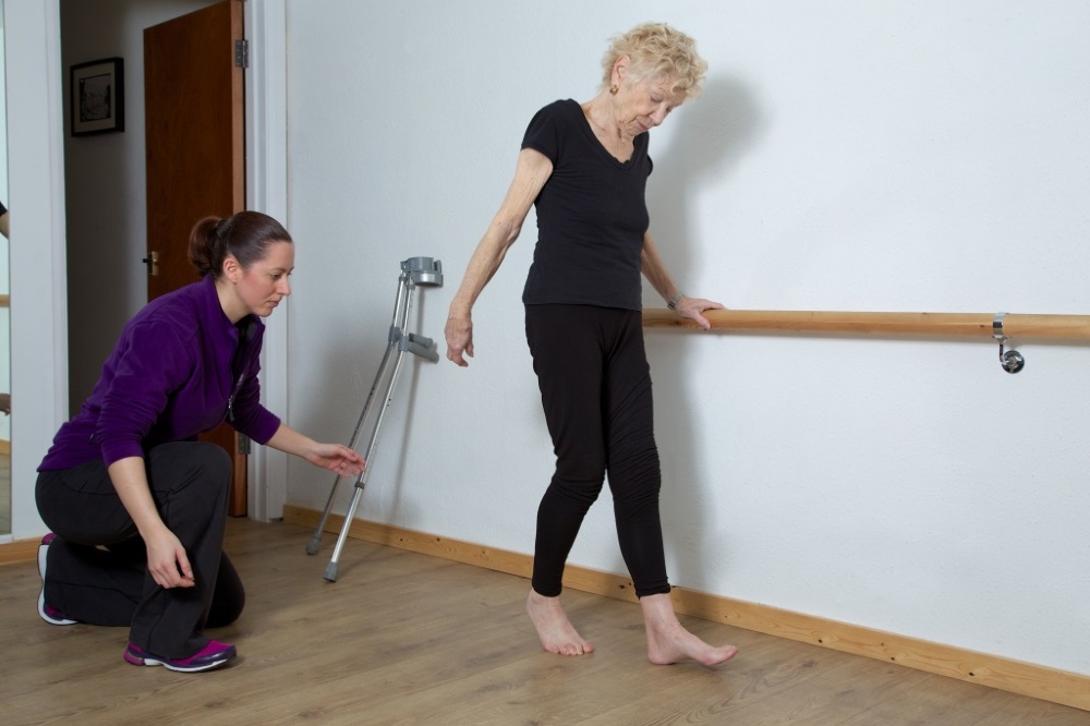
- Learning to walk with crutches and choosing comfortable crutches
- Walking on crutches without putting any weight on the leg
- Gradual loading (25-50%) up to 4-6 weeks
- No weight bearing until consolidation of the fracture is visible on the X-ray (4-6 weeks)
- Movement without load depending on pain and risk of displacement of the bone fragment
Mobilizing the soft tissues around the hip can be helpful in relieving muscle spasms and pain and increasing the range of motion of the joint.
However, in stress fractures of the femur, mobilization techniques should NOT be used as they may impair the healing of the fracture. The range of motion of the hip joint is rarely impaired in stress fractures of the femoral diaphysis.
Stretching of the tendons, quadriceps, hip flexors, adductors and soleus flexors with light to moderate intensity.
Online protocol
In addition, we can offer you a protocol with a video demonstration of all exercises for hip fractures without surgery!
Other treatment areas from week 4-6:
- Easy freestyle swimming
- Running in deep water with a swimming belt
- Cycling with low to medium resistance
- General strengthening of the shoulder
The medial vastus femoris and adductor shortus insert at a site that is common in stress fractures of the femoral diaphysis (the junction between the proximal and medial thirds), so exercises for these muscles should be kept to a minimum of intensity.
- Hip stretch in the side position
- Hip stretch while standing on the calves
- Hip stretch in supine position with support
- Hip stretch while standing against a wall
- Splits on all fours
Criteria for moving to the next phase:
- Pain-free weight bearing without antalgic gait
- Radiological signs of hip fracture healing
- Full range of motion of the hip joint
Consequences after a leg fracture
The consequences of a broken leg can be varied: the circulatory and lymphatic systems, nerve fibers, joints and muscles are damaged. This can lead to lameness, stretching of the limbs, destruction of the large and small joints, contractures, etc.
Let’s look at the most likely consequences after a broken leg – these are:
- Contractures and ankylosis of the joints immobilized during the plaster cast;
- Atrophy of muscle fibers and loss of tone;
- Thinning of the cartilage layer in the large joints of the lower limbs (knee, hip and ankle) and development of osteoarthritis;
- shortening of the ligaments and tendons;
- Impairment of blood flow and development of varicose veins in the lower limbs;
- Entrapment of nerve fibers and development of neuropathy and neuralgia;
- wrong joints.
Contracture and ankylosis are related conditions. They occur when a type of protein called fibrin is deposited in the joint and forms bands that prevent the joint from moving. The joint capsule, ligaments, tendons and muscles are gradually drawn into this process. Due to their secondary deformation, the joint can lose its mobility with discomfort. This stage is called contracture.
Muscle atrophy is a natural process when the limb is immobile, the muscles are not physically stimulated and become dystrophic due to lack of capillary supply.
Osteoarthritis in the knee or hip after a hip fracture is the most common complication of an improper rehabilitation period. What is the cartilage and synovial layer inside the joint? It is a covering made of hyaline cartilage. When the joint is in use, it is compressed and stretched to properly distribute the cushioning load. During this time, the cartilage tissue is nourished. If the joint is immobilized for a long period of time, the cartilage is not compressed and stretched. Atrophy of the cartilage tissue occurs.
How to properly stabilize the leg after a fracture
Before stabilizing the leg after a fracture, you need to check whether the callus layer has fully developed and stabilized. To do this, it is necessary to make an x-ray. They should be taken to an orthopedist who will develop an individual program for the rapid recovery of the leg after the fracture. This program includes a set of gymnastic exercises that the patient can perform independently at home.
However, it is better to start rehabilitation under the guidance of a specialist. There are several ways to treat the leg after a fracture with positive results, including chiropractic, osteopathy, kinesiotherapy and physiotherapy. We will discuss each method separately later in the article.
In the meantime, a few words about how to train the foot after a fracture and fully restore its correct position. The problem with bone fractures in the foot is that they heal very difficult and a thickened callus can form. This deforms the foot, disrupts the blood supply to the muscle arches and obstructs the passage of nerve fibers. This is very unpleasant for the patient.
Rehabilitation should be started as early as possible. The osteopath's work improves blood supply to the area and reduces the risk of callus formation. Special exercises accelerate the recovery of muscle flexibility. Massage and physiotherapy restore the regeneration processes in the tissues.
Recovery in specialized centers
Immediately after surgery, many people seek a supervised rehabilitation program to help them regain lost hip function. Specialized centers that offer a comprehensive program are suitable for this type of treatment:
- Temporary physical therapy using exercise equipment;
- Supervised massage;
- kinesitherapy;
- a selection of physiotherapeutic methods;
- Working with a psychologist.
In addition, there is the possibility of undergoing sanatorium treatment. These treatments can be organized at a later date and are recommended for almost all patients.

recovery time.
How long does rehabilitation take after a femoral neck fracture? - Much depends on the severity of the fracture, the age and health of the patient. Mild fractures in young people heal within a few months if the doctor's recommendations are followed. In elderly patients with osteoporosis, healing can take a year or more and requires a strict regimen and supportive therapy.
Please check the quality and level of detail of the information provided
Useful tips
- Advice on rehabilitation of an inguinal hernia
- Which specialists are involved in the rehabilitation?
- Rehabilitation after peptic ulcer disease
- Coma after brain injury: consequences and length of stay
- What is a urinary tract infection according to ICD 10?
- What should you do if you have a stroke and the right side is paralyzed?
- Autism: What are the symptoms and at what age does the child show them?
- Hand hygiene for people using a wheelchair or other mobility aids
- Is my leg pain sciatica or something else?
- Risk of falling
- Rehabilitation after a fracture of the radius bone
- Instructions for discharge from hospital after a stroke
- Rehabilitation after a hip fracture
- How does rehabilitation work for children with cerebral palsy?
- Lower back pain: Is it just pain or a sign of a serious problem in the body?
- Stroke on the right side: consequences and treatment
- Hand rehabilitation after stroke: How to restore hand movement
- Why are my legs and arms going numb?
- Nutrition after a stroke: principles and contraindications in the menu
- ICD-10 code for osteochondrosis
- What is cerebrovascular disease?
- Why do my muscles all over my body hurt?
- Osteochondrosis of the spine (M42) – ICD-10 code
- Main phases of rehabilitation after spinal fractures
- Rehabilitation of the hip joint
- Wandering pain throughout the body: causes and treatment
- Speech disorder after stroke
- Diagnosis of cerebral palsy during pregnancy
- What is the rehabilitation and treatment of sports injuries?
- Hemorrhagic stroke – what is it?
- What is rehabilitation for children with delayed language development?
- What is a stroke?
- What factors increase the risk of stroke?
- Travel to Thessaloniki
- Neuromuscular stimulation after a stroke
- How do you get out of bed after a fall?
- ATHASIA
- Lymphedema – useful tips for treatment
rehabilitation
Recovery from a radius bone injury is complete, both structurally and functionally. Even if treatment was successful and the bone has healed well, if an elderly person is not active for a long time, it may be difficult to move their arm as usual. The joints harden and the muscles atrophy. A long rehabilitation period is required to achieve a full recovery.
To achieve a full recovery, an elderly person needs patience, organization and the will to work towards it. Often it is difficult to do this alone because the elderly are afraid and forget how to do this or that exercise correctly, or because it must be done in principle. The ideal solution is therefore to turn to specialized centers that not only know the effects of complex injuries and can deal with them, but also know how to work with the elderly.
If conservative treatment is recommended, work on the fingers can begin on the fifth day after the swelling has subsided. If the patient has been immobilized, repositioned or operated on, the rehabilitation period is shorter and can only be started if the doctor allows it.
The movements are first performed with the healthy hand. Take the fingers of the healthy hand one by one and slightly bend each finger for 5 minutes. This procedure is repeated three times a day. The thumb is not involved in this exercise. Practice for a week, after which you will be able to move your fingers independently. Repeat the same bending and stretching procedures, but do it very carefully and without haste. If pain or swelling occurs, the exercise is stopped.
The shoulder and elbow joints are also trained from the first day if the doctor allows rehabilitation. The pensioner is advised to raise, lower and bend the arm at the elbow.
Features of the problem during pregnancy
In addition to the pain symptoms, there are other dangers lurking for women in this position:
- A fracture can be accompanied by a traumatic shock, which always has a negative impact on the child;
- An X-ray examination of the rupture site may be harmful to the fetus;
- Treatment often involves painkillers, anti-inflammatories and antibiotics, which can be very harmful to the fetus.
It is therefore important that the treatment process is accompanied by observation and consultation with the gynecologist, who regulates the administration of certain medications.
Characteristics of fractures in children
Fractures are common in children and adolescents. They are the result of increased physical activity. Emergency treatment consists of splinting before taking them to the doctor or hospital.
If the fracture is open, the wound must be covered with a sterile cloth. The child must not eat or drink, as the pediatric traumatologist may recommend surgery based on the examination and X-rays.
During treatment, the child suffers from both pain and limited mobility, which can be psychologically damaging. Therefore, children need to be treated creatively to dispel boredom and organize their recovery time.
Bone fractures in children heal much faster than in adults.
The opinion of the doctors
In addition to the usual exercises, doctors recommend using a ball, sponge or other objects held in the hand during physical therapy. The sponge should be squeezed in the hand. Other objects or light plastic dumbbells will help the shoulder to regain mobility faster. It is recommended to do all of the above exercises at every opportunity. Over time, a wrist expander can be used after the sponge. If you experience pain and discomfort, it is better not to do the exercises, as this can only aggravate the situation. The success of the treatment largely depends on exercising at home and carrying out rehabilitation measures, so be patient and follow your doctor's instructions daily.
Remember that healing a humerus fracture is quite simple. The most important thing is to do the exercises prescribed by your doctor, as well as massage and physiotherapy. If you follow the simple rules of recovery, your shoulder will heal faster and you will not experience any discomfort for the rest of your life.
gallery


Please register online and we will be in touch shortly to answer any questions you may have.
Evaluation of the results of your treatment
There are standards that, when met, indicate the completion of a patient's rehabilitation.
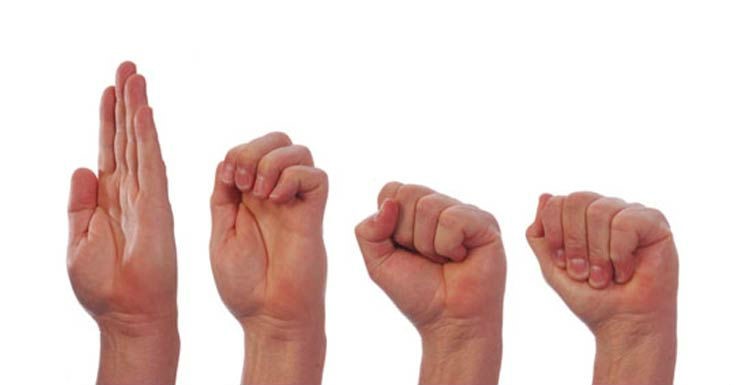
Back muscle strength test
Exercise: 'Swallow', lie on your back and stretch your arms backwards while simultaneously lifting your head, shoulders and straight legs off the level of the bed.
Standard: Hold the position for 2-3 minutes.
Abdominal strength test
Exercise: While lying down, lift your legs and keep them stretched at a 45° angle.
Standard: Hold the position for 2-3 minutes.
Checking the willingness to sit on a chair
Exercise: Continuous walking for 1.5-2 hours.
Normal: Perform exercise without pain or discomfort in the area of injury
Tested three months after injury. A positive result allows the patient to sit in a chair with a bolster under the lower back.
Functional spine test
- Throw your shoulders up and bend backwards;
- Tilt your upper body to the side;
- Straighten your shoulders and lean forward with your back straight;
- Lean forward and touch the floor with your palms.
Standard: Perform all exercises without pain or discomfort at the injury site
Four months after injury, if X-ray and clinical examination are positive.

Please note that this website is for informational purposes only and the information materials and prices provided on the website in no case constitute a public offer within the meaning of Article 437 of the Civil Code of the Russian Federation.
Thank you, your request has been sent successfully!
Rehabilitation after a tibia fracture
A broken tibia is an injury that requires a long and difficult recovery.
The most important treatments during rehabilitation:
Rehabilitation after a shin fracture is primarily aimed at eliminating the dystrophic changes in the soft and hard tissues in the area of injury. It is necessary to restore joint function and eliminate congestion. For this purpose, massage, physiotherapy, exercise therapy and the Ilizarov apparatus should be used. After the cast is removed from the lower leg, the first stage of rehabilitation is to restore mobility to the leg and eliminate swelling. During this period, it is necessary to work on the injured limb. Physiotherapeutic procedures and massage promote good blood circulation, and therapeutic exercises are designed to keep the leg in good condition. During this difficult period, it is advisable to walk as much as possible. Exercises for turning the ankle, rolling from the heel to the foot and vice versa, and scissor exercises are also very helpful. The most important thing is to do this cycle of exercises every day. Do not overexert yourself, that is, do it in moderation. The diet should not be lacking in vitamins and nutrients so that the human body feels good and can recover quickly. Cheese, milk, cabbage, currants, bran, beef, fish - foods that promote complete and rapid healing of damaged tissues. In some cases, the patient needs the help of a psychologist. Sometimes after an injury there is a fear of a re-fracture, so there is no desire to work on the leg. The moral attitude is very important, and we must do everything to make it positive.
Rehabilitation after a pelvic bone fracture
Injuries to the pelvis cause bone compression in two planes:
Most pelvic bone fractures are caused by traffic accidents and falls from great heights. Like all bone fractures, pelvic bone injuries can be open or closed. They also differ in the degree of damage to the organs surrounding the hip.
In cases where the pelvic ring joints remain intact and there are no static or dynamic abnormalities, pelvic bone fractures are considered non-threatening. Since the nervous system is not damaged, the patient can recover quickly using conventional methods. In this case, rehabilitation after a pelvic fracture is also short.
Pelvic fractures with complications
In addition to pelvic ring fractures, complicated fractures are also common. In this case, blood vessels and nerve endings are damaged. For this reason, the symptoms, nature of the disease, treatment and rehabilitation take much longer. The front parts of the pelvis are most vulnerable to injury.
There are several types of fractures, namely:
- marginal. In this case, the injury occurs outside the pelvic ring (ischial tuberosity, ilium, sacrum);
- transverse;
- caudal bone;
- Injuries with and without interruption (Malgen fracture);
- Injuries to the floor and roof of the acetabulum;
- a combination of different fracture types.
In addition, pelvic fractures can be combined with injuries to internal organs, such as urethral rupture, intestinal trauma, vaginal trauma and others. It is obvious that treatment and rehabilitation in such cases takes a lot of time. Fractures of the pelvic bones mostly occur in elderly patients, especially women. Of course, age and chronic diseases significantly complicate rehabilitation. Such patients need constant presence and care, and this care can only be provided by a professional rehabilitation center. You will not be able to carry out rehabilitation at home, even if you wanted to. With pelvic bone fractures, the patient often loses all movement and self-care, so care requires patience and professionalism. However, with the combined efforts of the patient, his relatives and the staff of the rehabilitation center, full rehabilitation after a pelvic bone fracture is possible.
Treatment methods and prognosis of femoral neck fracture in the elderly
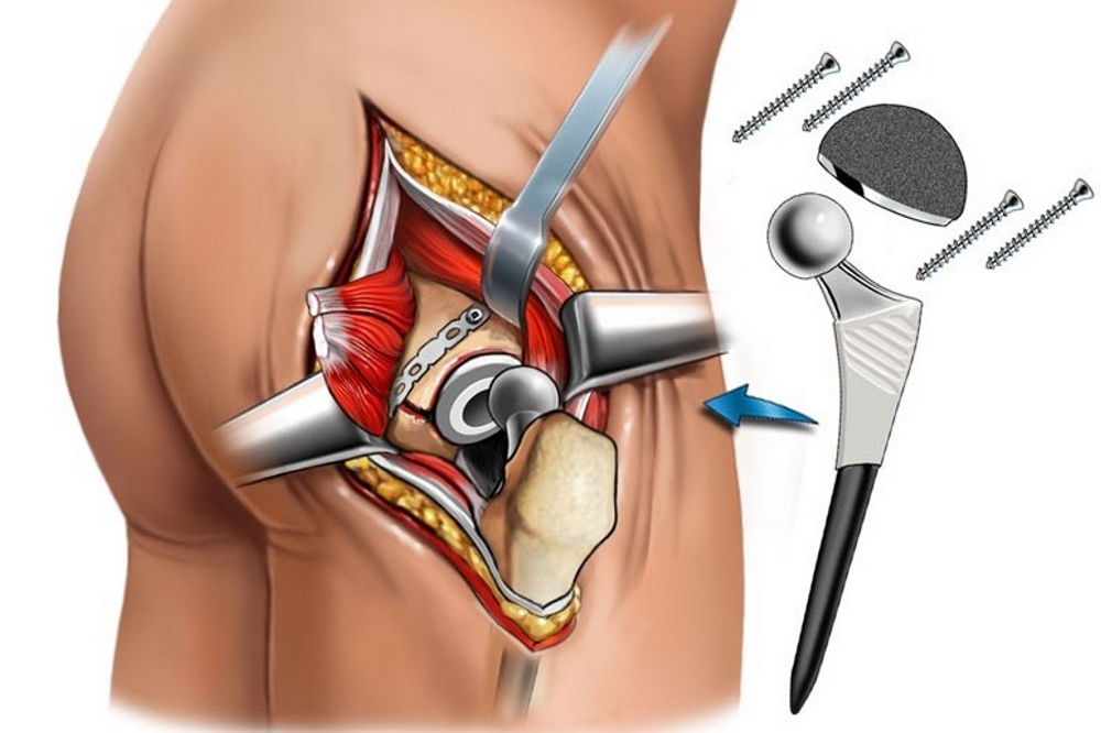
Fractures are treated conservatively or surgically. In conservative treatment, the patient is placed in bone traction for one to two months and then a cast or bandage is applied. The operation consists of osteosynthesis (bone fragments are fused and fixed with metal screws and plates) or an endoprosthesis. After the endoprosthesis is inserted, the foot can be loaded again within a week.
Rehabilitation can vary greatly (from three months to two years) and depends on a number of factors:
- Nature and severity of injury;
- the treatment method (after surgery, bone healing and recovery occurs much faster than after conservative treatment)
- the age;
- Presence of other diseases;
- physical and emotional condition of the patient
- individual characteristics of the body;
- Follow the doctor's instructions.
Goals and methods of rehabilitation
- Preventing possible complications that may lead to death;
- restoration of motor activity;
- Regaining the ability to care for oneself and live independently.
Medical therapy
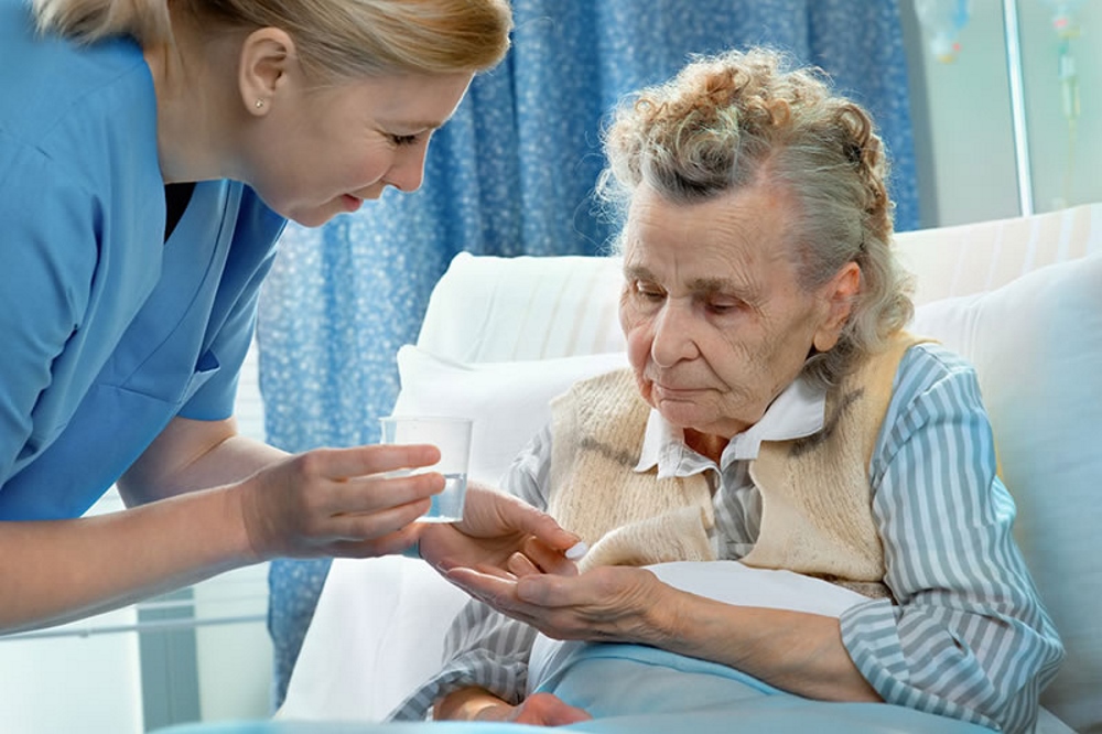
If necessary, the doctor prescribes the patient:
- Painkillers – relieve pain;
- Anticoagulants – to prevent thrombosis;
- Calcium supplements – strengthen bones and activate the healing of bone fractures;
- Chondroprotectors – rebuild cartilage tissue;
- Sedatives – calm the nerves;
- Vitamin complexes – supply the body with vitamins and minerals, activate metabolism and improve immunity.
How can complications be avoided?
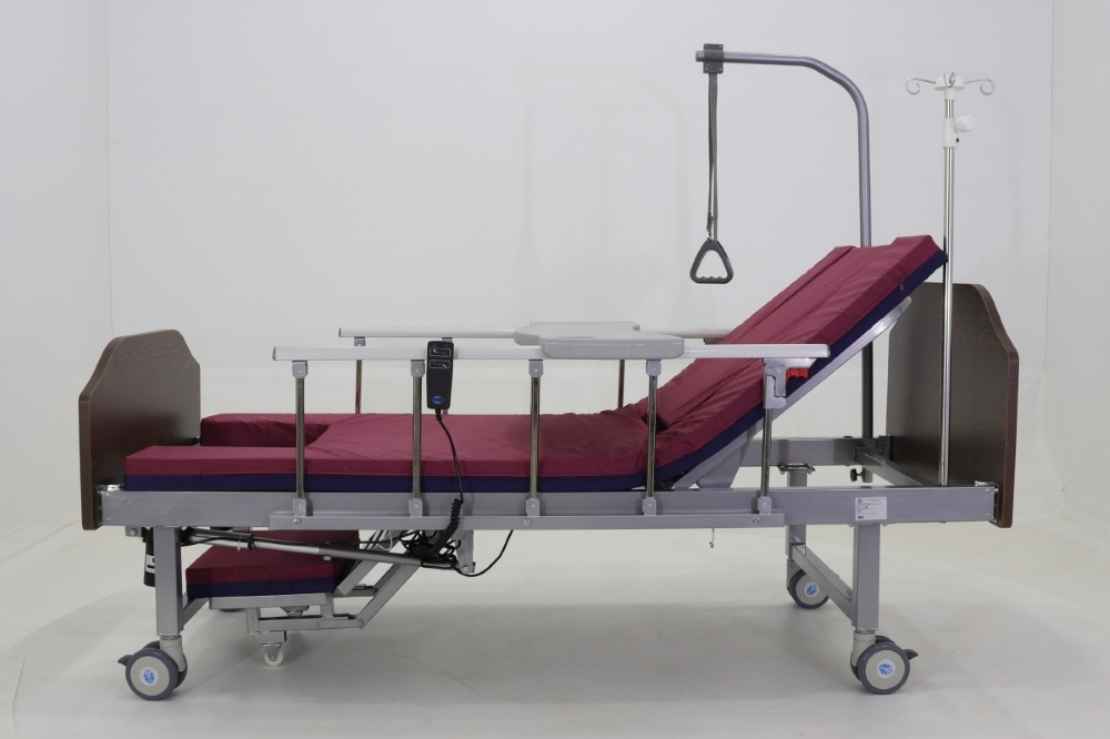
The patient should prepare a bed with a thick and dense foam mattress. It is also advisable to purchase a 'Balkan frame' so that the elderly person can sit up independently. If such a purchase is not possible, a strong rope or a twisted sheet should be tied to the headboard.
To prevent complications, it is recommended:
- have the patient do breathing exercises (breathing in and out with outstretched arms or inflating balloons) to avoid pulmonary congestion;
- Massage and bandage the lower limbs or wear compression garments to prevent thrombophlebitis and varicose veins;
- to prevent pressure ulcers, turn the patient over every 2-3 hours and rub the skin with camphor or salicylic alcohol twice a day;
- Use ointments and drying powders to prevent diaper rash;
- Include vegetable oil and dairy products in the diet to improve intestinal peristalsis and avoid constipation;
- Drink plenty of fluids to avoid dehydration;
- Change diapers more frequently if incontinence occurs (do not use diapers as turning diapers causes severe pain).
In order for the rehabilitation of an elderly person with a hip fracture to be as quick and positive as possible, it is important not only to strictly follow the doctor's recommendations, but also to surround the loved one with care and love so that he does not feel like a burden. It can be very difficult to provide the elderly person with round-the-clock care at home and in the family, offering him dietary and recovery measures. If this is a problem for your family, consider placing your elderly relative in a nursing home where he will be cared for by professionals and the pleasant atmosphere and company will speed up his recovery.
Read more:- Dislocation of a bone in a joint.
- Photo of an ankle fracture.
- Corset for broken fibula.
- Fracture of the lateral condyle.
- What is the connection between the femur and tibia?.
- external rotation of the hip.
- Rupture of the radial-clavicular joint.
- Photo: Outer malleolus fracture.