In some cases, the neoplasm grows out of the cartilage tissue. However, as it grows, it hardens into a spongy bone, typical of this anomaly. The growth area of the osteochondroma grows into the hyaline cartilage. Given these facts, it is incorrect to refer to this type of disease as cartilage. The term 'osteochondral cartilage exostosis' is more appropriate.

- X-ray of the heel bone
- What an x-ray of the heel bone shows
- pharmacokinetics.
- contraindications
- Indications for an X-ray of the heel
- Preparing for surgery
- what is so special about that?
- About the manufacturer
- diagnosis
- treatment methods
- conservative therapies
- removal of the tumor
- When should vitamin D be taken?
- When should you take Omega-3?
- Treatment of heel spurs
- Treatment of heel spurs with folk methods
X-ray of the heel bone
- Prices
- |
- doctors
- |
- addresses
- |
Bilingual service: Russian, English.
Leave your phone number and we will call you back.
The information contained in this section should not be used for self-diagnosis and self-treatment. In case of pain or other exacerbations of the disease, diagnostic tests can only be prescribed by a doctor. Consult your doctor for a diagnosis and appropriate treatment.

X-ray of the heel bone is an indispensable and easily accessible examination method, widely used by traumatologists and orthopedic surgeons in the diagnosis of anomalies of the heel bone. The heel bone is the largest bone of the foot and due to its anatomy, it carries most of the load and can cause injury. X-rays of the heel bone are essential for diagnosing and evaluating the effectiveness of treatment for heel spurs (plantar fasciitis), and the examination is also used to detect tumors and injuries to the heel bone.
- Non-invasiveness and diagnostic accuracy
- Minimal radiation dose and high image quality thanks to our radiology system
- Possibility to save the image on the patient's electronic data carrier (digital equipment).
What an x-ray of the heel bone shows
With the x-ray of the heel bone, the doctor can determine the following, among other things
- the condition of the heel bone (integrity, shape);
- Damage to the heel bone (subluxations, fractures and breaks);
- congenital structural abnormalities;
- Benign and malignant tumors of the bones and joints;
- flat feet
- heel spur;
- osteochondropathies (Haglund-Schinz disease);
- osteoarthritis (arthrosis) and arthritis;
- synovitis (joint inflammation);
- Gout;
- Degree of pathological changes in the heel bone.
pharmacokinetics.
Absorption. After injection of Cmah of the drug in the plasma is reached after 1 hour and then gradually decreases over 2 days. With a single intra-articular administration of 200 mg, the Cmah of chondroitin sulfate in the plasma after 1-2 hours and is 52.5-86.9 ng/ml.
distribution. After injection, chondroitin sulfate is rapidly distributed. It is detected in significant concentrations in the blood as early as 30 minutes after the injection. Chondroitin sulfate accumulates mainly in articular cartilage. The synovial membrane does not pose an obstacle to its entry into the joint cavity. Experiments have shown that chondroitin sulfate can be found in the synovial fluid 15 minutes after i/m injection and then penetrates into the articular cartilage, where its Cmah reached after 48 hours. After intra-articular administration, chondroitin sulfate is retained in joint tissue and gradually released into the bloodstream.
Excretion. t1/2 with intra-articular injection is 2.5 hours.
contraindications
Hypersensitivity to the product or to any of the excipients listed under 'Composition';
bleeding and bleeding tendency;
presence of an active inflammatory or infectious process in the joint, presence of an active skin disease or skin infection in the area to be injected (intra-articular injection);
Pregnancy (no safety data are currently available);
lactation (there are currently no data on the safety of using the drug);
Child and young person (under 18 years of age).
Indications for an X-ray of the heel
Due to its special anatomical structure and high daily loads, the heel bone is subject to frequent trauma and pathological processes. An X-ray of the heel bone is the easiest, fastest and most meaningful way to diagnose diseases at an early stage.
An appointment for an X-ray examination should be made if the following symptoms are present:
- burning or severe pain in the heel
- limited joint mobility in the foot
- Pain and discomfort when walking
- Changes in gait, shifting the center of gravity from the heel to the toes
- Swelling, redness and local temperature increase in the area of the heel bone protrusion.
X-rays are taken after trauma to the foot when fractures, breaks, or dislocations are suspected. If there is bony hypertrophy in the heel area, the diagnostician can assess the development of a spur. X-rays help doctors assess the effectiveness of treatment for inflammatory diseases, bone healing after fractures, and the causes of chronic pain.
X-rays are required if there is any suspicion
- osteoporosis
- flat feet
- osteitis
- Achilles tendon rupture and heel sprain
- Arthritis, arthrosis, osteochondropathy.
X-ray shows signs of inflammation characteristic of bursitis, plantar fasciitis, and tendinitis. When describing the x-ray, attention is paid to the integrity of the bony structures, the shape of the heel shaft and its position in relation to the rest of the tarsal area. If abnormalities are found, additional investigations, including examining the other foot, may be needed to make an accurate diagnosis.
Preparing for surgery
X-ray examination of the heel bone for fracture, spur or inflammation is performed without preparation. A referral can be made to a traumatologist, orthopedist, podiatrist, rheumatologist, or surgeon. It is advisable to bring the results of previous examinations so that the radiologist can check the progression of the pathological process.
Immediately before the X-ray, the specialist will ask you to take off your shoes and free your foot from socks, jewelry and foreign objects.
what is so special about that?
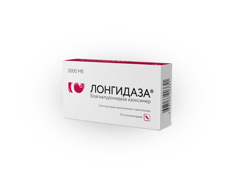
- The most prescribed drug for fibrosis and adhesions in the Russian Federation 1 .
- The originality of the molecule has been recognized by the World Health Organization 9 .
- It increases the effectiveness of comprehensive disease therapy 2, 3, 5, 6 .
- It has a long-lasting effect: it is used 3 times a week.
- It can be used in both acute and chronic forms of the disease 7 .
- Proven efficacy and favorable safety profile 2, 3, 5, 6, 7 .
- Does not damage normal connective tissue.
- Well tolerated by patients - the allergenic properties of hyaluronidase are reduced by conjugation.
In the program 'On the Essentials', experts spoke at length about inflammatory diseases in men and women, explained what fibrosis is and shared their choice of product for adhesions and fibrosis.
About the manufacturer

The drug Longidaza® is manufactured by the Russian pharmaceutical company NPO Petrovax Pharm according to international and Russian GMP standards. The drug was launched in 2005 and is now widely used in medical practice in Russia and other countries.
The company has been developing and manufacturing medicines and vaccines since 1996. The Company's portfolio includes multiple patents on molecules and drug manufacturing technologies. 'NPO Petrovax Pharm' exports medicines to EEU, EU and Iran. The official website provides detailed information about the company and a description of its development.
* Number of prescriptions for Longidase® (7 specialties) issued by physicians 2017-1HY 2020. Data from Proxima Pharma_RX Test Russia.
** In the treatment of diseases associated with adhesions, according to Proxima Pharma_Rx Test Russia.
1 Priindex_Comcon, 2017-1HY 2020, Proxima Pharma_RX Test Russia, 2017-1HY 2020.
2 Zaitsev AV, Khodyreva LA, Dudareva AA, Pushkar D.Yu. Current view of the use of enzyme preparations in patients with chronic prostatitis. Clinical Dermatology and Venereology, No. 2, 2016.
3) Troshina NA, Dolgushin II, Dolgushina VF et al. Microbiological efficacy of hyaluronidase-based preparation in patients with chronic endometritis and uterine myoma. Gynecology, 2015, vol. 17, № 6
4 Petrovich Ye.A., Manukhin IB An innovative approach to the treatment of tubal-peritoneal infertility. Voprosy of Gynecology, Obstetrics and Perinatology, 2010, vol. 9, no. 6, pp. 5-10
5 Khodyreva LA, Dudareva LA, Karpov VK. Longidase® in the treatment of chronic prostatitis
6 Avdoshin VP, Mikhailikov TG, Andryukhin MI et al. Evaluation of the effectiveness of treatment of patients with chronic prostatitis with Longidase® 3000 ME. Effective pharmacotherapy in urology № 4, 2010.
diagnosis
- Primary: after visual inspection and palpation of the suspect area.
- An x-ray is taken to confirm the suspicion, showing the actual location, size, shape, stage, and other signs of growth. Sometimes patients undergo this procedure for other reasons and tumors are found.
Because the outer layer of cartilage is not visible on the X-ray, the actual size of the exostosis is always larger than the examination reveals.
treatment methods
Patients whose tumors do not grow in size, are not enlarged and do not interfere with daily life are examined regularly. No therapeutic measures are carried out.
Physiotherapeutic treatment of osteochondromas is strictly prohibited. Like any benign growth, it can develop into a malignant tumour.
conservative therapies
This type of therapy does not treat the disease itself, but relieves symptoms when a bony overgrowth, particularly in the hip or foot, begins to cause regular discomfort or interfere with daily life. Medicines used:
- NSAIDs are the most commonly used drugs. All forms and types of drugs are used: injections, capsules, ointments. They inhibit the development of inflammation in the tissues around the tumor that are irritated by its presence and pressure.
- Dipropane helps when anti-inflammatory drugs are ineffective. It has a long-lasting pain-relieving effect when placed in the bone cavity.
- Therapeutic exercises are recommended to improve tissue metabolism around the tumor.
removal of the tumor

Doctors advise against an operation in underage patients, since the growths can regress spontaneously at this age.
Surgical intervention is required when:
- rapid growth of the anomaly is noted;
- the exostosis has reached such a size that it is visible to the naked eye on the surface of the skin;
- the structures of the mass affect the nerve fibers or blood vessels.
The surgical procedure is performed under general or local anesthesia depending on the location and size of the piece to be removed. The piece of bone is splinted with a chisel and the area is carefully ground down.
When should vitamin D be taken?
Vitamin D is a complex of fat-soluble substances. Vitamin D3 is the most abundant. It is believed to be better absorbed. Vitamin D is important for strong bones, a good immune system and the functioning of the entire body. Vitamin D sensitive receptors are found in almost all organs and systems.
Vitamin D should be taken year-round, especially if you live in northern regions where there aren't many sunny days. But vitamin D deficiency is also widespread in southern regions, since not all sunlight is equally good and contributes to vitamin D synthesis in the human body.
- prevention and treatment of rickets;
- prevention and treatment of osteoporosis;
- thyroid dysfunction;
- low immunity of the body;
- frequent colds, viral and infectious diseases;
- frequent fatigue, systematic malaise;
- autoimmune diseases
- oncological diseases;
- bowel diseases;
- allergies.
When should you take Omega-3?
Omega-3 fatty acids are polyunsaturated fatty acids. With them we can keep all our organs working properly and improve our overall health. Omega-3 fatty acids are part of the membranes of all body cells and are good for the functioning of the brain, cardiovascular system and immune system.
Omega-3 fatty acids should be taken daily. Here are some indications for taking omega-3:
- Elevated cholesterol level;
- disorders of the nervous system;
- deterioration of hair and skin;
- brittle nails;
- visual impairment;
- sudden increase or decrease in body weight;
- occurrence of allergic reactions;
- Heart problems;
- pregnancy and breastfeeding;
Treatment of heel spurs
Treatment Heel spur treatment begins with conservative measures. This includes various types of physical therapy, massage, mud treatments, etc. Their goal is to reduce inflammation and restore normal tissue metabolism. In the early stages of heel spurs, these measures can restore health.
They also help in relieving heel. In severe cases, this means bed rest. In rather soften Heel spur shaping – it is necessary to wear special insoles and other devices that shift the center of gravity.
Painkillers and anti-inflammatory drugs can be used. In some cases, a drug blockade is used. This completely eliminates the sensitivity of the heel. heelThis completely eliminates the sensitivity of the heel, so the patient no longer experiences pain.
The most difficult treatment of heel spurs is. the distance of. However, such operations Not everyone is prescribed such an operation - there are many contraindications and possible side effects.
Therefore, you should seek treatment as early as possible See your doctor as soon as possibleIf you have pain while walking, you should see your doctor as soon as possible. If you know that you have flat feet, obesity, rheumatic diseases and other such pathologies, you should be extra careful and visit a orthopedic surgeons.
Treatment of heel spurs with folk methods
Before describing the different ways to eliminate heel spurs, it must be said that there is often a clear misunderstanding of this pathology. Perhaps what people understand by a 'spur' is as follows. a bubblePerhaps people think of a 'spur' as a blister that impedes walking and causes pain.
Therefore, some suggest using different baths and steaming the feet. feet. They suggest using different herbal decoctions, such as B. buttercup to use as a bath additive.
Various lotions and compresses are also very popular.
However, while you are performing all of these measures, your condition may worsen. Therefore, it is best to first visit a surgeon, get a diagnosis and with the appropriate one therapy. U Physician You can also find out about the effectiveness of some folk remedies.
This article is for informational purposes only.
Read more:- Anatomy of the heel bone x-ray.
- Heel bone human anatomy photo and description.
- Heel bone tendon sac in Latin.
- Pain in the heel bone of the foot.
- Heel spurs and Achilles spurs.
- Treatment of plantar fasciitis ointment.
- heel bone injury.
- bones in the heel.
