Immediately after a knee injury, before a doctor arrives on site, first aid should be administered using a few simple steps:

- Acute joint injuries
- symptoms and signs
- species
- Symptoms and signs.
- Symptoms of a joint bruise
- Types of skeletal joint contusions
- Method of diagnosis
- Which doctor should be consulted?
- Possible complications
- Congenital joint anomalies
- ICD-10
- Bruise of the knee joint
- AdobeStock_156955086-1.jpg
- Conditions that can cause a knee injury
- Conservative therapy
- surgical treatment
Acute joint injuries
Acute joint injuries are a large group of injuries that are classified according to the severity of the injury and the expected consequences. Statistically speaking, injuries to the bony and soft joint structures account for more than 60% of all musculoskeletal injuries.
The main cause of acute joint injuries is physical stress caused by overexertion or inadequate adherence to safety measures. Isolated injuries are most commonly caused by impacts, falls, or subluxations of limbs, while combined injuries occur from falls from heights, accidents, traffic accidents, etc.
In addition, joint injuries are more common in people exposed to:
– Overweight.
– Innate body characteristics.
– Unstable joint tissue.
– Unequal length of the lower limbs.
– Excessive physical exertion.
– Tendonitis (dystrophic changes in muscle tissue).
– Tendonitis (inflammation of the tendons at their attachment points).
symptoms and signs
Pain and swelling at the injury site are typical symptoms. Bruising may also occur. The mobility of the affected body part is directly related to the severity of the injury - the larger the injury, the more restricted the movement.
Important: However, some types of joint injuries result in increased mobility. This could be an indication of torn ligaments, fractures or similar injuries.
Dislocations are particularly noticeable. This type of injury is characterized by significant curvature of the limb, atrophy of the natural bone projections in the joints, and sometimes the appearance of a protruding joint end of the distal or proximal segment. An additional sign of damage is often the accumulation of blood in the joint (hemarthrosis). In these cases, the joint is significantly enlarged, has a spherical shape and fluctuates.
The characteristic symptoms therefore include:
- Pain that increases with physical exertion.
- Limited range of motion.
- The appearance of bruises.
- Swelling of the injured part of the body.
- numbness.
- Crunching or clicking in the joint tissue.
species
There are 3 main groups of knee joint injuries.
– Damage to the bony structures of the joint in the form of fractures.
– Soft tissue injuries such as sprains, strains, ligament and meniscus tears.
– Combined injuries.
– A bruise is the simplest type of injury.
– Strain of the tendons and ligaments that support the kneecap.
– Meniscus tear – occurs when the knee is suddenly twisted while the foot is stationary. This type of injury always requires surgery.
– Complete and incomplete rupture of the ligaments of the knee joint – caused by violent impacts on the knee: traffic accidents, sports, falls, etc.
– Cartilage damage – caused by intra-articular bruises or fractures.
– Fractures or breaks in the bone in the knee.
– Dislocation of the kneecap – this injury usually occurs in youth, between 13 and 18 years of age.
Symptoms and signs.
– swelling;
– Limitation of mobility, inability to perform certain movements;
– crunching, cracking;
– Increase in temperature in the area of injury;
– Inability to lift a heavy object;
– Acute pain – occurs within seconds of injury;
– Persistent, nagging pain;
– loss of sensation due to damage to nerve endings;
– Hemarthrosis – accumulation of blood in the joint cavity.
Important: At the time of injury, the injured person has the feeling that the knee is 'bouncing, popping and falling back'. Mobility is restricted and limping occurs.
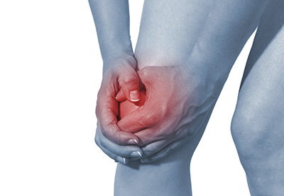
Symptoms of a joint bruise
The main symptom is pain. Patients complain of a stabbing, dull, or aching pain that increases with movement and touch. Pain syndrome is also sometimes caused by an accumulation of blood in the joint cavity.
- Redness and swelling in the ankle area
- limited mobility of the leg or arm due to pain
- Forced posture of the limb
- local impairment of superficial sensation
The symptoms largely depend on the cause of the injury and the area of tissue damage.
Types of skeletal joint contusions
The main criterion for classification is the location of the pathological impact. The most common types are right or left forearm, shoulder, pelvis or lower extremity.
- Shoulder contusion
- hip bruise
- Elbow injury
- ankle injury
- Injury to the knee joint
Ankle injuries are common among athletes. Elbow and pelvic injuries are more common after a fall.
Method of diagnosis
Joint injuries are diagnosed based on the patient's characteristic complaints, external changes and the results of diagnostic tests. The doctor carefully examines the surface of the injured area to detect swelling, bleeding and other pathological signs. The mobility of the bone joint is assessed.
X-ray. Using images in two projections, the doctor can identify the fracture and determine its type.
Ultrasound examination. With the ultrasound examination, the doctor can visualize the soft tissues in real time.
MRI. This is a more powerful imaging technique that creates volumetric images of the joint layer by layer to assess the condition of all structures.
Arthroscopy. An endoscope is inserted through an incision/point into the joint cavity to examine the tissue and provide treatment.
MRI is the preferred examination method. Radiologists in MRI clinics use specialized, state-of-the-art equipment to provide meaningful and clear images for accurate diagnosis.
Which doctor should be consulted?
Joint injuries are diagnosed and treated by orthopedic traumatologists. In some cases, the help of a surgeon or neurologist (if nerve damage is possible) may be required.
The diagnosis is based on anamnesis, an interview and an external examination, which is supplemented by instrumental examination methods. The doctor will inquire about the nature of the injury - what, how and under what conditions it was sustained, whether there are other injuries, what sensations occurred during and after the injury (pain, crunching, discomfort, dizziness). It is also important to indicate whether there were previous problems with the injured joint (osteoarthritis, trauma, damage to surrounding tissues).
The doctor examines the injured area and gently palpates it to assess the range of motion and alignment of the limbs.
In the case of large joint bruises where movement is impaired and pain is severe, more serious injuries must be ruled out. X-rays, ultrasounds, CT or MRI scans of the injured area are performed. This means that sprains, joint fractures, ligament tears and meniscus damage can be ruled out when it comes to the knee joint.
Possible complications
Minor bruises usually do not cause complications. They heal quickly and without leaving any trace. Severe leg injuries can result in large bruises and soft tissue necrosis. Injuries to large joints, especially the knees, often result in hemarthrosis (bleeding into the joint cavity) or synovitis (inflammation of the joint tissue). In rare cases, tendon or muscle tears may occur, requiring surgery.
Long-term consequences without appropriate treatment can include fibrosis in the muscle area and the development of ossifying myositis. Fibrosis of the muscle leads to muscle sheath syndrome, in which the fibers are replaced by connective tissue. In ossifying myositis, ossifications form in the area of the muscle fibers. The quadriceps muscle is most commonly affected.
Congenital joint anomalies
Among the most common congenital joint anomalies, hip dislocation or dysplasia is the most common. The diagnosis is usually made after an initial examination in the delivery room, during an outpatient examination by a trauma surgeon or a neonatologist at 1 month of age. Treatment is selected individually based on examination and x-ray findings.
The second most common form is subluxation or dislocation of the atlas or first cervical vertebra. This serious diagnosis is usually the result of a traumatic birth or the use of instrumental birth methods. Treatment is also recommended based on x-ray findings and clinical presentation and may last several months.
ICD-10
Trauma to the knee joint is one of the most common injuries due to the high load on this segment and its anatomy. Most injuries are minor, occur at home (e.g. as a result of a fall on the street) and can be treated on an outpatient basis at a trauma center. In addition, it is not uncommon for the knee joint to be injured during various sporting activities, and the severity and type of injuries can vary greatly.
Knee injuries resulting from falls, road traffic accidents and work-related injuries are less frequently treated in traumatology. In these cases there is a higher proportion of intra-articular fractures with injury to the joint structures. Treatment usually takes place in a trauma unit. A combination of other injuries may be present: head trauma, fractures of the trunk and limbs, and tears of hollow and parenchymal organs.
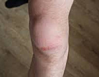
Bruise of the knee joint
A knee joint contusion is a soft tissue injury in which no anatomical structures were injured. However, at the microscopic level, not only the skin and subcutaneous tissue, but also intra-articular elements are damaged, resulting in reactive inflammation, hemarthrosis or synovitis. The symptoms of a bruise are non-specific and can be associated with other injuries, so the diagnosis is made after excluding other injuries.
The injured person complains of pain. The joint is slightly to moderately swollen, and bruising to the skin is not uncommon. The footing is usually maintained, but there may be restricted movement and a slight limp. Pain in the bruised area can be determined by palpation. Palpation of the ligaments and bony elements is painless and there are no signs of abnormal mobility. Fluid often accumulates in the joint (blood in the first few days, effusion after 2-3 weeks).
To rule out other injuries to the knee joint, the affected person is referred for an X-ray examination. Sometimes an MRI, ultrasound or CT scan of the knee joint or an arthroscopy is ordered. Treatment takes place in a trauma center. For hemarthrosis and synovitis, a joint puncture is performed. For minor bruises, rest is recommended, while for severe bruises, a plaster cast is applied for 2-3 weeks. Cold treatment is recommended for the first 24 hours, and patients are referred to the NFZ on the third day. Regular follow-up examinations and repeated LPs are carried out as necessary. The duration of incapacity to work varies between 2 and 4 weeks.
AdobeStock_156955086-1.jpg

AdobeStock_156955086-1.jpg
Knee injuries related to various traumatic events are the most common type of injury. These usually involve situations such as a blow to the knee joint, bending the leg too much and forcefully, twisting the limb or falling on the knee.
Within seconds of the traumatic impact, severe pain, swelling, bruising, and nerve and blood vessel damage can occur. The patient may experience sensations ranging from numbness in the knee area to general weakness to a color change from blue to pale skin.
In any case, you should not wait and see a doctor immediately.
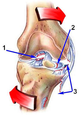
1 bruise. The least serious injury to the knee joint. A bruised knee joint can have many different causes, but the most common is direct trauma to the knee from a fall or blow.
2. Strains of the tendons and ligaments that support the kneecap.
3. Tears of the meniscus. This problem can be caused by a sudden twisting of the knee with a locked foot. This is not always a diagnosis in professional athletes. We often see meniscus damage in normal people in everyday situations. The only option for treatment is usually surgery.
4 A complete or incomplete tear of the ligaments in the knee joint can be caused by a violent blow to the knee. This can be caused by sports activities, traffic accidents, falls, etc.
5. Cartilage injuries. This is often an accompanying symptom of a knee bruise or intra-articular fracture.
6. the fractures or breaks of the bones belonging to the knee joint in one way or another. Fractures can be caused by excessive stress on the knee.
Conditions that can cause a knee injury
In addition to extreme stress or sudden trauma, certain conditions can also lead to damage to the knee joint:
– Osteoarthritis leads to pain that is particularly severe in the morning. The pain is usually limited to the injury site. Pain and stiffness in the knee area can also be caused by many other conditions, from gout to lupus to arthritis;
– Popliteal cyst. Characterized by swelling in the back of the knee;
– Delaminating osteochondritis. Characterized by a pain syndrome and limited mobility;
– Problems in the hip joint, pinched nerve. Also leads to a pain syndrome.
Conservative therapy
Non-surgical treatment is carried out separately for incomplete meniscus tears. The most important therapeutic measures are:
- Prescription of drugs from various pharmacological groups, which include analgesics, edema inhibitors and chondroprotectors.
- Physiotherapy with drug electrophoresis, mud baths and magnetic therapy.
- Creating functional rest for the knee joint.
Conservative therapy is a long-term process. It takes time for the cartilage and ligaments in the knee joint to fully recover.
surgical treatment

meniscus surgery
Surgery is the most common method to repair a torn meniscus in the knee joint. Different surgical techniques are used. The main groups are:
- Open surgery – the skin, subcutaneous tissue and capsule of the knee joint are cut, then access is created to manipulate the structures of the joint.
- Arthroscopy – is a diagnostic and therapeutic procedure in which an arthroscope using small surgical instruments is inserted into the joint cavity through small incisions.
The extent of the procedure depends on the severity of the injury or pathology. If a tear in the medial meniscus of the knee joint is diagnosed, the operation consists of removing it through an open approach. If there is damage to two menisci, the extent of surgical intervention depends on the extent of the tear and is usually performed through an open approach. For small tears, arthroscopy with plication and restoration of the anatomical integrity of the meniscus and its supporting ligament can be performed.
Read more:- Closed Foot Injury.
- joint injuries.
- contusion of the ankle.
- Femoral collateral ligament.
- Dislocation of a bone in a joint.
- tibial ligaments.
- recurrence of joints.
- tibia and fibula.