Injuries to the joints – A large group of injuries that vary in their consequences and severity. These include bruises, ligament injuries, sprains and fractures within the joint. They can be caused by a domestic or sporting injury, a motor vehicle accident, a criminal incident, an industrial accident, or a natural disaster. The most common symptoms are pain, swelling and restricted movement. In some joint injuries, deformities, hemarthrosis, and abnormal mobility are noted. X-rays, CT scans, MRIs, ultrasounds, arthroscopy and other tests are used to confirm the diagnosis. Treatment can be conservative or surgical.
- Closed knee injuries
- Short description
- Clinic automation: fast and cheap!
- Clinic automation: fast and cheap!
- classification
- How does a joint work?
- Injuries and their classification
- How dislocations occur: the role of injury
- Symptoms of injury: pain and functional limitations
- Treatment
- complications
- prevention
- What questions should you ask your doctor?
- Advice for the patient
- Symptoms of joint injuries
- Types of joint injuries
- Fractures of the knee joint
- ICD-10
- anatomy
- types of injuries
- Symptoms and localization of pain
- Symptoms of wounds and joint injuries
- Causes of joint tears and injuries
- Symptoms and treatment
- Torn hip ligaments
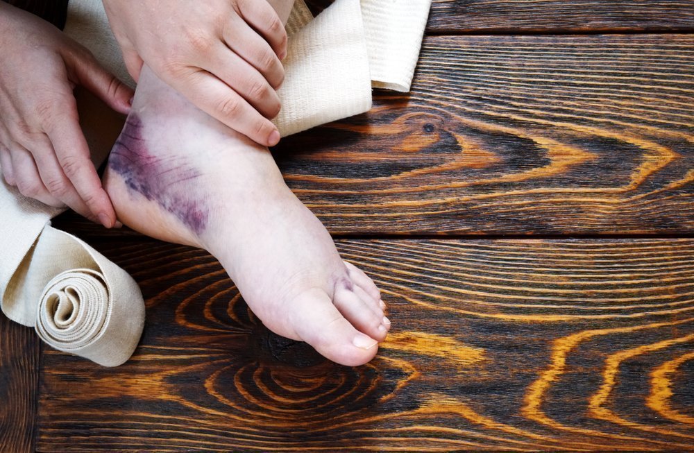
Closed knee injuries
ICD categories: Fresh meniscus tear (S83.2), Fresh knee cartilage tear (S83.3), Strain, tear and strain of the (external) (internal) collateral ligament (S83. 4), Sprain, tear and strain of the (anterior) (posterior). ) cruciate ligament (S83.5), sprain, tear and strain of other and unspecified parts of the knee joint (S83.6), injury to several structures of the knee joint (S83.7), contusion of the knee joint (S80.0).
Short description
Closed knee joint injury – Injury to structures of the knee joint without skin injury caused by mechanical energy [1].
User of the log: Orthopedic traumatologists, surgeons, general practitioners, emergency doctors, paramedics.
Class I – the usefulness and effectiveness of the diagnostic method or treatment measure has been proven and/or generally accepted
Class III - the available evidence or general consensus suggests that the treatment is not useful/effective and may even be harmful in some cases
B - Results from a single randomized clinical trial or large non-randomized trials
C – General expert opinion and/or results of small studies, retrospective studies, registries.

Clinic automation: fast and cheap!
– 800 RUB / 5500 KZT / 27 BYN – 1 order per month

Clinic automation: fast and cheap!
classification
II. DIAGNOSTIC AND THERAPEUTIC METHODS, APPROACHES AND PROCEDURES
Magnetic resonance imaging of the knee joint (indications: torn, dislocated and damaged knee ligaments)
Minimum list of examinations to be carried out upon referral for a planned hospitalization: No planned hospitalization.
1. Magnetic resonance imaging of the knee joint (indications: ligament tears, sprains and injuries of the knee)
Collection of complaints and anamnesis, physical examination.
Medical history: Presence of trauma with a direct (severe blow to the knee, fall on the knee) or indirect (acute rotation of the trunk with a standing foot) mechanism of injury.
3) gross pathological mobility in the area of the knee joint.
– two-dimensional x-rays of the knee joint: no bony pathology, but secondary signs of soft tissue damage: widening of the joint space, spreading of the syndesmosis.
– MRI: There are signs of damage to the capsular ligaments and tendons.
How does a joint work?
A joint is a movable connection between two bones. It is needed to redistribute the loads acting on the limbs. The ends of the bones, the epiphyses, converge: where one bone has a bulge, the other bone has a depression. The caps are covered with hyaline cartilage. The joint is surrounded by a dense fibrous membrane. The cavity is filled with a highly viscous synovial fluid. This fluid is used to supply the joint with nutrients. There are ligaments around the joint that reinforce it on all sides. The movements carried out in the joint are precisely defined: flexion and extension, adduction and retraction, rotation and rotation.
Correct joint geometry is extremely important for joint health. If this geometry is disturbed at the slightest sign - a torn ligament, an inconspicuous misalignment of the joint surfaces - the rest of the joint becomes chronically overloaded and wear accelerates dramatically. There are many reasons for this: micro-injuries, monotonous everyday stress, jerky sports or excessive physical activity. If the joint remains untreated, the biomechanics and structure of the articular cartilage change irreversibly. In order to stabilize the joint, the body forms osteophytes, i.e. bony outgrowths that change the joint so much that movement within it becomes impossible.
Injuries and their classification
An injury is the action of a mechanical force that has a different direction. The tissue of the joint, the ligaments and the surrounding muscles counteract this force. Depending on the mechanism and strength of the impact, these types of injuries are distinguished:
- Bruise. The soft tissue is damaged.
- Incomplete and complete ligament tears, more commonly known as sprains.
- Intra-articular fracture. Depending on the fracture line, it may be through the adjacent surfaces, outside the adjacent surfaces of the bone, or splintered.
- Periarticular fracture.
- Contortion. Often accompanied by trauma or stretching of the capsule.
- Fracture-dislocation.
- Meniscus tear. This injury only occurs in the knee joint, where there is a special layer of cartilage.
How dislocations occur: the role of injury
There are two types of injuries - congenital sprains and acquired conditions - the causes of which are very different. If we are talking about congenital pathologies, then they are based on unfavorable heredity, genetically determined connective tissue defects or a violation of the intrauterine development process, as well as the influence of external environmental factors.
In acquired lesions, the most important factor is trauma. Of all possible injuries, the most common are sports and household injuries involving falls, sudden unnatural movements, and hyperextension of the limbs. Numerous dislocations and fractures are not uncommon in traffic accidents or technical disasters, natural disasters and work accidents. Injuries to the cervical spine are possible due to whiplash, ie due to a sudden hyperextension of the neck during an emergency stop in traffic. Whiplash can be caused by diseases of the skeletal system or strong contractions of muscle groups in the area of tetanus or by changes in the nervous system.
Symptoms of injury: pain and functional limitations
The most common symptom of a sprain is a clicking or popping sound followed by a stabbing pain. This pain is localized directly at the site of the injury. The pain is accompanied by swelling of the tissues, the limb looks unnatural, and the surrounding tissues may lose sensitivity. Due to the pain and the altered position of the bones, movement of the limbs is severely limited or completely impossible, and any attempt to move the limbs increases the pain. A crawling feeling, pale skin and bruising due to damage to blood vessels are also typical. Depending on the site of the injury, other symptoms may occur.
Most dislocations are closed, meaning the skin and underlying tissue over the dislocation site are not torn. The swelling is severe and the joint is immobilized. Open sprains with torn ligaments and muscles, skin injuries, wound formation and severe swelling of the surrounding tissue as well as bleeding are possible.
- Dislocations with complications in the form of intra- or extra-articular bone fractures and damage to muscle fibers, nerves or large blood vessels can occur. Crippling contortions lead to nerve damage and paralysis of the limbs.
- A special variant is habitual sprain, which affects the same joint due to a weakening of the ligaments. It can be caused by mild force, the surrounding tissue is slightly swollen and can repair itself.
Treatment
Injuries to the meniscus and ligaments of the knee joint must be treated immediately. If only the cruciate ligaments are partially damaged, instability of the knee joint will develop over time. If the affected person is not very active and the instability does not bother him too much, it is sufficient to wear a joint stabilization device with metal inserts to keep the joint stable and fix it in the longitudinal plane.
In other cases, or if the leg can no longer be supported at all, surgical treatment is necessary. There are various types of operations and plastics to strengthen the ligaments of the joint capsule. If these measures are not effective, knee replacement surgery is performed. In the event of a torn meniscus, surgery is the only way to preserve the function of the joint. In addition to open surgery, small incisions can also be made through an arthroscope.
complications
Impairment of knee joint function (limited mobility), development of osteoarthritis (dystrophic joint disease), muscle loss in the thigh.
prevention
To prevent injuries to the meniscus and ligaments of the knee joint, the following measures should be taken. Avoiding knee injuries at home and during sports. The extent of physical stress must be adapted to the level of training and age. Wearing protective equipment during sporting activities. Comfortable footwear should always be worn.
What questions should you ask your doctor?
How does damage to the menisci and ligaments of the knee joint occur?
Does surgery always have to be performed?
Advice for the patient
If you suspect that the meniscus and ligaments of the knee joint are damaged, you should see a trauma surgeon. Avoid hard work, direct hits and falls on the knee to avoid such injuries.
Symptoms of joint injuries
Pain is the main nonspecific symptom of a pathological condition. The pain can vary in intensity and localization. The pain often spreads to neighboring areas of the body. The bony junctions of the skeleton are swollen and enlarged. Deformation is observed. The motor function of the affected joint is often partially or completely impaired.
- Shortening or curvature of the affected limb
- Abnormal bony protrusion
- Enlargement of the bone joint with flattening of the contours
- increased pain with movement
- Appearance of a visible bruise
Swelling is not the only cause of an enlarged bone joint. Bleeding into the joint cavity often occurs. The bone joint then takes on a spherical shape.
Types of joint injuries
According to the classification used in trauma surgery and orthopedic practice, the following main pathologies are distinguished:
Bruise. There are no open wounds and significant damage to the joint components. The main symptom is a hematoma.
Ligament involvement. These connective tissue structures are completely torn, torn or overstretched.
Fracture of the joint ends of the bones or outside the bone joint. May be closed, open and/or splintered.
Dislocation or dislocation with fracture. The articular surfaces of the bones are separated, which leads to a loss of motor function in a certain area of the skeleton.
The severity of the injury and treatment options depend on the type of injury.
Fractures of the knee joint
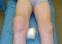
A patellar fracture is caused by a fall on the front surface of the knee. It is accompanied by severe pain, swelling, hemarthrosis and the inability to keep the leg straight in an elevated position. Support is difficult or impossible. When palpating the kneecap, a 'gap' may be visible - diastasis between fractures caused by contraction of the quadriceps muscle. The diagnosis is confirmed by an x-ray of the knee joint. Treatment of fractures without displacement is conservative – immobilization for 6-8 weeks. For fractures with displacement, surgical treatment is performed, during which the fractures are fixed with a special wire and fused. Physiotherapy, massage, physical therapy and painkillers are then prescribed. The recovery time is two to three months.
Fractures of the tibial and femoral condyles are among the most serious injuries to the knee joint. They are caused by a high-energy impact. They are usually associated with damage to other structures (ligaments, meniscus). They often occur as part of a combined injury. They manifest themselves as acute pain, significant swelling, hemarthrosis and deformity of the knee. Movement is impossible. Crepitus is sometimes noted on palpation. Treatment is usually conservative: skeletal traction or casting. In the case of severe displacement, osteosynthesis is carried out using screws, plates or staple screws. The patient is prescribed exercises and physiotherapy, and rehabilitation measures are carried out during the recovery period.
ICD-10
Joint injuries are injuries to the bony structures and soft tissues of the joints. They account for approximately 60 % of all musculoskeletal injuries. They can be diagnosed in people of all ages and genders. They often occur in the home environment. They are common in athletes, and a pattern has been identified between exercise and the occurrence of certain joint injuries. The ankle and knee joints are most commonly affected, less commonly the joints of the upper limbs (shoulder, elbow and wrist).
Joint injuries can occur in isolation or in combination with other injuries: limb bone fracture, pelvic fracture, rib fracture, spinal fracture, CMT, blunt abdominal trauma, kidney injury, bladder rupture, etc. Isolated joint injuries are most often caused by an impact, fall, or falling foot. Joint injuries are caused by falls from heights, workplace accidents, traffic accidents, and natural and industrial disasters. The treatment of joint injuries is carried out by trauma surgeons.
anatomy
A joint is a movable connection between two or more bones of the skeleton. Joints that consist of two bones are called simple joints, and those that consist of three or more bones are called complex joints. The articular surfaces are separated from each other by a gap and are connected by the joint capsule. The capsule is reinforced in places by thick and strong ligaments, which additionally fix the joint and also act as a kind of guide, restricting some movements and allowing others. The following movements are possible in the joints: extension, flexion, adduction, adduction, pronation, supination and rotation.
The articular surfaces of the bones are covered by hyaline cartilage and are located in the joint cavity, which contains a small amount of synovial fluid. The smooth cartilage slides easily against each other and, thanks to its elasticity, absorbs shocks when walking and moving, thus acting as a kind of shock absorber. The outer part of the joint is covered by the joint capsule, which is attached to the bone near or just below the edge of the articular surfaces. The strong outer part of the capsule protects the joint from external damage, while the thin and delicate inner membrane secretes synovial fluid that nourishes and lubricates the joint and reduces friction of the articular surfaces.
Surrounding the joint are the periarticular tissues: ligaments, tendons, muscles, blood vessels and nerves. Damage to these structures negatively affects the joint itself by impairing movement, affecting the amplitude and direction of movement, affecting nutrition, etc. The joints are supplied with blood by an extensive arterial network of 3-8 arteries . All components of the joint, with the exception of hyaline cartilage, have a large number of nerve endings. In the event of an injury, these nerve endings can become a source of pain.
types of injuries
The most common wrist injuries include the following types:
- Fractures. The spoke bone is particularly often injured. There are two types of fractures of the radius bone of the wrist:
- Smiths fracture (volar fracture). This injury is caused by a fall onto the outstretched hand, onto the back of the hand. There is a fracture of the bone and displacement of bone fragments towards the palm.
- Colles fracture (extensor fracture). This injury occurs when a person falls on the hand and the bone fragments are displaced toward the thumb and back of the hand.
- Sprain of ligaments. When the ligaments are stretched during a sprain, the tissue remains intact, but the function of the joint is impaired for a period of time.
- Sprains. When a wrist is sprained, the ends of the joint become dislocated so that they completely lose contact with each other. Sprains can be pathological (due to bone and joint disease) or traumatic (due to trauma).
- Inflammatory diseases. They can be caused by trauma, endocrine disorders, overuse of the joint, infections, etc.
- Tendinitis is an inflammatory lesion of the tendons and surrounding membranes in the wrist.
- Styloiditis is an inflammation of the ligaments that attach to the spinous processes of the radial or ulnar bones.
- Synovitis is an inflammatory disease of the synovial membrane of the joint.
- Bursitis is an inflammatory disease of the joint capsule with the formation and accumulation of fluid in its cavity.
Symptoms and localization of pain
Each of the above types of injuries has its own symptoms, based on which a trained specialist can make an accurate diagnosis and prescribe appropriate treatment. The main signs of a wrist injury are.
- Pain in the joint with varying intensity and localization
- Swelling and loss of sensitivity at the site of injury
- Changes in the appearance of the hand and wrist
- Redness of the skin at the site of injury
- Bleeding at the injury site
- Joint contracture (acute limitation of joint mobility)
- Increase in general temperature
- Weakness, malaise, chills
- nodules (in the hierogons)
Symptoms of wounds and joint injuries
Common symptoms of wounds and other joint injuries include.
- Painful sensations that worsen with any physical exertion;
- Restricted range of motion in the affected joint;
- bruising;
- swelling around the joint and a feeling of numbness;
- Cracking or grinding of the joint and instability of the joint.
The symptoms of the disease depend on the affected area.
For example, if the shoulder ligaments are damaged, movement in the shoulder is restricted. The ligaments of the elbow are torn, causing pain when moving the forearm. The ligaments of the hand are torn in the elbow area and cause discomfort in the elbow area. The torn ligaments in the fingers are accompanied by a lateral deviation of the finger bones.
Symptoms depend on the extent of the injury:
- In the first stage, part of the ligament fibers are torn. There is usually no swelling, swelling, or bleeding at the injury site. The patient complains of moderate, tolerable pain.
- In second-degree injuries, most of the fibers are torn. In this case, swelling and bruising occur. The mobility of the joint is limited and the patient complains of pain in the joint. In some cases, slight instability is noted.
- In the third stage the ligament is completely torn. The joint becomes unstable and bruising and swelling occur. Significant pain occurs.
Causes of joint tears and injuries
The main cause of such a joint injury is strong physical pressure applied to the joint or non-observance of safety precautions when performing violent movements.
It should be noted that the following factors significantly reduce the strength of the tapes:
- Certain congenital diseases;
- overweight (including obesity);
- Frequent physical activity;
- Instability of tissue due to inflammatory processes;
- tenosynovitis with tendonitis;
- Tenosynovitis with inflammation of the tendon;
- Tendinosis with damage to muscle tissue.
Injuries to the ligaments of the leg joints can be caused by different limb lengths or a high arch of the foot. There are also other factors that can be indirect causes of injuries to the ligaments in the joints.
Symptoms and treatment
In order to recognize the injury and initiate treatment, it is important to know the main symptoms:
- Pain in the gluteal region, in the lower back, extending to the groin: it sometimes occurs immediately after the event, but more often with repetitive exertion (walking or squatting);
- Slight rigidity of movement – this is limited by reflex spasms;
- Discomfort when sitting or walking: numbness or tingling;
- Difficulty moving hips to the side;
- A clicking or grinding sound can be heard when a bent limb is rotated - accompanied by pain.
If all of these symptoms occur together, a trauma surgeon should be consulted immediately to recommend treatment.
First and foremost, the damaging effect on the ligaments must be eliminated. For this purpose, orthoses selected by the doctor are indicated. The duration of their use (usually two weeks) depends on its recommendations. This period of time is just enough for the joint to heal. During this time you should lie on your back more. To limit mobility, an elastic bandage (spike bandage) can be placed around the upper part of the hip.
For a mild sprain (first degree), cold compresses may be applied the first day after the injury to reduce swelling and pain. The next day, warming compresses, UHF and other physiotherapeutic measures to improve blood circulation can be applied. Special ointments and nonsteroidal anti-inflammatory drugs (NSAIDs) may also be used to relieve pain and swelling, but note that these medications have many side effects. You can start exercising a week later.
If first aid is provided in a timely manner and rehabilitation is monitored by a doctor, conservative treatment will allow the ligaments to function normally even with a second degree sprain. However, this requires a lot of time (about two months) and effort. The specialist may prescribe muscle relaxants, painkillers, calcium supplements, etc. In some cases, surgery may be necessary.
Torn hip ligaments
The third degree of sprain is a complete tear of one or more ligaments. This happens very rarely. Sometimes severe tears are associated with other injuries to the joint. In this case, surgery is the only treatment option. A tear can occur as a result of a fall from a great height (even slightly larger than your own height), a traffic accident, a blow with a heavy object or a sports injury.
The main symptoms are sharp, throbbing pain that spreads to the hip and groin, swelling and bleeding. Freedom of movement is restricted and the affected person has difficulty tilting the trunk from side to side. It is also not possible to stand on the injured leg. This type of injury is very painful and is characterized by destruction of the integrity of blood vessels, so healing will take several months.
During the examination, the doctor will look at the injured area, assess the mobility of the joint and the condition of the muscles, and carry out diagnostic tests. The diagnosis is usually made based on the following criteria.
- Examinations: to detect signs of inflammation, infections, bone and cartilage abnormalities;
- X-rays: to rule out bone sprains and fractures, osteoarthritis;
- Ultrasound: Allows visualization of inflammation and soft tissue damage;
- MRI: allows tissue sections to be displayed at any level.
The traumatologist can also perform a puncture to diagnose internal bleeding. If there is a lot of blood in the hip, especially a few days after the injury, urgent arthroscopy is indicated - a visual diagnostic method to assess the condition of the hip from the inside. A specialist will take a close look at the tissue and perform surgical procedures using small instruments to restore the integrity of the tissue. On the first day after surgery, cold should be applied for twenty minutes every three hours. In case of a hematoma, a cream and ointment with anticoagulants are prescribed.
After the operation, a detailed rehabilitation plan is drawn up for each patient. It should be borne in mind that physical activity during rehabilitation reduces the risk of blood clots.
- Ankle ligament strain, ICD.
- Sprained ligaments in the metatarsal area.
- How to distinguish a fracture from an ankle sprain.
- tibia and fibula.
- How do you treat a sprained ankle?.
- Dislocation of a bone in a joint.
- Femoral collateral ligament.
- tibial ligaments.