And our proprietary treatment regimen is capable of not only replacing numerous medications, but also reversing the most complicated pathologies and preventing complex endoprostheses.
- Treatment of temporomandibular joint dysfunction
- Causes of jaw joint dysfunction
- Who suffers from this disease?
- Emotional symptoms of rheumatoid arthritis
- The principle of the method
- Plasma lifting
- Rheumatoid factor
- Norms for rheumatoid factor in the blood
- Other causes of increased rheumatoid factor
- ADCP
- What the ADC test shows
- Test procedure and meaning
- How to choose chondroprotectors for joints in adults?
- What else should adults know about chondroprotectors for the joints?
- Osteoarthritis and coxarthrosis
- Causes of pain associated with coxarthrosis
- Symptoms of osteoarthritis and coxarthrosis
- Treatment blocks for the pain associated with coxarthrosis
- causes of gout
- symptoms of gout
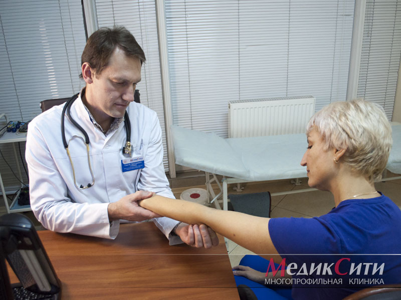
- What does a gnatologist do?
- How to find out if pain and spasms in the jaw area are related to the TMJ?
- WHAT DR OST OFFER TO TREAT JOINT PAIN
- Physiotherapeutic treatments for joints
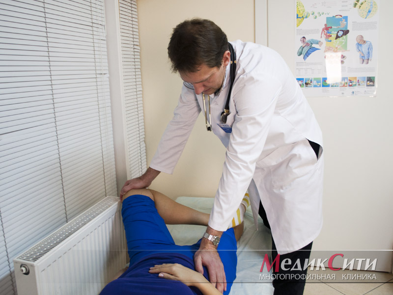
Treatment of temporomandibular joint dysfunction
Temporomandibular joint dysfunction (TMJD) describes the disruption of the jaw joint and the partial or complete loss of its function. It causes pain and limited jaw movement when opening the mouth, chewing and speaking. It is difficult to treat due to the variety of symptoms and causes. This complex problem is successfully treated in our center by the following doctors, among others. DD, specialist doctors Training in gnathology and have 11 years of practical experience.
The temporomandibular joint (TMJ) connects the movable lower jaw to the immovable temporal bone of the upper jaw. There are two such joints, located on either side of the skull in front of the ears. They each consist of the temporal fossa above and the articular head (mandible) below. In between there is an articular disc that alleviates the friction of the articular surfaces when the lower jaw moves.
The most important helpers of the joints are the masticatory muscles, which help to move the lower jaw relative to the upper jaw and keep it in the correct anatomical position to relieve pressure on the joint.

The joints are sufficiently mobile and work synchronously:
- Jaw movements characteristic of speaking;
- the jaw movements when chewing;
- the maximum opening of the mouth, for example when yawning.
Under normal conditions, all components of the system provide smooth, smooth and silent movement. However, if there are changes to the joint surfaces and/or the muscles that attach to the joints, the entire system fails. There is an impairment of joint mobility or a dysfunction of the joint (one or both).
Causes of jaw joint dysfunction
The main causes of TMJ problems can be divided into several categories:
They lead to dysfunction of the joints due to physiologically abnormal clenching of the jaws (occlusion) and their interaction in general:
They lead to increased joint stress and the development of jaw joint dysfunction on the part of the jaw muscles (muscular dysfunction):
- Long-term mechanical stress on the facial muscles
- Hypertonicity of the masticatory muscles as a result of bruxism
- Excessive muscle tension as a result of facial nerve entrapment
Progressive pathology affects the SSJ and impairs its function:
Increased likelihood of temporomandibular joint dysfunction:
- Congenital anomalies of the lower jaw, abnormally low alveolar processes.
- Anatomical misalignment between the temporal fossa and the condyle, abnormal position of the intervertebral disc at its junction
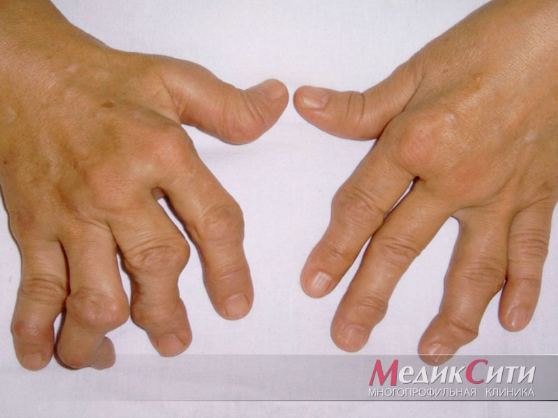
Who suffers from this disease?
To date, the exact cause of rheumatoid arthritis has not yet been clarified. Theories include genetics, environmental influences and hormonal imbalances. Statistics show that women are three times more likely to develop rheumatoid arthritis than men. The average age of onset of the disease is 40-60 years.
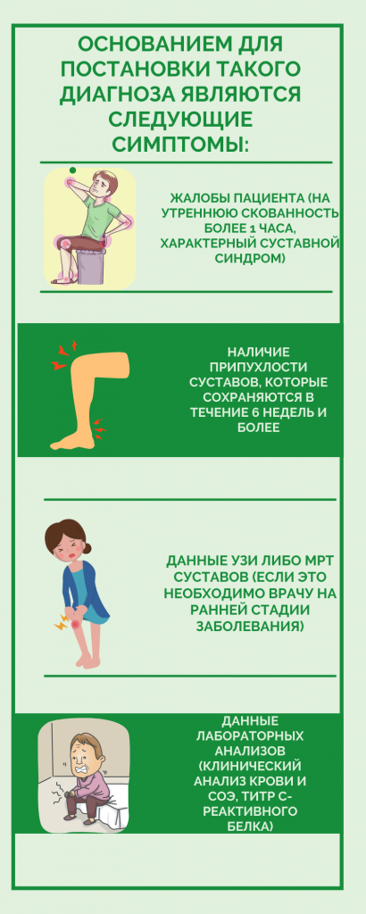
In the early stages of rheumatoid arthritis, the fingers, feet, knees and ankles can be affected. As the disease progresses, the number of affected areas increases. The main clinical manifestations of rheumatoid arthritis are:
- Pain syndrome The severity varies depending on the progression of the pathology.
- Stiffness. Particularly pronounced in the morning hours.
- Subfebrile body temperature. Lasts throughout the entire period of exacerbation.
- weight loss. Occurs against the background of loss of appetite.
- Functional impairment of the joints. Pain and stiffness interfere with usual activities.
- General tiredness. Has nothing to do with work or sleep.
Emotional symptoms of rheumatoid arthritis
In addition to physical symptoms, patients with rheumatoid arthritis suffer from emotional disturbances. Associated symptoms may include:
- Depression;
- increased anxiety
- disturbed sleep;
- daytime sleepiness;
- low self-esteem;
- Feelings of helplessness.
As the disease progresses, emotional tension increases. The disease affects both productivity and family relationships. It is important to seek help in a timely manner and follow the doctor's recommendations.
The principle of the method
The PRP method for treating knee joints promotes the regeneration of intra-articular tissue. Thanks to the platelet-rich plasma, cartilage, meniscus, ligaments and other tissues regenerate faster.
Even in ancient times, doctors pointed out that traumatic injuries accompanied by the formation of hematomas heal much faster. The reasons for this have only been identified in the last few decades. It was discovered that platelets secrete a number of substances that accelerate regeneration. These are growth factors:
These substances have a polypeptide structure (protein). They have different functions. But all of the substances mentioned are involved in tissue regeneration. They stimulate the mitotic activity of cells and accelerate their growth and division.
PRP therapy of the knee joint offers the opportunity to significantly increase the intensity of the reparative processes of both soft and bone tissue. Platelet-rich plasma is increasingly being used in medicine. It is used for all types of injuries: soft tissue injuries, muscle and ligament tears, bone fractures. All types of tissue regenerate faster when there are enough platelets.
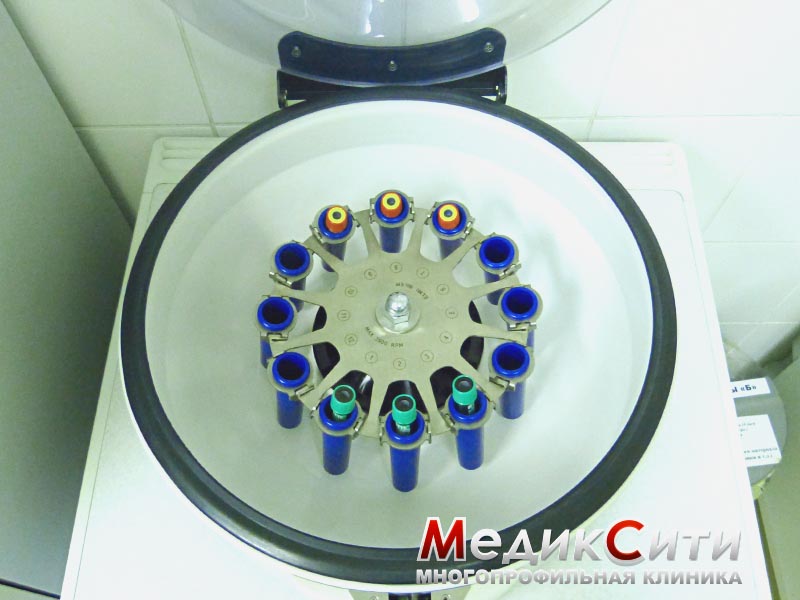
Plasma lifting

Plasma lifting
The following diseases and conditions are considered indications for the use of PRP for knee joint diseases:
Arthritis of the knee or gonarthritis. Osteoarthritis with progressive loss of articular cartilage. The use of PRP in stages I – II can improve the regeneration processes in cartilage tissue. Due to this effect, the development of the pathological process is significantly delayed. Thanks to the use of PRP, doctors can eliminate many symptoms of osteoarthritis, postpone the need for knee arthroplasty and even avoid surgery altogether if an effective conservative treatment regimen, in which PRP is a component, is used.
Injuries. The knee is often injured by athletes. Ligament tears and meniscus injuries are the most common. Intra-articular fractures can also occur. If the doctor determines that conservative treatment is sufficient, he or she may use PRP to speed healing of the injuries. Otherwise, they may take a long time to heal. This process can take weeks or even months.
Surgery. After arthroscopic surgery on the knee, a phase of rehabilitation follows. This lasts until all tissue damage has been repaired. PRP can be used to speed up this process.
Damage to the joint surface. PRP is used for cartilage defects on the articular surface of any origin. The cause can be trauma, arthrosis, inflammatory diseases of the knee joint, metabolic diseases, psoriasis, systemic connective tissue lesions and other diseases. If the defect is small, the regeneration processes can be supported with PRP. If the cartilage abrasion is extensive, surgical treatment is indicated.
Rheumatoid factor
Blood test for the presence of Rheumatoid factor aims to detect specific antibodies of the IgM to IgG class.
The rheumatoid factor laboratory test is a screening test used to detect autoimmune diseases. The main purpose of the rheumatoid factor test is to detect rheumatoid arthritis, Sjögren's syndrome and a number of other autoimmune diseases.
A rheumatoid factor test may be necessary if the following symptoms occur:
- pain and swelling in the joints;
- Limited mobility in the joints;
- feeling of dryness in the eyes and mouth;
- rashes in the form of hemorrhages;
- Weakness and loss of strength.
Norms for rheumatoid factor in the blood
Theoretically, there should be no rheumatoid factor in a healthy body. However, some even healthy people have low titers of this factor in their blood. Depending on the laboratory, the upper limit for rheumatoid factor varies between 10 and 25 international units (IU) per milliliter of blood.
Rheumatoid factor is equally high in men and women. Older people have slightly higher rheumatoid factor levels.
In children, rheumatoid factor should normally be 12.5 IU per milliliter.
The Rheumatoid Factor Test (Rheumatoid Factor Test) is used in the diagnosis of the following diseases:
Other causes of increased rheumatoid factor
Other causes of increased rheumatoid factor can be:
- Syphilis;
- Rubella;
- Infectious mononucleosis;
- Malaria;
- Tuberculosis;
- Flu;
- Hepatitis;
- Leukemia;
- Cirrhosis;
- sepsis
ADCP
The blood test for ADCP involves the determination of antibody titers to cyclic citrullinated peptide and is one of the most accurate methods for confirming the diagnosis of rheumatoid arthritis. With its help, the disease can be detected several years before symptoms appear.
What the ADC test shows
Citrulline – Citrulline is an amino acid formed by the biochemical conversion of another amino acid, arginine. In a healthy person, citrulline is not involved in protein synthesis and is completely excreted from the body.
However, in rheumatoid arthritis, citrulline is incorporated into the amino acid chain of peptides of synovial membrane proteins and articular cartilage. The 'new' modified citrulline-containing protein is perceived as 'foreign' by the immune system and the body begins to produce antibodies against the citrulline-containing peptide (ACPP).
ADCP – is a specific marker for rheumatoid arthritis, an early precursor of the disease, with high specificity.
Antibodies against cyclic citrullinated peptide can be recognized long before the first clinical symptoms of rheumatoid arthritis appear and persist throughout the course of the disease.
Test procedure and meaning
An enzyme immunoassay is used to detect ADCP. The blood test for ADCP is carried out using the in vitro method (Latin for 'in vitro'), examining serum from venous blood. The blood test for ADCP can take up to 24 hours (depending on the type of laboratory).
The detection of ADCP in rheumatoid arthritis may be an indication of a more aggressive, so-called erosive form of the disease, which is associated with a more rapid resolution of joint lesions and the development of characteristic joint deformities.
If the ADC test is positive, the prognosis for rheumatoid arthritis is considered less favorable.
How to choose chondroprotectors for joints in adults?
When choosing medicines to treat a disease, one should primarily pay attention to the composition. This also applies to chondroprotectors.
– Clinical guidelines clearly state that chondroprotectors contain the most important active ingredients: collagen hydrolyzate, glucosamine and chondroitin sulfate. Each of these ingredients has an important effect,' emphasizes the rheumatologist. rheumatologist Rheumatologist Sergei Kozlov.
– Collagen hydrolyzate has been proven to stimulate collagen synthesis in chondrocytes. Although it is weak, it helps slow down degenerative changes in cartilage and promotes the regeneration of cartilage and bone tissue in the musculoskeletal system. Glucosamine can increase the production of cartilage matrix and provide non-specific protection against chemical damage to cartilage.
Chondroitin sulfate maintains the viscosity of the synovial fluid, stimulates the repair mechanisms of cartilage and inhibits the activity of cartilage-degrading enzymes (elastase, hyaluronidase).
The composition is obvious. But the range of pharmacies is so large - what should you pay attention to when choosing chondroprotectors for joints for adults?
According to our expert, the Russian pharmacological classification divides chondroprotectors into 3 generations:
- 1 generation.: products of natural origin based on animal or vegetable cartilage extracts;
- 2nd generation.Monotherapies: Monotherapies consisting of purified hyaluronic acid, collagen, chondroitin sulfate or glucosamine;
- 3rd generation: Combination products containing both collagen and glucosamine with chondroitin sulfate, sometimes in combination with other active ingredients (e.g. additional vitamins, minerals, or polyunsaturated fatty acids).

What else should adults know about chondroprotectors for the joints?
Our expert points out that the form in which the preparation is presented and the substrate from which it is made are important.
– Drugs obtained from animal or plant tissues do not show a clear protective effect on chondrocytes. Semi-synthetic or synthetic chondroprotectors have a more pronounced effect when used correctly, explains Sergei Kozlov.

It should also be noted that chondroprotectors in the form of ointments and gels are a myth. Each ointment penetrates the skin by 1-2 mm, while the joints, as we know, are much deeper. Therefore, chondroprotectors can only help in the form of tablets and injections.
Osteoarthritis and coxarthrosis
The best doctors of the Elena Malysheva Medical Center will help eliminate the symptoms of the disease and its causes. Delaying treatment can make the situation worse, so don't hesitate to make an appointment. Telephone: 8 (495) 268-12-12
Coxarthrosis is a pathology of the hip joint caused by disruption of local blood circulation. It is a dystrophic process in the joint cavity that leads to degeneration of cartilage tissue. In coxarthrosis, the synovial fluid in the joint cavity becomes less viscous and can no longer adequately lubricate the hyaline cartilage. The cartilage is damaged and microfractures occur. Due to the reduced functionality of the cartilage, the bones rub against each other and become damaged. An inflammatory process develops.
Causes of pain associated with coxarthrosis
Causes of secondary coxarthrosis:
Symptoms of osteoarthritis and coxarthrosis
- The main symptom of coxarthrosis in the hip is pain in this area;
- stiffness in movement;
- lameness and changes in gait;
- Muscle wasting in the thighs, buttocks and lower legs and shortening of the affected lower limbs.
Treatment blocks for the pain associated with coxarthrosis
Specialists prescribe different treatments at different stages of the disease. In the early stages of coxarthrosis, conservative methods are used. Surgical methods are used when previous methods do not help or when the disease reaches the third stage.
A conservative treatment method is a hip joint block.
A joint block is a medication that relieves the pain associated with coxarthrosis. These involve injections of medication into the problem area that temporarily 'switch off' the pain reflex. For example, if a joint hurts, the medication is injected into the joint capsule, joint capsule, or ligament area.
The blocks relieve pain syndromes mainly related to diseases of the spine, joints, muscles and tendons and nerves. The dosage is calculated individually. The manipulation is carried out under X-ray control.
The blockade relieves not only pain, but also muscle spasms, edema and inflammation, helping to stop the pathological process and achieve the maximum therapeutic effect. Multicomponent injections, in which several drugs are combined, are carried out as part of complex therapy.
causes of gout
The main cause of gout is a disorder of uric acid metabolism, which leads to an excess of uric acid in the blood - hyperuricemia. This condition occurs when the intake of purines with food increases, when their metabolites do not have enough time to be excreted in the urine, or when the excretory function of the kidneys is impaired and when the cells producing the adenine and guanine -Nucleosides contained in the RNA and DNA from which uric acid is formed in the human body are quickly destroyed. The uric acid salts (urate) accumulate in the soft tissues of various organs, especially in the joints, and cause inflammation there.
Gout is also known as the 'disease of the festival kings'. It was so named because it was believed that its main cause was excessive consumption of food and alcoholic beverages.
- Consumption of foods rich in purines - meat, fatty fish, legumes, caffeinated products, by-products (kidney, liver, brain), seafood and large amounts of alcohol;
- consumption of carbonated drinks, sweetened drinks and fruit juices;
- Obesity;
- Hemolytic anemia, leukemia, lymphoma;
- Genetic defects (most often in men) and a reduced number of enzymes involved in uric acid metabolism;
- Taking medications such as diuretics, cytostatics, salicylates.
In women, gout usually develops after menopause, so there is a connection between the disease and hormonal changes in the body.
symptoms of gout
Translated from Greek, gout means 'pinched foot', as those affected usually experience severe pain in the first metatarsophalangeal joint of the foot, knee or ankle. Oligoposis or polyposis (inflammation of two or more joints) can also occur in the hand. The joint swells and the skin over it becomes red and shiny. This is called acute arthritis with gout.
The first clinical signs of pathology may be preceded by a long symptomless period of hyperuricemia, during which abnormalities are detected only by laboratory blood tests.
The first attacks of pain occur suddenly, usually early in the morning or at night, worsen over the course of the first day and disappear completely within a few hours or a day. If gout worsens, symptoms of intoxication such as increased body temperature, chills and weakness may also occur. Once the inflammation has subsided, gout will recur, usually within six months to two years. As the disease progresses, the symptom-free periods become shorter and the joint pain becomes more frequent and severe.
With chronic arthritis, the joints are deformed and mobility is limited, and pain of varying intensity occurs constantly. Uric acid crystals later become visible. Tofu appears under the skin, often in the joints, as white or yellow nodules with crumbly, curdled contents. Ulcers or purulent sores can form over them.
Hyperuricemia is often accompanied by an exacerbation of concomitant diseases - coronary heart disease, diabetes, hypertension, arteriosclerosis. When complications of gout develop, specific symptoms arise. In urolithiasis, these include a pulling pain in the lower back, occasional appearance of blood in the urine and frequent urination at night. In the long-term course of chronic gout, the articular cartilage is destroyed, the limitation of joint mobility persists beyond the periods of exacerbation, and complete closure of the joint cavity with the development of ankylosis (complete immobilization of the joint) can occur. The consequence of persistent pain syndrome can also be depression.
What does a gnatologist do?

A gnathologist is a specialist whose job it is to diagnose and treat diseases of the temporomandibular joint and dysfunctions of the entire dental system. It is important to understand that gnathology in dentistry is appropriate when there is a functional disorder, but not a specific disease. If a joint is severely damaged and there is an organic change, a surgeon should be consulted. The surgeon is able to determine the cause of the anomaly and choose a treatment method.

The doctor focuses on the condition and treatment of the pathology of the temporomandibular joint. This consists of two parts with which the lower jaw is attached to the skull bone. The main function of the temporomandibular joint is to enable the lower jaw to function properly when opening and closing the mouth and grinding food. Accordingly, functional disorders can lead to joint pain and tooth decay.
However, joint diseases or congenital anomalies are not always the cause of dysfunction. High levels of stress lead to muscle tension, including in the chewing muscles. As a result, the jaws are constantly under pressure. This leads to increased wear on the teeth and tooth displacement. The patient can no longer chew food freely because this process causes acute pain. However, the cause is not in the teeth, and its treatment is rarely successful until the muscle spasms are eliminated. Patients often become aware of a problem with the chewing system during a visit to the dentist. You will then be referred to a gnathologist who will advise you.
How to find out if pain and spasms in the jaw area are related to the TMJ?
There are several reasons why you should see a doctor. First of all, there is unexplained toothache. If there is no caries and the dentist has not found any abnormalities in the root area, it is worth visiting a gnathologist. Teeth in this situation can only be treated if the cause has been eliminated.
While the dentist should be visited regularly even if there are no symptoms, the gnatologist is usually referred by another specialist. The visit is usually necessary during the planning phase of orthodontic or prosthetic treatment. The doctor conducts an examination, assesses the condition of the joint and other elements of the masticatory apparatus. On this basis he will make recommendations. This allows the selection and fabrication of dentures so as not to overload the jaw joint and to ensure that the prosthesis or orthodontic device does not cause permanent discomfort.

If abnormalities are detected, the doctor conducts a comprehensive diagnosis and then prescribes treatment. For diagnosis, special devices are used to assess the function of the jaw muscles and the joint. X-rays of the jaw and joints can also be taken.
The gnatologist uses special devices to relax the tension of the chewing muscles. Trapezius traps and mouth guards are also used for this purpose. The latter are primarily used in dental procedures before orthodontic treatment or after the removal of a restoration. They not only distribute the pressure on the tissue appropriately, but also shorten the treatment time. Braces and veneers are traditionally used, especially for crowded teeth. At the same time, the braces allow the muscles to relax so that nothing stands in the way of the jaw adapting to the size of the teeth. In other words, normalizing the jaw muscles allows the dentist to avoid more serious dental problems. This applies not only to children but also to adults.
WHAT DR OST OFFER TO TREAT JOINT PAIN
With Dr. East we primarily use hardware and regenerative therapies to treat joint pain. The advanced technology is able to stop an attack of joint pain immediately and in just one session. The treatment has a long-lasting effect as it reverses inflammatory and degenerative processes deep in the tissue. When joints hurt, the surrounding soft tissues and nerve endings become traumatized. A pain attack is essentially a defense mechanism. The body tries to limit mobility in such a way as to prevent further wear and tear on the damaged joint itself and the surrounding muscles, ligaments and vessels.
As an alternative to traditional medications, Doctor OST uses technology that stimulates healthy cell growth and tissue renewal. In some cases of severe pathology, this treatment can even replace surgery.
Advanced joint treatment with stromal cells. Allows stimulation and regeneration of damaged joint tissue without chemotherapy or surgery. Read more
For the first time and only on Doctor OST! The latest technology to treat acute pain caused by damage to the musculoskeletal system and joints and ligaments. The effect starts tomorrow! Read more about this.
The latest method of treating joints now at Doctor OST! Completely without chemicals, synthetic foreign materials, surgery and prostheses. Read more here
Revolutionary cell technology! Even makes bedridden people able to walk again! Indicated for advanced osteoarthritis, non-healing bone fractures, bone prolapse. Read more
PRP therapy is a state-of-the-art treatment method that repairs damaged tissue using platelet-rich plasma. Read more here
Physiotherapeutic treatments for joints
We have high-performance physiotherapy facilities. Many of the technologies are completely unique or only available in a handful of clinics in Russia and Kazakhstan.
It immediately relieves acute pain, stimulates the regeneration of healthy cells and can replace prosthetic procedures. Read more
Relieves acute pain quickly and without drugs, restores blood circulation, improves lymph flow and accelerates healing after injuries and surgeries. Read more here
The UHT method effectively treats a variety of spine and joint problems and avoids surgical treatment. Read more here
A safe technique that accelerates the return to normal motor function and relieves pain. Read more here
Allows you to administer drugs in a concentrated form directly to the site of pathology. Read more
Read more:- Which doctor treats the hip joints.
- Shapar joint.
- Which doctor do you go to if you have joint problems?.
- There is abduction and reduction of the joints.
- Ellipsoid in knee osteoarthritis.
- heel strike hurts.
- What is joint rotation?.
- The key to a chopper joint is.