Treatment focuses on eliminating the underlying cause of nerve fiber damage, restoring normal function, and relieving symptoms that are uncomfortable for the patient.
- Femoral nerve neuropathy
- Background information.
- Anatomical features of the femoral nerve
- Types of nerve damage
- What triggers the destructive process?
- Types of cramps
- Causes of cramps in different areas of the body
- species
- symptoms
- Alcoholic polyneuropathy
- Diabetic polyneuropathy
- Causes of pathology
- Treatment
- PHYSICAL THERAPY
- prevention
- Diagnostic errors in radicular syndrome
- Treatment of radicular syndrome
- Benefits of treatment at the Spina Zdorova clinic
- Causes of pathology
- Symptoms of severe abnormalities
- Causes of peripheral nerve injuries
- Clinical manifestations
- Magvit – the perfect remedy for cramps
- Advantages of Minskinterkaps preparations
Femoral nerve neuropathy

Neuropathy of the femoral nerve (femoral nerve) – Damage to the femoral nerve of various etiologies, leading to impairment of nerve impulses. The clinical symptoms depend on the subjects of the lesion and may include pain and sensory disturbances on the anteromedial surface of the thigh and lower leg, difficulty in walking due to impaired extensor movements at the knee, etc. Diagnosis of femoral nerve neuropathy is based on ultrasound examination of the nerve and EMG. Treatment strategies include elimination of nerve compression, metabolic, vascular, anti-inflammatory, analgesic and antiedema therapy, physical therapy and electromyostimulation.

Background information.
Femoral neuropathy was first described in 1822 as 'anterior sacral neuritis'. Today it is one of the most common variants of mononeuropathies of the lower limbs. Although femoral neuropathy has been studied for nearly 200 years and is quite common, in some ways it is still an unknown condition. Lack of awareness among both general practitioners and some neurologists means that femoral neuropathy is often treated as a vertebrobasilar pathology (root syndrome, myelopathy, etc.) or as a manifestation of polyneuropathy. This is favored by a variety of symptoms, ranging from purely sensory disturbances to predominantly motor dysfunction, depending on the topography of the lesion.
Anatomical features of the femoral nerve
The femoral nerve (N. femoralis) arises from the three lumbar vertebral roots L2, L3 and L4, which unite to form a single nerve trunk. It runs between the iliopsoas major muscle and descends to the inguinal ligament, under which it exits at the front of the thigh, where it divides into a cutaneous (sensory) and a muscular (motor) branch as well as a popliteal nerve. In the area of the thigh bone, the femoral nerve innervates the muscles between which it runs. Their job is to flex and supinate the hip and, when the hip is stationary, to flex the lumbar spine so that the body bends forward.
The muscular branches that arise from the femoral nerve after passing under the inguinal ligament innervate the muscles responsible for hip flexion and knee extension. The cutaneous branches supply sensation to the anterior and slightly inner surface of the thigh. The saphenous nerve separates from the n nerve. The femoral nerve runs anteriorly along the thigh near the inguinal ligament, then passes medially and enters the intermuscular Gunther's canal (adductor canal), from which it exits at the medial edge of the knee joint, where it gives off a popliteal branch that innervates the anterior surface of the patella. The saphenous nerve then runs along the medial edge of the tibia and foot, reaching the base of the big toe. It provides sensitivity to the skin of the tibia on the anterior and medial surface as well as the skin on the medial edge of the foot.
Types of nerve damage
Damage to the medial saphenous nerve is rarer due to its location and usually occurs in old age or old age (up to 60 years) in people with inflammatory processes in the lumbosacral area, in people with a chronic history of recurrent lumbar spine diseases and often in athletes .
The etiology of the development of tendon pathology of the tibial nerve is mainly associated with the following factors:
There are three types of median nerve injuries:
More details about each type of injury:
- A condition in which there is compression (damage) to the internal cervical nerve is called neuralgia. neuralgia. It is caused by.

All processes differ in their etiology, course, treatment and effects.
What triggers the destructive process?
There can be many causes of median nerve damage; In addition to the usual ones – trauma, inflammation, compression – there are the following.
- poor blood circulation due to varicose veins;
- Valgus flatfoot;
- Rheumatoid arthritis;
- long periods of sitting from leg to leg;
- wearing shoes that are too tight;
- Musculoskeletal changes
- Musculotonic reflex disorders caused by curvature of the spine (osteochondrosis, scoliosis, spondyloarthritis);
- surgical procedures that cause limb misalignment;
- Diseases that cause overgrowth of connective tissue (gout, polymyositis)
- Metabolic disorders (diabetes, obesity)
- intoxication (alcohol, drugs);
- local tumor processes.
Types of cramps
When the involuntary contraction of muscle fibers is caused by excessive stimulation of the cerebral cortex, these are epileptic convulsions. Non-epileptic convulsions are triggered by CNS diseases, unbalanced diet or unfavorable environmental conditions.
The involuntary muscle spasms can also be classified according to their type:
- tonic – long-lasting; they become tense;
- myoclonic – short;
- Clonic – jerky, cyclical, alternating with relaxation.
Depending on the localization, these phenomena are divided into generalized and localized. The former extend over a large part of the body (arms, legs, face, neck, side, trunk and sometimes extend into the respiratory tract). The latter occur in certain places (e.g. only in the back or only in the buttocks).
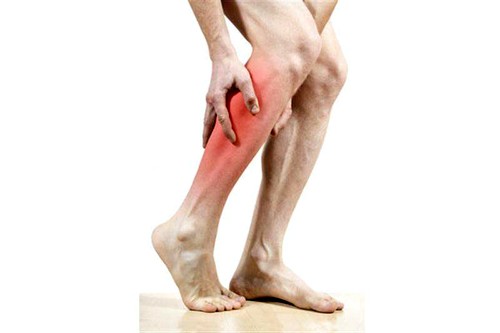
Causes of cramps in different areas of the body
Muscle cramps in a specific area of the body are caused by certain factors. It most commonly occurs in the legs. Overexertion (including through intensive training), varicose veins and hypothermia can be to blame.
Not only the calf muscle, but also the thigh and even the gluteal muscles can contract. Sometimes the symptoms can spread to the entire leg.
Abdominal cramps are also common because most abdominal organs are made of smooth muscle cells that can contract severely. The most common are cramps of the digestive system. This leads to the well-known abdominal pain. In women, uterine cramps (during menstruation, pregnancy or gynecological diseases) cannot be ruled out.
Muscle cramps in the back indicate problems with the musculoskeletal system. These may include osteochondrosis, intervertebral fractures or dystrophic changes in the spine.
species
Polyneuropathy is divided into several types depending on the type of fibers affected
- Sensory: The nerves that convey sensations are affected;
- Motor: The motor nerves are affected;
- Motor-sensory: a mixed form in which motor disorders predominate;
- Sensory-motor: mixed neuropathy in which the sensory disorders are more pronounced;
- Autonomous: disorders of internal organs, changes in vascular tone and associated trophic disorders.
Significant differences in symptoms can only be seen in the early stages of the disease. In the later stages, all nerve fibers are affected and the symptoms become mixed.
There are also other classifications of the disease:
- according to the cause: alcoholic, diabetic, idiopathic, etc;
- according to the localization of the focus: polyneuropathy of the upper or lower limbs;
- according to the type of course: acute, subacute, chronic;
- according to the mechanism of formation: demyelinating (destruction of the membrane covering the nerve fibers), axonal (due to the death of nerve branches - axons), axonal-demyelinating.
symptoms
The symptoms of polyneuropathy depend on its cause and the characteristics of the damaged fibers. The most common signs of pathology include:
- Decreased sensitivity in the feet and hands, resulting in a subjective feeling of wearing socks or gloves;
- Pain in the affected limbs (sharp or dull, brief or aching, usually worse at rest and at night);
- spasms and twitching of muscle fibers;
- pain when touched or pressure on the affected limb;
- decreased reflexes;
- excessive sweating of the extremities;
- trophic disorders: swelling, discoloration and dryness of the skin, trophic ulcers;
- muscle weakness of the arms and legs, slight tremors;
- Paresthesias: abnormal sensations of tingling, burning, cold;
- Muscular dystrophy;
- Restless legs syndrome.
Alcoholic polyneuropathy
The lower limbs are most commonly affected. It develops with prolonged uncontrolled alcohol consumption and is primarily manifested by burning and tingling in the legs. As the disease progresses, numbness and muscle cramps occur, which are due to a simultaneous deficiency of B vitamins.
Diabetic polyneuropathy
Elevated sugar levels affect the nerves in the feet (distal variant) and in the legs (proximal variant). In the first case, characteristic symptoms appear:
- numbness and reduced sensitivity to pain;
- intermittent burning pain;
- muscle weakness;
- decreased reflexes;
- Impaired motor coordination, manifested by a wobbly gait.
The combination of polyneuropathy and small vascular lesions leads to trophic ulcers on the lower tibia. The proximal variant is characterized by severe pain in the buttocks and thigh and gradual muscle loss in this area.
Causes of pathology
Inflammation of the tibial nerve can have a variety of causes, resulting from both external and internal factors.
Fractures of the tibia, tibial bone
Tumors of the tibia bone
Neurodystrophic processes in the ligamentous muscles of the joints in connection with spinal diseases
Prolonged standing in awkward positions and continued pressure on the nerve can lead to femoral neuropathy.
Degenerative changes in the tibial nerve are not uncommon in people whose jobs involve heavy physical work while standing. This is due to poor blood supply due to the constant spasms that cause narrowing of the capillaries.
This leads to secondary trophic neuropathy of the tibial nerve, which, although it does not cause severe pain, swelling and redness, reduces the sensitivity and strength of the muscles.
Make an appointment for an online consultation if you have mild symptoms and are still living a normal life. Early treatment is faster and more effective. Our neurologists will advise you personally, help you interpret tests and answer all your questions.
Treatment
If neuropathy is caused by a general illness or joint disease, it is primarily corrected. If the joint disease worsens (in rheumatoid arthritis), non-steroidal and basic anti-inflammatory drugs are prescribed. The therapy can be supplemented by wearing orthopedic shoes and knee orthoses.
The pain of pressure lesions is relieved by therapeutic blockade with a combination of hormone and anesthetic. As a rule, it is lidocaine in combination with triamcinolone, dipropane or hydrocortisone.
Case example:
A 52-year-old woman visited a neurologist because she was experiencing pain and partial numbness in her left leg. Examination revealed neuropathy of the tibial nerve. Concomitant diseases: diabetes mellitus type 2, gonarthrosis. Antidiabetic drugs, intramuscular injections of diclofenac and milgamma were prescribed.
Treatment of inflammation of the tibial nerve is impossible without medications to improve its nutrition and blood supply. For this purpose, patients receive injections of B vitamins, nicotinic acid, pentoxifylline and alpha lipoic acid systems.
Other medications can also be included in the therapy regimen:
- anticholinesterases – ipidacrine, neostigmine;
- Tissue regeneration stimulants – Actovegin, Solcoseryl;
- Anticonvulsants – carbamazepine, pregabalin;
- antidepressants.
Physiotherapy can help eliminate the symptoms of neuropathy:
Atrophied muscles are rebuilt with massage and complex therapeutic exercises.
PHYSICAL THERAPY
The therapeutic effect of physiotherapy is mainly based on the activation of blood and lymphatic circulation at the site of the injury. The increased blood flow to the nerve fibers stimulates cell regeneration and accelerates healing.
Training the neuromuscular system supports the activation of adaptive reserves through the dynamic remodeling of the nervous system and muscle function. The developed compensatory functions can be gradually expanded through the use of muscles that continue to be innervated.
prevention
If you have undergone effective treatment, this is not a reason to rest. To prevent the numbness from recurring, specific prophylaxis is recommended. This includes:
- Diet – exclude fatty and salty foods from your diet; ask your doctor for a list of allowed and prohibited foods;
- Healthy lifestyle to improve blood circulation and muscle function;
- Physiotherapy 2-3 times a week - shoes with special insoles that alternately inflate to trigger tactile sensitivity in the toes are particularly helpful;
- Avoid uncomfortable footwear – get good quality shoes or sneakers that are comfortable for your feet.
It is strongly recommended to give up unhealthy habits that make numbness worse.
Numbness in the toes can be caused by physiological abnormalities in the body. If the symptom persists for a long time, one should suspect a chronic disease in the body. At the first signs of weakness, urgent examination and treatment is required.
Diagnostic errors in radicular syndrome
A common mistake is to confuse radicular syndrome with pseudofascial syndrome.
Pseudofascial syndrome is pain caused by muscle soreness rather than spinal nerve compression.
There is a muscular disorder called myofascial syndrome. There is a detailed article on this topic on our website. In short, with myofascial syndrome there are small spasms - trigger points - in the muscles that cause very severe pain. Sometimes the area of myofascial pain can extend over quite large areas of the body and resemble the zone of segmental innervation in radicular syndrome. For example, pain can spread from the lower back to the upper and lower leg. In such cases the term 'pseudo-pain syndrome' is commonly used. The image below shows the area of the L4 radial complex - shown in blue - and the area of the myofascial complex of the gluteus medius - shown in red. There are two variants of myofascial pain of the gluteus maximus.
Areas of pain. Radial – colored blue. Musculo-fascial – orange.
Isn't it the case that the radial pain zone of L4 and the myofascial pain zone of the gluteus medius muscle are very similar? That is why inexperienced practitioners often confuse the symptoms of radial and pseudofascial (myofascial) syndromes in this and many other cases. Just as an inexperienced mushroom hunter runs the risk of confusing real and false honeydew.
It is important to be able to distinguish between radial syndrome and pseudosyndrome!
- We determine whether the symptoms are due to radicular syndrome or 'pseudoradicular' myofascial syndrome and examine the muscles for active and latent trigger points.
- The diagnosis time is 30 minutes. This is a comprehensive examination and not a 2-minute 'palpation' with pliers.
- The diagnosis is made by Dr. Vlasenko AA, a doctor with 30 years of experience and an expert in the treatment of myofascial and radicular syndromes.
Treatment of radicular syndrome
Treatment for radicular syndrome depends on the cause.
- If radicular syndrome is caused by a tumor, cyst, or fracture, these problems can only be solved through surgery. The neurosurgeon must then take care of these problems.
- If radial vascular syndrome predominates in the clinical picture, then medication is the priority. In this case, your doctor is a neurologist.
- In cases where osteochondrosis - radicular syndrome - is diagnosed, as well as in cases where radicular syndrome is accompanied by a herniated or bulging disc, the attending physician should be - a chiropractor.
It goes without saying that if pain occurs or if radicular syndrome is suspected, it is necessary to immediately consult a doctor who will take all possible measures to treat the disease and prevent its exacerbation. It only remains to add that this should be an experienced and knowledgeable doctor.
When choosing a clinic, the most important thing is to find an experienced and knowledgeable doctor.
He will be able to understand the symptoms of your disease, make an accurate diagnosis and eliminate not only the disease, but also its causes.
Benefits of treatment at the Spina Zdorova clinic
- Guarantee of complete and competent treatment. The word 'competent' is the key word in our work.
- Highly qualified and extensive practical experience – 30 years.
- Each case is treated individually and comprehensively - no formalism.
- Synergy effect.
- Guarantee of a fair approach and a fair price.
- Two steps from the metro in the heart of Moscow.
Causes of pathology
- Previous injuries to the spine and neck.
- Poor posture, curvature of the spine.
- Sedentary lifestyle.
- Chronic intoxication with alcohol, drugs, vapors of toxic substances (e.g. from working in harmful conditions), heavy metal salts.
- Hormonal disorders, endocrine imbalance.
- Obesity, overweight.
- Wrong attitude to sports training.
- Execution of repetitive (especially jerky) movements.
Another cause of musculoskeletal disorders that lead to loss of sensation in the arms and legs is excessive physical exertion. Sometimes the innervation of the arms and legs is impaired after a traumatic brain or spinal injury.
Numbness in the limbs is not only caused by abnormalities in the joints, bones or vertebrae. Numbness in the limbs occurs with stroke, heart attack, abdominal aortic embolism, hyperkalemia, and Sjögren's syndrome. Numbness in the hands and feet often occurs as a result of iron deficiency anemia, in which the hemoglobin level is reduced. In order for the doctor to establish a causal connection with the deterioration, he must know at what age the symptoms appeared and what factors preceded the deterioration.
Symptoms of severe abnormalities
Sensitivity disorders in the limbs are accompanied by few symptoms. They affect the color of the skin on the limbs, the temperature of the skin on the hands and feet - the epithelium becomes pale and cold to the touch. Depending on the cause of the innervation disorder, the patient is accompanied by the following symptoms
- Back and leg pain that occurs when bending or rotating the trunk.
- Paresthesias (when the patient feels that goosebumps are crawling over their skin).
- Decreased muscle tone, reduced strength in the limbs – difficulty standing up or making fists with your hands.
- The feeling of a foreign body in the back.
- Impairment of movement coordination (gait becomes wobbly, unsteady).
- The feeling that weak electric shocks are acting on the muscles of the limbs.
Other symptoms include pale skin, dizziness, drowsiness, apathy, nausea and motion sickness during transport. If the legs and arms do not feel nerve impulses and this condition is not caused by diabetes, constant thirst is annoying and wound healing is delayed. Due to the reduced innervation of the limbs, the patient does not feel the effects of temperature and is therefore susceptible to burns or frostbite.

The success of the treatment depends to 90 % on the doctor's experience and qualifications.
Causes of peripheral nerve injuries
When it comes to peripheral nerve injuries, a distinction is made between closed and open injuries.
- Closed injuries: Compression of the soft tissues of the arm or leg, e.g. B. due to improper application of a pressure bandage in the event of bleeding, severe bruises or impacts, prolonged forced positioning of the limbs with external pressure, after bone fractures. As a rule, the nerve is not completely severed in such cases, so the outcome is usually favorable. In some cases, e.g. B. in the case of dislocations of the hand, foot or large joints as well as in closed fractures of limb bones with displacement of fragments, the entire nerve trunk or even several nerves can be severed.
- Open injuries are caused by broken glass, knives, metal sheets, mechanical tools, etc. In these cases, the integrity of the nerve structure is always violated.
Unfortunately, nerve damage is also often the result of surgical procedures.
Depending on the extent of nerve damage, the type of injury, or the duration of exposure to the damaging substance, the appearance of lesions manifests as a dysfunctional syndrome.
Clinical manifestations
In a closed trauma With a bruise or concussion, there are no changes in the internal structure of the nerve trunk; the impairment of sensory organs and limb functions is temporary, fleeting and usually completely reversible. With a bruise, functional impairment is deeper and more permanent, but complete recovery is noted after 1-2 months. However, the sequelae of such injuries should not be underestimated and self-diagnosis and treatment is unacceptable as the effects of 'self-medication' may be irreversible. A trauma surgeon, neurologist or traumatologist should be consulted immediately. If necessary, the doctor may recommend additional tests to clarify the extent of nerve damage - electromyography, ultrasound examination of the nerve pathways and sometimes even CT or MRI scans. Only a qualified doctor can recommend appropriate treatment.
Open peripheral nerve injuries. The fibers of all peripheral nerves are of mixed type – motor fibers, sensory fibers and autonomic fibers; The ratio between these types of fibers varies from nerve to nerve, so that in some cases the motor disorders are more pronounced, in others there is a reduction or complete absence of sensation, and in still others autonomic disorders occur.
Motor disorders are characterized by paralysis of muscle groups or individual muscles, accompanied by atrophy of reflexes and atrophy of the paralyzed muscles over time (1-2 weeks after injury).
Sensitivity disorders such as reduced or absent sensitivity to pain, temperature and touch are present. Pain that worsens with a delay.
Autonomic symptoms - in the first period after the injury the skin is hot and red, a few weeks later it becomes pale and cold (wasculopathy), swelling, sweating disorders, trophic skin disorders - dryness, scaling, sometimes even ulceration, deformed nails.
Magvit – the perfect remedy for cramps
One of the cheapest, safest and most effective medicines for the treatment of convulsions – the drug. 'Magvitis manufactured by the Belarusian pharmaceutical company 'Minskinterkaps'.
It is the first remedy completely developed by domestic pharmacists that has a positive effect in the treatment of convulsions.
'Magvit' effectively combats both primary and secondary magnesium deficiency, while pyridoxine (vitamin B6) improves the absorption of magnesium in the blood and its uptake into cells.
The combined use of vitamin citrate and magnesium is justified by the following clinically proven data:
– Magnesium and B6 enhance and complement the pharmacological effects;
– The vitamin reduces the amount of magnesium excreted by the body and increases its content in red blood cells;
– the absorption and assimilation of magnesium are improved;
– Pridoxine, which is influenced by magnesium, is converted by the human liver into the active metabolite pyridoxal-5-phosphate.
Advantages of Minskinterkaps preparations
The company has earned a well-deserved reputation for producing affordable, high-quality medicines. All formulations comply with the GMP standard and their quality and composition are tested in a chemical and biological laboratory accredited according to the requirements of STB ISO/IEC 17025.
The medicines are made from raw materials supplied by world-leading companies. Production is controlled by pharmacovigilance and complies with the requirements set by the European Parliament.
Read more:- Syndrome of the tibial nerve.
- hip pronation.
- lower leg muscles.
- The lateral side is.
- Long fibula muscle.
- Calf muscle cramps when walking.
- Charcot-Marie foot.
- median soleus nerve.