Copyright © 2023 Merck & Co, Inc, Rahway, NJ, USA and its subsidiaries. All rights reserved.

- 3 effective exercises for heel pain
- Unique advertising
- 'Regain Youthfulness Your Feet'.
- Frequently Asked Questions
- Causes and symptoms of a bone fracture
- Principles of Diagnosis
- Diagnosis of fractures of the heel bone
- Treatment of heel bone fractures
- How is the investigation carried out?
- Direct x-ray
- Stress radiography
- evaluation of results
- Symptoms of a heel bone fracture
- Society and culture
- species
- symptoms
- signs and symptoms
- complications
- Ground
- osteoporosis
- diabetes
- Diagnosis of a stress fracture of the heel bone
- Causes of stress fractures of the heel bone
- Damage to the heel tendon
- Symptoms of a heel tendon injury
- Achilles tendon bursitis and posterior bursitis
- Achilles tendinitis
3 effective exercises for heel pain
The most effective treatment for heel pain (plantar fasciitis) is exercise. Your goal is to make the plantar fascia more flexible so it doesn't tear and cause pain.
The exercises should be performed three times a day for 10-15 minutes for at least a month. If pain persists after a month of intense exercise, additional treatments may be performed.
This video shows the three most effective exercises for treating plantar fasciitis or heel spurs. You can easily do it yourself at home.
Unique advertising
'Regain youthfulness
Her feet'.
There are now modern, safe and effective treatments to help you regain the health of your feet. Some of them are completely free, others are very affordable. Unfortunately, many older people don't know about it.
That's why I'm launching a new unique campaign for beautiful older generation ladies called 'Reclaim your feet's youth'.
I invite you to my office for a FREE consultation where I will examine your feet.
I will also tell you what modern, safe and effective treatments are available for your situation and how you can get them for free or at the lowest possible cost. We will talk to you about all the options available and choose the one that suits you best.
1. Any person over the age of 55 is eligible to participate.
2. You can apply with any foot and ankle problem.
3. The promotion runs from August 1st to September 30th, 2019.
4. You can personally come to a consultation in St. Petersburg at Yaroslavsky pr. 66 come. 1, or you can get advice online.
Call your mothers, grandmothers, relatives, friends - anyone who my guidance can help. Tell as many people as possible, share it on your social networks.
Old age is a wonderful time to enjoy life. So don't let pain and discomfort in your feet interfere with your quality of life. Let's get your feet back in shape together so walking can become one of your favorite pastimes again.
If you would like to know more about the Rejuvenate Your Feet campaign, send an SMS to WhatsApp +79219651182
or call us 8 (812) 336-60-22
Frequently Asked Questions
The operation itself is no different. In all the operations I perform, I use state-of-the-art medical equipment, the best imported implants, high-quality consumables, the necessary medications and regional anesthesia (a type of local anesthesia - two injections are given into the foot). There are only two differences in free surgery: 1. If you want to operate on both feet at the same time, you have to carry out the operation on two different days. The other foot can only be operated on after 2 weeks. 2. If you have a quota, you will not receive follow-up care. Aftercare is always included in the price of the operations I pay for. This way I can monitor how you are feeling, how your bone is healing and how your joint has regained mobility. This way I can prevent possible postoperative complications. This always leads to excellent results and satisfied patients. Postoperative management can be purchased separately.
Causes and symptoms of a bone fracture
Most fractures of this type usually occur due to falling from a height, falling on the foot, traffic accidents, poor bone health, osteoporosis and excessive exercise.
Fractures of the heel bone are usually divided into several groups:
In extra-articular trauma, the heel shaft presents different fracture levels and accounts for approximately 20 % of trauma, consisting of:
- burst injury;
- trauma to the styloid process of the heel bone;
- anterior process of the biceps ligament;
- Tear of the tuber of the calcaneus.
Intra-articular deformities occur in the joint cavity of the bone and account for approximately 80% of all calcaneal bone injuries. These injuries include:
- Fracture of the posterior lamina of the calcaneus with lingual view;
- Fracture of the posterior lamina with sagittal alignment
- Fracture of the middle part of the posterior surface, imitating the letter 'Y';
- Multiple fracture of the outer and middle part of the posterior surface of the calcaneus, with possible 'subsidence' of the middle part;
- a displaced fracture with multiple injuries to the posterior surface of the calcaneus.
A fractured heel bone can be recognized by characteristic symptoms that cause discomfort. A person begins to worry about symptoms such as:
- Pains;
- Swelling;
- inability to stand on foot;
- Severe limb deformity;
- Bone splinters in the wound.
Principles of Diagnosis
If you suspect a fracture, you should consult a specialist at the sports injury rehabilitation center. The doctor will perform a visual and tactile examination and examine the skin around the heel for dislocations or skin damage. The specialist will also order additional examinations, such as: B..
- Radiological examination. This is done in 4 projections and helps the doctor determine the exact location of the dislocation or the position of the fragment.
- CT scan. This allows the specialist to recognize and assess the extent of damage to parts of the leg.
CT scanning is the gold standard for traumatic calcaneal injuries.
Diagnosis of fractures of the heel bone
If the doctor suspects a heel bone fracture, he or she will prescribe x-rays, on which the fracture (if present) is usually clearly visible. However, sometimes a computed tomography (CT) scan is necessary. A CT scan combines X-rays with computer technology to provide a more detailed, three-dimensional image of the injured area.
Since heel bone fractures are caused by high force, the doctor also rules out other injuries, e.g. B. a knee or spine fracture.
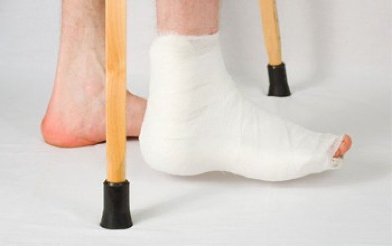
Treatment of heel bone fractures
Sometimes protection (usually with a splint), rest, ice, a pressure bandage and elevation of the injured limb are necessary.
Doctors consult a specialist (orthopedist) to choose the best treatment for the heel bone fracture. Depending on the type of fracture, surgery may be necessary.
If the fracture does not involve a joint, treatment includes protection (usually with a splint), rest (do not put weight on the feet or use crutches), ice, a pressure bandage, and an elevated position (PRICE PRICE PRICE A fracture is a break of a Bone or a broken bone. Most fractures result from force on the bone. Fractures most often occur as a result of trauma or excessive stress. Read more ). Once the swelling has subsided, the patient can be referred to an orthopedist who will apply a plaster cast.
If the heel bone fracture affects a joint, surgical intervention is considered. If surgery is required, open repositioning with internal fixation (ORVF) is performed.
Patients are advised not to put any weight on the heel until the fracture heals. The duration of this measure depends on the injury and can be several months. Doctors often recommend that patients move the foot and ankle and sometimes shift their body weight to the ankle until the pain is as tolerable as possible.
Therapeutic exercises are required. This includes specific exercises to improve the range of motion of the injured foot and ankle and to strengthen the supporting muscles.
How is the investigation carried out?
X-rays of the heel bone can be taken in different projections.
Direct x-ray
For a direct projection x-ray, the patient must lie on their back on the x-ray table. The legs are bent at the knees and the feet are resting on the table.
Stress radiography
The heel bone is stressed when standing. To perform a stress radiography, the patient must stand up and put weight on the foot.
evaluation of results
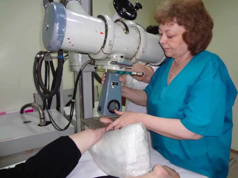
Immediately after the X-ray images have been taken, the radiologist carries out the evaluation. The X-ray will help clarify the cause of the pain and determine the nature of the pathological process. For an accurate interpretation of the result, the doctor can compare the images of the healthy and diseased heel.
If plantar fasciitis (also called heel spurs) is diagnosed, the specialist will prepare a detailed description of the X-ray image, on which he will note the presence of osteophytes, indicating their length and location. Osteophytes are usually visible in the superior projection of the posterior tuberosity of the talus.
Symptoms of a heel bone fracture
Patients with a heel bone fracture initially complain of pain. These can occur when moving or at rest. Examination may reveal swelling extending to the Achilles tendon. Symptoms of a fracture also include flattening and enlargement of the heel and a hematoma in the middle of the sole bone.
When palpated, the patient feels a sharp pain at the site of the injury. It is difficult to support your foot while standing.
Society and culture
title Lover's foot broken derives from the fact that a lover can jump from a great height when trying to escape the lover's spouse. [18]
- ^ abcdежplayTimeyajkлmпопqr'Fractures of the calcaneus - OrthoInfo - AAOS'. orthoinfo.aaos.org. January 2016 Archived from the original on June 18, 2017 . Retrieved October 12, 2017 .
- ^ abcdеж Palmersheim, K; Hines, B; Olsen, B.L. (April 2012). 'Fractures of the heel bone: an update on current treatment options'. Clinics of Orthopedic Medicine and Surgery.. 29 (2): 205-20, vii. doi:10.1016/j.cpm.2012.01.007. PMID22424485.
- ^Fractures of the heel bone (calcaneal bone fractures). In eMedicine.
- ^ Richman, J.D.; Barré, P.S. (1986). 'The plantar ecchymosis sign in calcaneal fractures'. Clinical orthopedics and related research (207): 122-5. Doi:10.1097/00003086-198606000-00022. PMID3720074.
- ^ Berry, G.K.; Stevens, D.G.; Kreder, H.J.; McKee, M.; Schemitch, E.; Stephen, DJ G. (2004). 'Open fractures of the calcaneus: a review of treatment and outcomes'. Journal of Orthopedic Trauma. 18 (4): 202-6. Doi:10.1097/00005131-200404000-00002. PMID15087962.
- ^ ab Heier, Keith A.; Infante, Anthony F.; Walling, Arthur K.; Sanders, Roy W. (2003). 'Open fractures of the heel bone: soft tissue injuries determine the outcome'. Journal of Bone and Joint Surgery. American Vol.. 85-А (12): 2276-82. Doi:10.2106/00004623-200312000-00002. PMID14668494.
- [permanent dead link ]
- ^ ab Soeur, Robert; Remy, Robert (1975). 'Fractures of the calcaneus with displacement of the thalamic part'. Journal of Bone and Joint Surgery. British Vol.. 57 (4): 413-21. doi:10.1302/0301-620X.57B4.413. PMID1194308.
- [permanent dead link ]
- ^ Stoller, D.W.; Tirman, P.F. J.; Bredella, M. (2004). 'The ankle and foot, bone fractures and heel bone fractures'. Imaging diagnostics: orthopedics . Salt Lake City: Amirsys. p.70 -4.
- ^ Salzler, Matthew J.; Bluman, Eric M.; Noonan, Samantha; Chiodo, Christopher P.; de Asla, Richard J. (2012). 'Observed Injuries in Minimalist Runners'. Foot & Ankle International.. 33 (4): 262-6. doi:10.3113 / FAI.2012.0262. PMID22735197.
- ^ Cheng, Sulin; Suominen, Harri; Sakari Rantala, Ritva; Laukkanen, Pia; Avikainen, Veikko; Heikkinen, Eino (1997). 'Heel bone mineral density predicts the occurrence of fractures: a five-year follow-up study in the elderly'. Journal of Bone and Mineral Research.. 12 (7): 1075-82. doi:10.1359 / jbmr.1997.12.7.1075. PMID9200007.
- ^ Cathol, M.N; el Khoury, G.Y; Moore, T.E.; Marsh, J. L. (1991). 'Heel fracture fractures in patients with diabetes mellitus'. Radiology.. 180 (3): 725-9. doi:10.1148 / radiology.180.3.1871285. PMID1871285.
- ^ ab Badillo, Kenneth; Pacheco, Jose A.; Padua, Samuel O.; Gomez, Angel A.; Colon, Edgar; Vidal, Jorge A. (2011). 'Assessment of calcaneal fractures using multidetector computed tomography'. RadioGraphics.. 31 (1): 81-92. doi:10.1148 / rg.311105036. PMID21257934.
- ^ abPage Rebuilder 562In Archived 2017-11-05 at the Wayback Machine at: Mark D. Miller; Stephen R Thompson (2015). Miller's review of orthopedics. (7th ed.). Elsevier Health Sciences. ISBN9780323390422.
- ^ Radswicki; et al. ' Fracture of the heel bone. Radiopedia. Archived from the original on October 14, 2017. Retrieved July 20, 2017 .
- ^ abFractures of the heel bone ~ treatment In eMedicine
- ^ Godes, Joe; Klingman, Robert. 'Heel bone fractures and rehabilitation' (PDF). Kaiser Permanente. Archived (PDF) from the original on 06/03/2016.
- ^ Hatzokos, I; Karataglis, D; Papadopoulos, P; Dimitriou, K; Christodoulou, A; Pournaras, J (2006). 'Treatment of intra-articular heel splinter fractures'. Orthopedics. 29 (1): 25-9. Doi:10.3928/01477447-20060101-15. PMID16429931. S2CID39928758.
- ^ Lee, Patrick; Hunter, Tim B.; Taljanovic, Mihra (2004). 'Skeletal muscular slang terms: How did we get these terms?'. Radiography.. 24 (4): 1009-27. doi: 10.1148 / rg.244045015 . PMID15256625.
species
There are several criteria for classifying a lesion. For example, fractures of the heel bone are divided into intra-articular (the fracture line is directed to the area of the subtalar joint) and extra-articular fractures, which, in turn, are characterized by a fracture:
There are also abnormalities in the structure of the heel bone:
In most cases, this injury is not isolated. It is often combined with fracture displacements and ankle injuries as well as with lumbar and thoracic vertebral fractures.
symptoms
The symptomatic picture is quite simple:
- painful sensations;
- Significant swelling in the affected area;
- The heel flattens and widens;
- Often a bruise appears in the middle of the sole;
- Palpation of the injured area causes acute pain.
Although the motor function of the ankle joint is preserved, support of the limb is not possible.
signs and symptoms
The most common symptom is pain in the heel area, especially upon palpation or pressure. Patients usually have a history of trauma to the area or a fall from a height. Other symptoms include inability to shift weight on the affected leg, limited foot mobility, and limping. During the examination, the doctor may notice swelling, redness and bruising. A hematoma that extends to the sole of the foot is called Mondor's sign and is pathognomonic of a heel bone fracture. [3][4] The heel may also widen due to displacement of the lateral edge of the heel bone, which is accompanied by swelling. Soft tissue involvement should be investigated as it is associated with serious complications (see below). [5] [6]
complications
Assessment of soft tissue involvement is the most important aspect of the clinical examination as it is related to the progression of the disease. [6] [7] If skin blisters are not treated in a timely manner, they can become infected, which can lead to necrotizing fasciitis or osteitis, causing irreversible muscle or bone damage. Involvement of ligaments and tendons should also be investigated. Damage to the Achilles tendon can occur in posterior fractures (Type C). Because calcaneal fractures are associated with falls, other associated injuries should also be evaluated. Vertebral compression fractures occur in approximately 10 % of these patients. [8] A trauma-informed clinical approach should be adopted; Injuries to the shinbone, knee, hip, thigh, and head should be ruled out by history and physical examination. [ Reference required ]
Ground
Heel fractures are often associated with shear forces combined with compressive forces combined with the direction of rotation (Soeur, 1975 [7] ). These forces are typically associated with fall trauma, a car accident, or muscle strain, and the resulting forces can result in a fracture injury. An overlooked aspect of what can lead to a heel bone fracture is the role of osteoporosis and diabetes. [ Link required ]
Unfortunately, the prevention of falls and traffic accidents is limited and only applies to certain circumstances that should be avoided. The risk of muscle stress fractures can be reduced by stretching and weight-bearing exercises, such as: B. strength training can be reduced. In addition, shoes can influence the forces that can cause a heel bone fracture and also prevent it. A 2012 study by Salzler [9] found that the increasing trend towards minimalist shoes or walking barefoot can lead to various stress fractures, including heel bone fractures. [ Citation required ]
osteoporosis
Bone mineral density decreases with age. Osteoporotic bone loss can be prevented by adequate intake of vitamin C and vitamin D in conjunction with physical activity and avoiding smoking. A study by Cheng et al. [10] from 1997 found that higher bone density was associated with a lower risk of calcaneal fractures.
diabetes
In 1991, Cathol [11] conducted a study that showed an association between tendon ruptures due to calcaneal insufficiency (a fracture in which the Achilles tendon detaches part of the bone during resorption) and diabetes. People with diabetes are at higher risk of fractures and possible healing complications, as well as infections that can lead to amputations. Diabetes can be controlled and prevented through diet and exercise. [12]
Diagnosis of a stress fracture of the heel bone

How is a heel stress fracture diagnosed?A stress fracture of the heel bone is a complete or partial fracture of the integrity of the heel bone when the load exceeds the strength of the damaged area of the skeleton. The initial diagnosis of a stress fracture of the heel bone requires an x-ray of the heel and subsequent consultation with a trauma surgeon. As an additional examination, the doctor may recommend a computer tomography of the foot.
Which doctor treats a stress fracture of the heel bone?: If you have symptoms of a stress fracture of the heel bone, you should first consult a traumatologist; Based on your initial examination, your doctor may recommend additional consultation with a surgeon.
A stress fracture of the heel bone is a bone fracture caused by a fracture as a result of repetitive stress. Stress fractures of the heel bone cause severe pain and make walking difficult due to small breaks in the bone tissue. Heel bone fractures most commonly occur in men between the ages of 30 and 50. The likelihood of developing a stress fracture of the heel bone is higher if the patient participates in sports that require repetitive movements, such as basketball, dancing, long-distance running, soccer, and track and field. Other factors that increase the risk of a stress fracture of the heel bone include:
Stress fractures of the heel bone are rare. All fractures of the foot only account for about 2 % of the fractures. Only half of them affect the heel bone.
Causes of stress fractures of the heel bone
Stress fractures of the heel bone occur when the body does not have time to recover from the stress. These injuries occur during sports or increasing activity.
To diagnose a stress fracture of the heel bone, a traumatologist examines your foot and ankle. He or she will ask you to wiggle your toes. Visual examinations are carried out, such as: E.g.:
- X-ray of the heel – the most common imaging test used to diagnose fractures
- CT or MRI are common follow-up tests after x-rays. They provide more detailed images of the structures of the heel.
Damage to the heel tendon
All iLive content is reviewed by medical experts to ensure it is as accurate and factual as possible.
We have a strict policy in selecting information sources, linking only to reputable websites, academic research institutes and, where possible, peer-reviewed medical studies. Please note that the numbers in parentheses ([1], [2], etc.) are interactive links to such studies.
If you think any of our material is inaccurate, out of date or otherwise questionable, please highlight it and press Ctrl + Enter.
Heel tendon injuries include inflammation of the loose tissue surrounding the tendon, a partial or complete tear.
The calf muscle is connected to the heel bone via the heel tendon. When running, the calf muscle helps lift the feet off the ground. The repeated loading of the tendon during running, coupled with inadequate recovery time, can initially lead to inflammation of the tissue surrounding the tendon (the fatty layer that separates the heel tendon from its covering). A complete tear of the heel tendon is a serious injury and usually occurs as a result of sudden, violent stress.
[1], [2]
Symptoms of a heel tendon injury
The first symptom of heel tendonitis is pain in the back of the heel, which increases in the first few minutes of weight-bearing and then often decreases as weight-bearing increases. A complete rupture causes severe, stabbing pain and impairs the ability to move normally after the injury.
On examination, an inflamed heel tendon is painful when pinched between the toes. If there is a complete rupture, a defect is palpated along the course of the tendon and compression of the calf muscle does not result in the expected normal soleus flexion (positive Thompson test).
Achilles tendon bursitis and posterior bursitis
There are two synovial capsules in the heel bone area that can become inflamed. The first is between the heel bone and the Achilles tendon and is called the heel tendon capsule, the second is between the Achilles tendon and the skin and is called the posterior heel capsule. In the latter case, bunions usually arise from friction from ill-fitting footwear and are particularly common in women who wear high-heeled shoes. The bunion is usually stretched due to fluid and inflammation. In chronic cases, the pad thickens along with the overlying skin; there is pain and swelling at the back of the heel where it rubs against the shoe. With Achilles tendonitis, the patient complains of pain when moving; Palpation reveals local pain immediately in front of the Achilles tendon.
Treatment for Achilles tendonitis includes rest, warmth, and elevation of the limb. Patients with posterior heel bursitis should wear appropriate low-heeled shoes. In acute cases, the back part of the shoe can be cut out. Puncture of the capsule and injection of hydrocortisone provide rapid relief of symptoms.
Achilles tendinitis
Achilles tendonitis is a condition that causes excessive strain on the muscles of the lower leg. Patients suffer from swelling in the Achilles tendon area, pain and crepitus that occur when moving the foot. The painful area is clearly localized and the patient must keep the foot in a balanced position to relieve the discomfort. Passive dorsiflexion of the foot increases pain.
Achilles tendonitis is the third most common condition among long-distance runners. Heel pain can be caused by inflammation of the Achilles tendon itself, inflammation of the tendon sheath, or inflammation of the tendon mesentery. Your doctor should differentiate this condition from Achilles bursitis or back heel inflammation. The pain increases when running and subsides with rest. As already mentioned, a thickening over the tendon or in the peroneal tissue is often palpable. Conservative treatment includes limiting movement time and placing a small felt insert under the heel of the shoe. This often relieves symptoms. The runner should be advised to perform stretching exercises for the Achilles tendon complex. Anti-inflammatory medications can be taken orally. After running, an ice pack should be placed on the Achilles tendon for 10-12 minutes. If the pain is acute and other therapeutic measures are ineffective, a short plaster boot is applied for 10 days. In some patients, surgery to remove the thickened synovial membrane is effective.
Read more:- Anatomy of the heel bone x-ray.
- Anatomy of the heel bone.
- Fracture of the heel bone.
- Displacement fracture of the heel bone.
- heel bone flexion angle.
- tarsal bones of the foot.
- Fracture of the calcaneus of the foot.
- Fracture of the articular process of the heel bone.