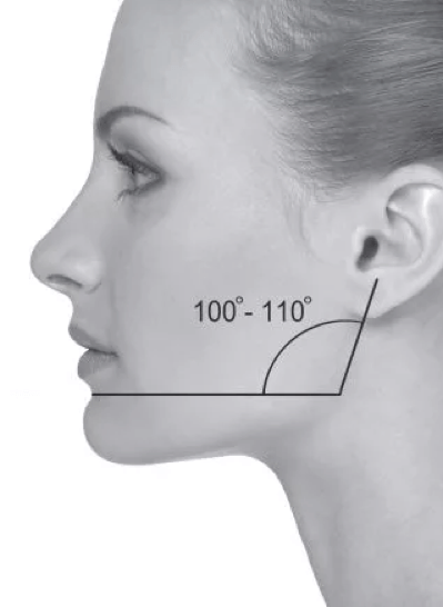
If you get into the habit of relaxing your mouth and looking at everything with a slight smile, after a month you will notice: even in the occasional photo taken from the side, you no longer look grumpy. This is my own experience.
- Anatomy: Superficial back muscles
- Anatomy: Superficial back muscles
- muscles
- How do the muscles work?
- How do the muscles make the body move?
- Features of muscle structure and function
- Changes in morphometric characteristics of uni- and bi-articular muscles during motor activities
- How can I improve the appearance of my neck and face with platysma?
- Platysma training
- Types of injuries: bruises, sprains, strains
- Symptoms of injury
- Muscles. Muscle types, structure and meaning
- Lesson summary 'Muscles. Types of muscles, their structure and their meaning'.
- Structure of the gluteus medius muscle
- Lipofilling of the buttocks photo.
- Feedback on lipofilling of the buttocks
- Structure of the gluteal muscles – evaluation criteria
- anatomy
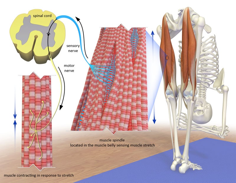
- What is flexibility?
- Structure of the knee joint
- The menisci of the knee
Anatomy: Superficial back muscles
The superficial muscles of the back have a flat shape and are arranged in three layers. The first (outer) layer contains the trapezius and the broadest back muscle; the second layer contains the rhomboid major and minor as well as the scapula muscle; the third layer contains the superior and inferior serratus muscles. The muscles of the first and second layers attach to the sternum and humerus; the third layer attaches to the ribs.
Trapezius muscle (t. trapezius) has a triangular shape, with the broad base of the muscle extending to the posterior midline and the laterally constricted part extending to the scapula. This muscle lies on the surface and occupies the upper back and the back of the neck. Together, the two muscles of the same name have the shape of a trapezius.
The trapezius muscle consists of three parts: the upper one - the so-called 'pars descendens'. the descending part (pars descendens) middle part transversal part (pars transvena) the lower - descending part (pars descendens) the ascending part (pars ascendens).
beginning, The trapezius begins with short tendons on the external occipital tubercle, in the middle third of the upper neck line of the occipital bone, at the base of the neck, on the spinous processes of the VIIth cervical vertebra and all thoracic vertebrae, and at the base of the supraspinatus.
Fixation: The upper bundles of the muscle run downward and laterally, attaching to the posterior surface of the outer third of the clavicle; the middle bundles run almost horizontally and attach to the supraspinatus of the scapula and the tip of the scapula; the lower bundles of the muscle run upward and laterally, attach to the DRAZIER plate and attach to the tip of the scapula.
At the level of the spinous process of the VIIth cervical vertebra, the two muscles form a quadriceps LAP, which in living humans has a clearly visible depression at this point. The upper lateral border of this muscle forms the posterior aspect of the lateral triangle of the neck. The lower lateral edge 'crosses' the dorsal broadest muscle and meets the medial edge of the scapula and also forms the medial side of the 'auditory' triangle. The lower side of this triangle corresponds to the upper edge of the dorsal broadest muscle and the lateral side corresponds to the lower edge of the biceps major muscle.
Anatomy: Superficial back muscles

The superficial back muscles attach to the bones of the axial skeleton and attach to the marginal bones and the free upper extremity. These include the trapezius and latissimus dorsi muscles. The former begins at the line of the upper udder and ligaments, at the spinous processes of the thoracic vertebrae and attaches to the shoulder blade and collarbone. This muscle raises and lowers the scapula, bringing it closer to the spine. The widest back muscle arises from the sacrum, the ilium, the spinous processes of the lumbar vertebrae and the 6 lower thoracic vertebrae and attaches to the humerus: it guides, stretches and rotates the upper extremity inwards.
The middle layer of the back muscles includes the rhomboidei major and minor muscles (Mm. rhomboidei major et minor), the serratus posterior superior and inferior muscles (Mm. serratus posterior superior et inferior) and the scapulae muscle (M. levator scapulae). The rhomboid muscles attach to the spinous processes of the last cervical vertebra and the five upper thoracic vertebrae and, together with the scapularis muscle, attach to the medial edge of the scapula. These muscles move the shoulder blade upward, bringing it closer to the spine. The superior dentate muscle extends from the C6-7 to T1-2 spinous processes to the four upper ribs, and the inferior dentate muscle extends from the T11-12 to L1-2 spinous processes to the four lower ribs. The superior dentate muscle raises the ribs and the inferior dentate muscle lowers them. The deep back muscles ensure movement and balance
the torso and the head. They are part of the 'muscular corset' that ensures the stability of the spine and the correct position of the internal organs. The muscles form two different pathways. The lateral tract is represented by the erector spinae muscle, which includes the longest muscle (M. longissimus), the iliocostalis muscle (M. iliocostalis) and the vertebral muscle (m. spinalis). The medial tract includes the transversospinalis muscle (m. transversospinalis), the bundles of which pass through a different number of vertebrae and form the semispinalis muscle (m. semispinalis), 5-7 vertebrae; the multifidi muscle (mm. multifidi), 2-4 vertebrae; and the rotator muscles (mm. rotatores), 1 vertebra.
muscles
Muscles are the tissue that causes various parts of the body to move by contracting. A distinction is made between the following muscle types:
Skeletal muscles. Skeletal muscles are connected to the bones and work in pairs, e.g. B. the biceps flexes the arm at the elbow and the triceps extends it. The contraction of skeletal muscles is voluntary (that is, you move them whenever you want).
Smooth muscles surround the arteries, veins and intestines. The smooth muscles of blood vessels contract and relax to regulate blood flow. The smooth muscles of the intestine contract to move food and feces through the digestive tract. You cannot control the smooth muscles. You don't have to think about them for them to fulfill their function.
How do the muscles work?
Muscles generate energy from nutrients and oxygen carried by circulating blood. The muscles use this energy to contract. The larger the muscle and the better blood supply it has, the more it can contract.
Without a good blood supply, muscles cannot work hard.
Some types of physical activity, like lifting weights, can make muscles bigger and stronger. Other types of physical activity, such as: Exercises such as running can increase the endurance of the muscles (they can work longer).
How do the muscles make the body move?
In order for the body to move, the muscles must connect one bone to another, forming a joint.
When the muscles contract, tendons pull on both bones, moving them in the direction that the shape of the joint allows. Since muscles can only contract, if a muscle can flex a joint, the other side of the joint must contract to straighten it.
Features of muscle structure and function
1) A muscle can have both unilateral and bipartite muscles. The quadriceps muscle consists of three unicompartmental muscles (lateral thigh muscle, medial thigh muscle, and middle thigh muscle) and one bicompartmental muscle (rectus femoris muscle). The triceps muscle of the lower leg consists of two two-part muscles (lateral head and middle head of the calf muscle) and one one-part muscle - the hamstring muscle. The muscles that allow the toes to move are multi-jointed.
2. The single-joint muscles are used to control the joint angle because only single-joint muscles can adjust a certain angle between the joints. The two-jointed muscles, because they 'cross' two joints, cannot give a specific value for the interjoint angle at a particular joint. However, they perform a number of other functions in human movement.
3. VM Zatsiorski, AS Aruin and VN Seluyanov, (1981) point out that:
- All two-jointed muscles produce opposing moments of force in the joints through which they pass (e.g. flexion of the knee joint and extension of the hip joint). Therefore, during natural movements such as walking and running, these muscles are active during the phases of movement that require such multidirectional forces.
- When two adjacent joints are simultaneously flexed or extended, the length of the biceps muscles changes very little (6-8 % of the original length). Therefore, these muscles can be considered to work in a near isometric mode.
- Bicompartmental muscle activity reduces the mechanical work associated with movement and thus reduces energy expenditure. Mechanical energy can be transferred from one joint to another.
Changes in morphometric characteristics of uni- and bi-articular muscles during motor activities
The morphometric characteristics are: the adhesion of the muscle to the bone, the length of the muscle, the pulling arm (the shortest distance between the axis of rotation of the joint and the line of action of the pulling force of the muscle), the moment of the pulling force of the muscle.
Kozlov (1984) showed that in single-joint muscular movement activities, a decrease in muscle length, which leads to a decrease in the pulling force of the muscle, is compensated for by an increase in the pulling arm. This allows the pulling moment of the muscle to remain constant relative to the axis of rotation over a long range of motion. In two-jointed muscles, a decrease in the tensile force moment relative to one joint is accompanied by an increase in the tensile force moment relative to the other joint.
How can I improve the appearance of my neck and face with platysma?
Of course, no creams and masks will penetrate the muscle tissue - this is simply unrealistic. But of course all products applied to the skin can improve it.
However, if we talk specifically about the muscles, there are 3 options:
✅ Surgical platysmoplasty. It is called platysmoplasty. The operation involves excising excess skin, removing fatty deposits, pulling the inner edges of the muscle inward, and performing other manipulations.

✅ Special exercises to improve platysma mobility. It is primarily a muscle. Therefore it can be trained.
Exercises for the front surface of the neck.
Biorevitalization, mesotherapy and the new PROFILO product can improve the appearance of the skin and also have a firming effect for a certain period of time.
Which of these methods you choose is at your own discretion.
Platysma training
Everything brilliant is simple. I never tire of repeating it.
'Do you want a good neck? Look at the stars more often!'
Katharine Hepburn. And she's not even sixteen anymore.
This woman's images speak for themselves. As long as we look down most of our lives (at our feet while walking, at a book/phone while traveling, at a plate of food, at other people figuratively), what Can we expect a beautiful neck line and a beautiful chin?
We rarely look up with our heads tilted back. And the platysma muscle actually only has the job of lowering the lower lip and the corners of the mouth onto the snout.
Types of injuries: bruises, sprains, strains
Classification of pectoralis major muscle injuries:
- Bruise or sprain of the muscle
- Partial rupture
- Complete rupture:
- Tear of the tendon from the attachment point to the humerus
- Tear at the junction of muscle and tendon
- Tear of the muscle fiber itself
- Tear of the tendon of the pectoralis major muscle with bone block
- Separation from the sternum, ribs
Symptoms of injury
- Pain, weakness and burning sensation
- Restriction of mobility of the shoulder girdle
- Swelling, swelling and hematomas appear
- The characteristic sound of a muscle tear is a cracking or popping sound (at the time of injury).
- Deformation of the muscle and disruption of its natural contours (in the case of a complete tear)
- Medical examination
- X-ray examination
- Ultrasound examination (USG)
- Magnetic resonance imaging (MRI)
Specialists you should see if you have any of these symptoms:
Muscles. Muscle types, structure and meaning

In this video lesson, students will learn about the structure and function of muscles. You look at the main groups of skeletal muscles. With the help of pictures, they learn about the location of the muscles in the human body. The lesson includes additional information about the largest and smallest muscles in the body. Students also learn about the history of muscle names.
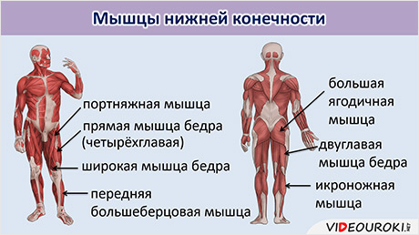
Lesson summary 'Muscles. Types of muscles, their structure and their meaning'.
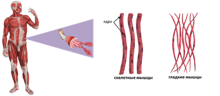
muscles consist of muscle tissuewhich consists of elongated, multinucleated cells that look like fibers with transverse contours. There are several Types of muscles – smoother i Skeletal muscles.
Smooth muscles are part of the walls of internal organs (heart, blood vessels, stomach and intestines). They play an important role in processes that occur independently of our consciousness, e.g. B. in the movement of food in the digestive tract. Smooth muscles are independent of human willThey contract slowly and can do so over a long period of time.
Skeletal muscles are Striated muscles muscles Of the head, the trunk and limbs. They contract quickly, allowing voluntary movements.
muscles consists of a large number of Muscle fibers …that are capable of contraction. They run parallel to each other and in bundles.
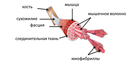
The muscle fiber consists of thin… fibers – myofibrilswhich in turn consist of the thinnest protein filaments. Through their interaction, the muscles contract and shorten.
Each muscle fiber is covered by a connective tissue sheath that merges into tendons at the ends of the muscle. tendons – The passive, nonconvulsive part of the skeleton through which muscles are attached to bone. They are firmly attached to the bone membrane (periosteum), which covers the bone from the outside. The tendons are very strong, practically indestructible and can withstand a load of up to 600 kg when stretched.
Structure of the gluteus medius muscle
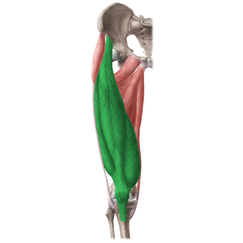
The gluteus maximus is the largest of all glute muscles. It consists of two parts – an upper (wide) and a lower (narrow) part. The gluteus maximus arises from the outside of the ilium, the back of the sacrum and coccyx, and the sacroiliac ligament. The muscle bundles of the upper part of the BLM end in the vastus femoris and attach to the greater trochanter, the fibers of the lower part attach to the gluteus tuberosity of the thigh. The maximum thickness of the gluteus medius muscle at its insertion on the sacrum is 6-7 cm, 2.5-3 cm in its upper outer part and 1.5-2 cm in its lower outer part. The gluteus medius muscle begins on the outside of the iliac crest and extends underneath the gluteus medius muscle, extending only in the anterior and posterior parts, where it is covered by a dense fascia. It is also connected to the trochanter of the femur. Interestingly, the BLM plays an important role in the aesthetics and volume of the buttocks, while the gluteus medius muscle has only a minor influence. The outer contour of the transition from one muscle to the other should be smooth, giving the impression that a single muscle covers the entire pelvic area. And that depends on the thickness of the fat layer above (Fig. 1).
Lipofilling of the buttocks photo.
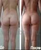
The before and after photos of buttock augmentation with lipofilling are the best indicator of the success of the procedure. You can see the fat grafting buttock augmentation before and after photos and examples of the results when you scroll down the page.
Feedback on lipofilling of the buttocks
'Now I could cut off the fat, and Maria Grigorievna had enough material to create a masterpiece, one might say, a work of art. Yes - yes, I've seen photos from the operating room. The butt looks like a ball, so round and firm! Oh, wow!!! There are no words, just emotions! Dear Maria Grigoryevna, thank you from the bottom of my heart! Beauty will save the world, and the beauty you create - a true master of your craft and a sculptor with a capital letter! Irina, 37 years old. For the sake of simplicity, in the further discussion we will use the word combination 'glutes' to combine both muscles - the large and the medium - so that it is clinically difficult to distinguish between their boundaries.
Structure of the gluteal muscles – evaluation criteria
One of the main tasks in assessing the shape of the buttocks is to determine the height, width and volume of the gluteal muscles, especially the gluteus maximus. However, sometimes (e.g. in obese patients) it is quite difficult to determine the size and volume. In such cases, an indirect indicator can be used: the ratio between the height of the sacrum and the length of the intercostal sulcus. Ideally, this ratio should be around 1:3. With a ratio of 2:1 the buttocks appear short, with a ratio of 1:2 they appear elongated. The height of the gluteus maximus in relation to the length of the intercostal sulcus must have a ratio of 2:1, and the upper and lower points of the intercostal sulcus must be equidistant from the respective edges of the muscle (Fig. 2).
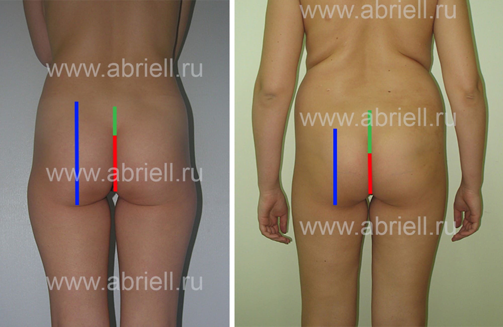
Fig. 2.
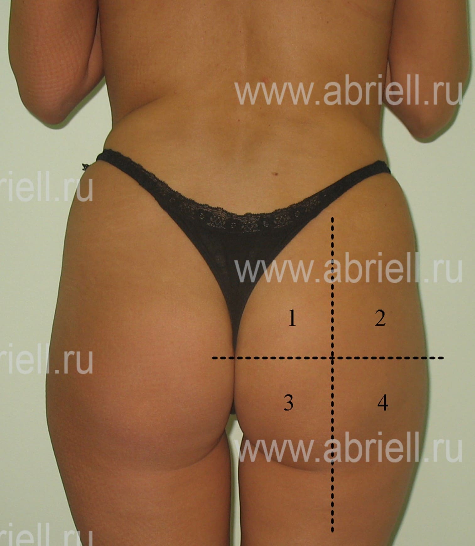
(a) Ratio of rump height (green) to length of gluteal sulcus (red) 1:3; Ratio of the height of the gluteal muscle (blue) to the height of the gluteal sulcus (red) 2:1 in patients with 'ideal' buttocks; (b) Ratio of sacrum height (green) to gluteal gap height (red) 1:1 in patients with short gluteus medius (blue) (explanation in text). This ratio is more important when planning buttock augmentation with implants, as it can influence the choice of implant shape. For gluteal fat grafts, the clinical relevance of the height and width of the 'gluteus maximus' is not as important. It is also important to assess the uniformity of the volume distribution of the gluteus maximus, which is usually divided into 4 quadrants by vertical and horizontal meridians. Ideally, as already mentioned, the buttocks have approximately equal soft tissue volume in the 4 sectors, with a certain tendency towards greater width in the lower quadrants. The shape of a beautiful buttocks is more like an oval than a circle. When doing the lateral projection, it is important to determine at which point the muscle has the greatest extent. Three types of volume distribution can be identified: a maximum in the upper, middle (central), or lower third of the buttocks (Figure 3).
anatomy
The muscle resembles an elastic band. It is connected to the bone at both ends and can expand and contract, causing joint movement. Changing the attachment points of the muscle is not possible. They cannot be moved a little further apart, either by stretching or by any other means (except surgery). In some people the muscles are naturally long, that is, their ends are far from the joint, in others they are short.
When the joint is fully extended, the muscle is at its longest and can no longer be lengthened. The exception is when the muscle is cramped and shortened from its normal resting state, i.e. the joint is not fully extended at rest.
What is flexibility?
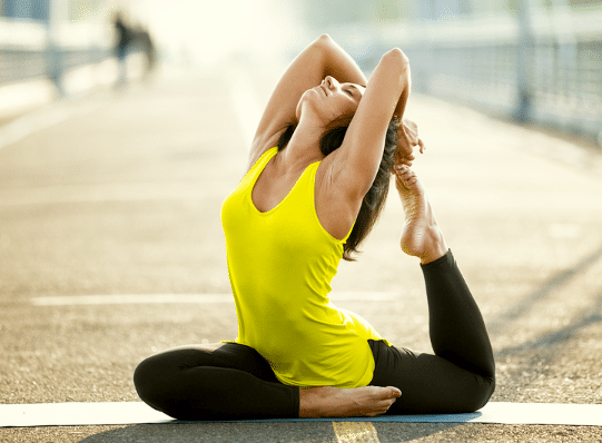
Anyone who has done stretching regularly knows that over time we go further. We become able to move with greater amplitude and we call this flexibility. But this is not because the muscle has been stretched and lengthened. The reason is adaptation, that is, the adaptation of the neuromuscular system to the load. What do you mean with that?
Our movements are controlled by the nervous system through electrical signals that travel through the nerve cells to the muscles. It determines our strength, flexibility and endurance. It is he who restricts them and prevents movement when he sees a risk of tissue damage. Todd Hargrove, author of 'A guide to better movement', gives this analogy:
Imagine you are the lucky owner of a sporty Ferrari. And imagine driving it with your mother. You accelerate hard, but your mother becomes restless and tells you to slow down. You've long since gotten used to obeying, turn up the music and give it full throttle. But for some reason the car won't accelerate above 65 km/h.
Suddenly you notice that mom has her own brake pedal. As soon as she thinks you're going too fast, she applies the brakes - meaning she has full control over the speed of the car. You realize that you can only accelerate properly if you convince your mother that you can drive safely at high speeds.
This analogy gives an idea of how the nervous system regulates the limits of our strength, endurance, or flexibility. If it perceives a movement as dangerous, the brakes are applied.
The nervous system is not overly interested in how good you look, how muscular you are, or how flexible you are. It wants to make sure you don't accidentally kill yourself and pass your genes on to the next generation. In the wild, even a minor injury plays a role in survival: a sprained ankle or torn tendon is a matter of life and death.
Structure of the knee joint
The knee joint is one of the largest joints in the human body. It allows people a wide range of movements, which is why the knee is always exposed to high levels of stress. The structure of the knee joint is very complex as it is a complex with many ligaments, muscles, nerves, blood vessels, cartilage and bones.
Bones of the knee joint
The first two bones are tubular, while the last bone is round and lies in front of them. A layer of cartilage provides cushioning and smooth movement.
The menisci of the knee
The menisci are cartilaginous structures that lie between the ends of the bones. They have the shape of a semicircular plate, the outer edge of which is thicker and gradually tapers towards the center. The structure of the knee joint is also such that the medial meniscus is larger than the medial meniscus. This is due to the nature of the bones that are attached to them. Their main task is to correctly distribute the weight of the human body and ensure stability.
The structure of the human knee joint is represented by a number of muscles surrounding it. These can be divided into different types:
- the front muscles, which include the hip flexors (with their help the upper and lower legs are moved):
- quadriceps (quadriceps);
- tailor muscle;
- Hip extensors (their main task is the extension and flexion of the hips as well as the lateral rotation of the lower limbs):
- biceps muscle (biceps);
- semitendinosus muscle (biceps);
- semitendinosus;
- Adductors of the thigh (their task is to adduct the thigh and flex the lower limbs):
- Thin muscle
- Adductor.
At the Garvis Clinic, qualified specialists offer treatment of knee problems of any complexity, from surgery to full recovery after surgery.
- The flexor muscles of the foot.
- The muscles that move the foot.
- Shin extensor muscles (tibialis extensor muscles).
- Tibialis posterior muscle.
- muscles in the legs.
- tibial fasciitis.
- Which muscles of the anterior tibial group do you know?.
- Pronator - what does that mean?.