Employees of the Department of Emergency and Urgent Treatment, Internal Diseases¹ 2 of the SD Asfendiyarov National Medical University of Kazakhstan: Candidate of Medical Sciences, Associate Professor Vodnev VP Candidate of Medical Sciences, Associate Professor Dyusembayev BK; Candidate of Medical Sciences, Associate Professor Akhmetova GD; Candidate of Medical Sciences, Associate Professor Bedelbayeva GG; Almuhambetov MK; Loykin AA; Madenov NN
- How to distinguish a fracture from a bruise
- Bruise or fracture: differential symptoms
- When are joint x-rays recommended?
- How they are made
- Do I need to see a doctor if I suspect a fracture?
- types of injuries
- signs
- For doctors
- Dislocated shoulder
- species
- symptoms
- classification
- Treatment
- diagnosis
- Treatment
- complications
- prophylaxis
How to distinguish a fracture from a bruise
The risk of injury exists everywhere, regardless of the environment. It can't always be avoided, so it's very important to know the symptoms of the two most common types of injuries - bruises and fractures. This will help you correctly analyze the situation and provide the necessary medical assistance.
The information in this chapter should not be used for self-diagnosis or self-medication. In case of pain or other exacerbations, diagnostic tests should only be recommended by the treating physician. A specialist should be consulted to make a diagnosis and prescribe appropriate treatment.
Bruise or fracture: differential symptoms
A fall or other mechanical impact on the body can cause severe pain, limb disorientation, and bruising. Soft tissue or bone injuries may occur. In the first case it is a bruise, in the second case it is a fracture or fracture. Symptomatically, these injuries differ primarily in the following features:
- If there is a bruise, motor skills are little or not affected at all;
- a fracture leads to partial or complete immobilization of the limb;
- Depending on the localization, a bruise can be accompanied by a concussion, minor damage to internal organs and dislocation of the joint;
- Fractures sometimes result in splinters, significant bone displacement, and open wounds;
- for a bruise, a cast is not applied, while for a connective tissue injury this is the standard treatment procedure;
- The overall condition is much more severe with fractures than with soft tissue injuries.
Both types are characterized by severe pain, swelling and bruising. The most common injuries are to the head, limbs and ribs. Fractures are often accompanied by externally noticeable deformations.
When are joint x-rays recommended?
X-rays are most often prescribed when strains, sprains, broken bones, tendon tears, and other injuries are suspected. This examination can also be used to detect changes in tissue structure and detect tumors, cysts, deformities, osteoarthritis and arthritis. This diagnostic method is therefore suitable for all musculoskeletal problems.
X-ray diagnosis is based on special X-rays emitted by a device. Soft tissues let them through, while hard tissues absorb them, which is why the former are darker in the picture and the latter are lighter in color. The bone tissue is most clearly visible in the images, which is why this method is used to examine the condition of bones and joints. The results of the examination are transferred to paper or digitally and stored on a computer hard drive.
Nowadays, X-ray images are also available in digital form.
How they are made
The X-ray examination is carried out without any preparation. The patient is placed on a special table. The groin area is protected from radiation with a lead apron. Children are protected in the eye and thyroid area, and in infants only the area to be examined, e.g. B. a limb, left open.
The doctor takes the image in one or more projections. In order for these to be clear, it is necessary to remain calm. The specialist determines the best projection depending on the segment to be examined: straight, lateral or combined.
If the patient is overweight, the image may be blurry.
Do I need to see a doctor if I suspect a fracture?
In most cases, minor fractures heal on their own without examination, plastering and medical attention. It is sufficient to protect the affected area and immobilize it. In the practice of trauma surgeons, it is not uncommon for a patient to discover, after an X-ray of a fresh fracture, that it is a fracture that has already healed and that he was previously unaware of. However, such injuries do not always go away without consequences.
Example. A lifeguard was trying to clear the ice from the steps when she slipped and hit her chest painfully. The woman's intuition told her that it was better to take precautions and call an ambulance. However, when the paramedic arrived on scene, he advised the man not to take an X-ray because he couldn't put the rib fracture in a cast anyway. He left everything as it was and walked away. In the morning, as the man awkwardly turned around, he felt a pang in his chest and began to inflate like a balloon. After a few minutes he found it difficult to breathe. Had another medical team not acted quickly, the lifeguard would have died of a pneumothorax, a lung injury caused by a splintered rib that damaged the soft tissues. After the operation, the lifeguard resumed his work in the spring when it was no longer necessary to clear the ice from the stairs.
An equally dangerous situation arises with embedded fractures of the femoral neck in the event of a fall on one's side with a swing. The external signs of the injury may be mild and the pain tolerable, allowing one to continue walking or even running. However, at some point the bone fragments can shift, making the condition significantly worse. On the one hand, they have to be joined together surgically, and on the other hand, the femoral head can be completely destroyed by interrupting the blood supply. The result is a prosthesis or complete immobilization.
A compression fracture of the spine can lead to chronic back pain, paresis and paralysis due to compression of the intervertebral nerves and even the spinal cord. A broken toe can lead to its subsequent deformity. In addition, you will no longer be able to wear modeling shoes.
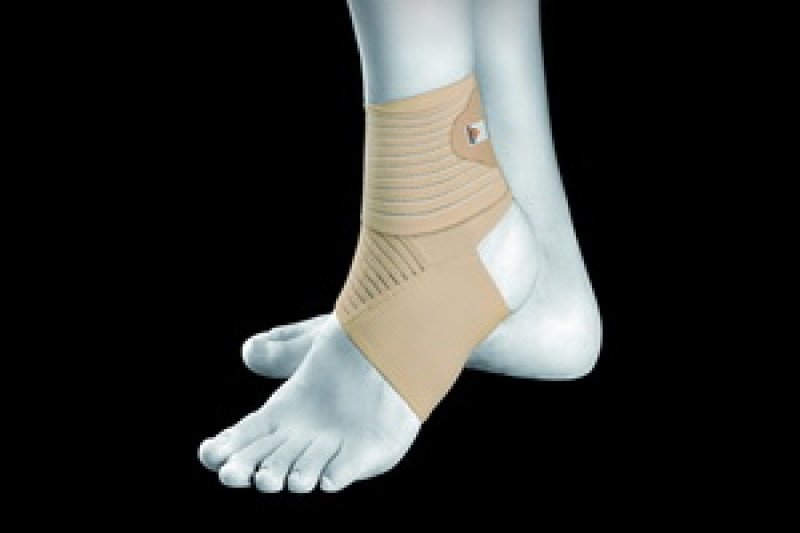
types of injuries
An ankle fracture can be open or closed. In the first case, the integrity of the skin is impaired, the soft tissues are damaged, bleeding occurs, and pieces of bone are visible from the wound on the leg. Such injuries are accompanied by severe pain. In such situations, surgical intervention is required. Open fractures, on the other hand, are rarer and usually occur in car accidents or other serious accidents.
The closed injury is often confused with a simple dislocation (when the bone has not been displaced) because the symptoms are very similar. For the most accurate diagnosis possible, an X-ray examination of the injured limb is indicated. With appropriate treatment, complications can be ruled out. In the case of a sprain, the situation is much more serious and improper treatment can have serious health consequences. There are several types of closed ankle fractures with dislocation:
- Medial ankle tear or dislocation of the joint inward or backward – this is an ankle injury with external rotation;
- Fibula fracture with lateral displacement – it is an injury to the abductor leg;
- Tear of the medial malleolus due to acute inward rotation of the foot is an adduction fracture that also involves the heel bone.

The main difficulty with this type of injury is that the fracture of the joint is usually combined with a dislocation, in which the bones are not only displaced, but also twisted around their own axis. In order for the fractures to heal properly and mobility of the leg to be restored, the bones must first be repositioned. Correct repositioning is crucial to the outcome of the treatment.
signs
In the case of an ankle fracture, the pathological symptoms appear immediately after the traumatic impact. The clinical picture is as follows:

- Severe pain in the area of injury that does not subside even when the leg is at rest.
- Inability to kick or move the injured leg and acute, increasing pain when doing so.
- Occurrence of swelling and its spread to the joint area of the lower extremity.
- Deformity of the limb (with significant displacement), unnatural positioning.
- Vitality of the skin in the area of the fracture, hematomas (bleeding due to damaged ligaments).
- Crunching noises when palpating and moving the leg.
- Bleeding, painful shock, open wound with visible bone fragments in an open ankle fracture.
To assess a closed fracture, bilateral x-rays and, if necessary, computer tomography are carried out. If there is suspicion of vascular damage, angiography is indicated. This diagnosis makes it possible to get the most accurate picture possible in order to prescribe the correct treatment and not to confuse the fracture with a simple sprain or twist.
For doctors
Ladies and gentlemen, just because something can't be repaired under local anesthesia doesn't mean you did something wrong. It doesn't mean you're a bad doctor. It just means that the patient's muscles are stronger than your hands. Additionally, there are certain types of sprains that can only be resolved conservatively, and if you don't know this, no matter how hard you try, it won't resolve. You have to operate. I personally observed a doctor with decades of experience suffering from a similar dislocation. And I was lucky, I knew how to do it. And that doesn't mean he's a bad doctor. And I might not be good at something else. And then there is Kaplan (for example here) [AV Kaplan, Bone and Joint Injuries, 1979. 428 p.]It's ancient, but useful. Not much has changed in this area since the days of Hippocrates (apart from anesthesia, of course). There are a variety of books on the Internet, here are the links. doc_zlo a whole bunch of them. I invite you to read, it is normal. Patients are somehow convinced that we know everything, and we know very little. In this way we are lifelong learners.
Dislocated shoulder
The most common contortion in nature. 50-60 % of all dislocations [Traumatology and Orthopedics: A Guide for Doctors / Edited by NV Kornilov: In 4 volumes - St. Petersburg: Hippocrates, 2005 - T.2 - P.54]. (Note – statistics from this source here and below). У morita The mechanism is described in great detail - falling on the shoulder. The position of the arm afterwards is called 'forced'. Every movement causes pain. The patient turns his entire body around the suffering arm and tries to find a comfortable position. It must be transported as it is and try to fix the arm in the position in which it is.
There are anterior-posterior dislocations (the vast majority) and posterior dislocations (rare but not uncommon) in which the head of the shoulder lies behind the socket of the scapula (also called horizontal dislocation). There are other forms (according to Kaplan's classification), but they only occur sporadically. Shoulder dislocations can have the following consequences complications Compression of the neurovascular bundle by the humeral head. The result can be (a) traumatic plexitis (or brachial plexus), in which the hand becomes numb and loses mobility (due to compression of the brachial plexus), a complication that requires very long (months) treatment by a neurologist can; and (b) compression of the axillary artery (fortunately rare), which in extreme cases (almost casuistically) can lead to gangrene of the hand if the dislocation is not quickly reversed. The latter can also manifest itself as numbness in the hand, albeit gradually, and above all as a lack of pulse in the usual place (on the radial artery).
species
- In the first form, some fibers of the ankle ligament are torn, causing swelling and discomfort, but mobility is still present.
- In the second, partial tear, the swelling spreads to the entire external area; Even in a static state, the tear site is painful and movement function is partially impaired but generally preserved.
- The third tear is complete, with acute persistent pain, severe swelling, and bleeding extending to both the external and plantar portions.
Lateral, anterior (rarely), posterior, habitual dislocations and subluxations may be present.
symptoms
- Severe pain syndrome;
- inability to stand on foot;
- Specific clicking sound in the affected area;
- Progressive swelling all around;
- deformation of tissue;
- abnormal angle, limited active and passive mobility;
- localized temperature increase.
If the pain occurs after a trauma, a trauma surgeon or orthopedist should be consulted.
classification
2 A cracking sound during the injury indicates a cruciate ligament tear; a tear of the cruciate ligament confirms abnormal movement of the joint in the anteroposterior plane.
4 Dislocations in the knee joint cause damage to the meniscus and joint capsule; Posterior dislocations can damage the popliteal vessels and the oculomotor nerve.
5 A patellar fracture often results in a rupture of the lateral epicondyle, causing the upper portion of the patella to displace upward. The knee joint is enlarged, pain occurs in the front area of the joint, and abrasions and hematomas are often found there. On palpation, a cavity can be seen between the patellar fractures.
– Severe pain in the knee joint, which increases with strain on the limbs and with attempts at active and passive movement;
– forced (upright) posture of the lower extremities with a significant increase in the volume of the knee joint (hemarthrosis);
– Bending and especially active stretching is painful; With the lower extremity extended, the patient can sometimes walk.
At the Fracture of the condyles of the tibia This leads to valgus deformity of the knee joint, hemarthrosis and impaired joint function.
On the website Fractures without displacement are characterized by pain in the knee joint, especially when the axis of the limb is loaded, and excessive lateral mobility of the lower extremity.
– Swelling (hematoma) and deformity of the shinbone (angular, rotational), often with shortening of the lower limbs;
– Local tenderness on palpation, associated with pain during axial loading, attempts to move the foot laterally, and anterior compression of the lower limb -. the symptom of 'radiation pain'..
Treatment
1 Pain relief - Lornoxicam 8 mg intramuscularly or intravenously, or ketorolac 30 mg intramuscularly, or 1 ml trimeperidine hydrochloride 2% intramuscularly.
Pain relief – Lornoxicam 8 mg intramuscularly or intravenously, or ketorolac 30 mg intramuscularly, or 1 ml trimeperidine hydrochloride 2% intramuscularly.
1. Analgesic effect - Lornoxicam 8 mg intramuscularly or intravenously, or ketorolac 30 mg intramuscularly, or 1 ml trimeperidine hydrochloride 2% intramuscularly.
Indicators of treatment effectiveness: Stabilization of the patient's condition.
diagnosis
There are numerous differentiation methods that can provide information about what exactly is present:
- On Fracture. It is impossible to stand on the leg and severe pain occurs. If the nerves have been hit, numbness may occur.
- Stretch is often accompanied by a slight crunching noise when moving. Swelling, instability, stiffness, or looseness of the knee joint may occur within the first few hours.
- On the side of torn ligament If the band breaks, the injured person may hear a popping sound. There may be bruising and a feeling of instability in the knee, sometimes even a hematoma. Mobility is severely limited, if not eliminated.
Treatment
First and foremost, the injured knee should be immobilized so as not to aggravate the pain and make the situation worse. Once the diagnosis is established, the bone and its fragments (if any) are repositioned. This can be done closed or surgically. After repositioning, the fracture site is fixed until healing. This can take several weeks or even months. After the bone has been restored, it is important that rehabilitation is carried out correctly to restore the normal function of the leg and allow a return to a normal lifestyle.
In any case, a knee injury requires special attention, because only a specialist can determine the severity, and delaying treatment can have tragic consequences. Enjoy life, not the knee pain.
complications
Serious injuries, untimely and inadequate treatment can lead to certain consequences of bone fractures in children:
- Damage to blood vessels and nervesresulting in restricted movement and dysfunction of the limbs
- Contractures – Restriction of movement in neighboring joints
- Ankylosis – complete loss of mobility
- Deformation, shortening of the limb due to abnormal bone connections
- Inconsistency of bone fragments and incorrect joint formation
- Interruption of bone growth in case of damage to the growth plates
- purulent and infectious complications In children with a fracture, symptoms are manifested by fever, severe pain, swelling and redness of the skin at the site of the injury.
prophylaxis
Parents should teach their child to follow basic safety rules and avoid situations where they could get injured, especially during outdoor sports activities. For young children, care should be taken to create a safe environment at home and in the garden (if the family lives in a private household). Of course, you can't keep an eye on everything and predict every situation - precautions must be taken within reason.
In most cases, the prognosis for bone fractures in children is favorable. They are treated successfully, and the bones in children's bodies heal quickly. However, to avoid complications, it is important to treat them as early as possible. The pediatric traumatologists at Our Time Clinic are always ready to help.
- Savchenko IV, Avtomonova TS, Martinen MS Peculiarities of traumatic injuries in children and factors determining them // Journal of Medicine: Theory and Practice, 2021, pp. 46-59.
- Khusainov NI, Vissarionov SV. Compression fractures of the spine in children: is it time for a change? // Journal of Spine Surgery, 2019, pp. 12-42.
- Kornilov NV Traumatology and orthopedics: a textbook // GEOTAR-Media, 2011, p. 592.
- Miroshnichenko VF, Kotelnikov GP Closed limb injuries // GEOTAR-Media, 2009, pp. 175-280.
- Closed Foot Injury.
- tibia and fibula.
- Dislocation of a bone in a joint.
- dislocation of the foot.
- The hock in which the person is located.
- Diagram of a joint with and without a dislocation.
- pelvic subluxation.
- This is what a dislocated leg looks like.