It is strictly forbidden to do this, since there is a risk of damaging healthy tissues and developing an infectious process.

- Calluses: what do they look like and how can they be treated?
- High-quality care: what modern diapers can do
- What is the difference between a wet callus and a dry callus?
- Causes of dry callus formation
- treatment of cornea
- Causes of corns
- Diagnosis of a cornea with nodules
- Treatment of pilar keratosis
- Symptoms of small cell keratolysis
- Diagnosis of punctate keratolysis
- Symptoms of follicular keratolysis
- complications
- Drug therapy in the fight against corns
- Plasters for removing calluses
- prevention
- Causes of plantar warts
- Which doctor can help?
- Our specialists
- Prices for Services
- causes
- symptoms
- Symptoms of a boil
- Treatment methods for boils
Calluses: what do they look like and how can they be treated?
Everyone has experienced blisters from wearing tight and uncomfortable shoes, especially women who like to walk around in stiletto heels. Men who are manual trades tend to get blisters on their hands, especially when wielding a shovel, axe, or saw. Dry and hard calluses do not develop overnight, but often remain for a long time. What do they look like and how to get rid of them?

High-quality care: what modern diapers can do
What is the difference between a wet callus and a dry callus?
If you have been rubbing your skin or applying pressure for a long time, you will experience a blister on your heel or other noticeable areas when you first step into your new shoes. The blister acts as a barrier that prevents further damage to the upper layers of the skin. If you take off uncomfortable shoes in time, the blister will heal on its own and the injured area will heal quickly. If it is not possible to take off the immobile shoes and you have to continue walking in them, the blister usually ruptures, the fluid drains, the integrity of the epidermis is disturbed, the subcutaneous tissue is further injured, blood and an increased risk of infection penetrate into the wound.
A dry cornea looks very different. The affected person often does not notice the negative changes at all, since he has become accustomed to the presence of the triggering factors and no longer perceives them. However, being indifferent to them does not mean that they stop doing their 'black' work. Over time, day by day, the skin gets thicker and thicker, developing hyperkeratosis, often referred to as a callus. In fact, corns can live for years without getting much attention. In rare cases, inflammation can develop around the callus, the most serious consequence of which is necrosis.
If you have dry calluses on your feet, it should be treated by a podiatrist. In no case should you seriously disturb the integrity of the skin, cut open the wart with a blade, etc. Also, such remedies may not be effective for callus horns for the following reason. This is because a cartilaginous cone forms under the thickness of the non-defoliated cells. Its peak lies in the deeper layers of the dermis. Such a core squeezes the vessels, spoiling the nutrition of the skin and causing the cone to grow even larger. Even if man removes the top, cornified layer, the core, ie the cornea, remains and after a while it reappears.
Causes of dry callus formation
Factors contributing to the formation of calluses:
- Rough seams on socks or tights that put a lot of pressure on the skin
- High foot load in athletes
- Walking up and down stairs daily
- Joint diseases of the foot
- Uneven distribution of body weight due to flat feet
- Frequent barefoot walking
- Nail fungus, skin damage on the feet
- Increased sweating
- Vitamin and micronutrient deficiencies
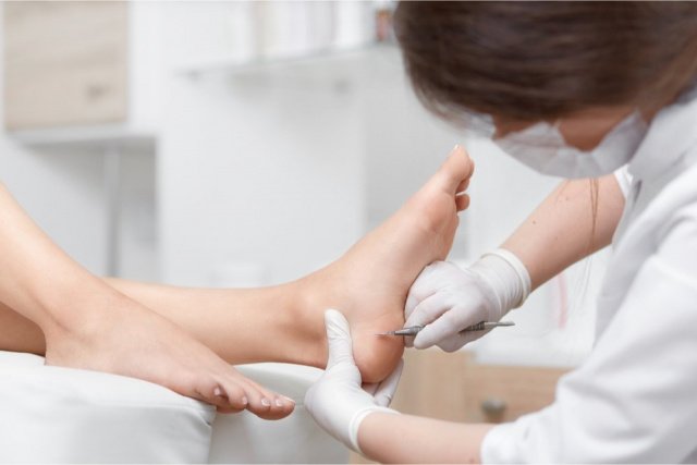
treatment of cornea
pedicure – is a method of effectively removing calluses. The root of the cornea is hollowed out with a special diamond or ceramic bur. The procedure is painless, sterile and doesn't take long in most cases, but once the callus root is removed, an antiseptic cream is poured into the area.
Laser pedicure is considered one of the safest and most comfortable procedures for the patient; healing occurs within two days.
laser therapy Effectively treats the affected area by removing the hardened seal layer by layer. Healthy tissue is not traumatized and the procedure lasts no more than 15 minutes. After the procedure, disinfectant solutions are applied to the skin to speed healing and prevent tissue infection. A protective cover forms at the site of callus formation, under which new dermis cells form. Complete healing after the procedure takes up to two weeks, and the laser leaves no traces on the skin.
The disadvantages of this method include:
- the high cost of the procedure
- The need for local anesthesia
- Unsuitable for people with diabetes and skin diseases
- Unsuitable for people with immune disorders and cancer.
cryoablation – Is a method of removing calluses or calluses using liquid nitrogen. The procedure must be performed by an experienced doctor to reduce the risk of injury. Callus removal by cryoablation takes up to two minutes. The procedure is painless and the healing process takes up to two weeks.
Disadvantages of cryoablation include:
- Long healing process - up to 14 days
- Risk of damage to healthy tissue
- Low effectiveness on calluses with deep pores
electrocoagulation – An effective method for removing calluses. The electric current destroys the proteins in the keratinized epidermis, the cells die, a protective crust forms, and the healing process takes one to one and a half weeks. The treatment takes about two minutes, is inexpensive and safe.
Causes of corns
In most cases, corns are caused by frequent pressure on the skin in a specific area. As a result, the horn particles are superimposed and can no longer detach themselves, which leads to thickening and roughening of the skin in this area. If you don't treat the calluses on your feet, they will begin to grow in the tissue. In this case, the callus area shrinks, and the shinbone takes on the shape of a cone.
The causes of calluses on the feet with a cone include:
– Wearing shoes with poor ventilation or shoes made of inferior materials.
– Frequent wearing of high-heeled shoes.
Other causes of calluses on the feet can be fungal diseases, the human papilloma virus and foreign bodies in the tissues of the foot.
On the hands, such calluses can be caused by:
– Sports activities on parallel bars, hoops and high bar.
– Frequent use of hand tools such as knives, hammers, axes, etc.
Calluses can also be caused by fungi, papilloma viruses, or foreign bodies.
Both men and women are at risk. However, the risk is higher in people with a compromised immune system, circulatory problems, and a history of diabetes. Most often, the neoplasm appears on the heels, palms or fingers.
Diagnosis of a cornea with nodules
In most cases, the patient will suspect that the growth is a cornea. However, an accurate diagnosis can only be made by a professional with a medical degree. Diagnosis usually involves visual examination and asking questions. If the doctor has doubts, they can issue a referral for tests or see another specialist. For example, to a dermatological oncologist if there is a suspicion that the tumor is malignant.
Treatment of pilar keratosis
Treatment of pilar keratosis requires its removal. The keratoma must be removed along its entire length, as removing just the top portion is ineffective.
Removal is done through the following procedures:
– Surgery. This method is considered outdated and is used in cases of severe corneal ingrowth. It is performed under local anesthesia. Using special instruments, the surgeon cuts away the top layer of the wart, and then removes the callus itself. Disadvantages of the procedure: painful, traumatic, risk of infection and bleeding, scarring, long recovery time.
– Machine pedicure. It makes it possible to eliminate dry calluses on the foot in one to five sessions with a stick. During the treatment, the specialist literally hollows out the root of the cornea using a special tool. In order not to injure healthy tissues, the specialist must perform hardware pedicure with great precision, changing phrases the deeper they penetrate into the body. Disadvantages are the inconvenience during the session and the relatively high cost of the procedure.
– cryodestruction. In this procedure, the dry cornea and its root are frozen with liquid nitrogen. The cells of the cornea and root then die, and the tissue is shed. After the procedure, a burn blister initially forms. After some time, it dries up, and a crust forms in its place. After about two weeks, the crust will peel off, leaving healthy skin. Disadvantages of cryoablation include discomfort during the session, the risk of freezing healthy tissue, and the appearance of a scar after the procedure.
– Laser removal. It is now considered one of the most effective methods of eliminating benign tumors. How to get rid of a keratoconus callus with the laser? The device generates a powerful laser beam that vaporizes both the cornea and its root in layers. Another advantage is that the laser beam immediately coagulates the vessels and disinfects the skin. There is no risk of bleeding or infection. In addition, laser treatment can remove a tumor in just one session. It is therefore worth taking a closer look at this treatment.
Symptoms of small cell keratolysis
The clinical symptoms of small cell keratolysis are very similar to those of fungal diseases of the feet. The main feature of the disease is a cone-shaped, pitted erosion, the depth of which depends on the thickness of the stratum corneum of the sole of the foot (usually 1-8 mm). The elements are located symmetrically in those places of the foot where rashes appear, in places of the foot that are subject to constant pressure, and on the friction surfaces between the toes. Sometimes the rashes are accompanied by itching and burning of the skin. If the erosions are accompanied by excessive sweating, that is, they are inside a protective aqueous layer, they become softened and have a whitish color.
Pitting erosions tend to coalesce and form erosive areas up to several centimeters in diameter over time. Skin erosions are usually not particularly uncomfortable as they do not cause pain. The reason for the report to the doctor is usually an unpleasant smell. Bacteria that are active on the surface of the skin in a warm and humid environment are responsible for the unpleasant odor. It should be noted that untreated fine keratolysis can last indefinitely. In the long term, case reports of skin lesions on the hands have been described.
Diagnosis of punctate keratolysis
The diagnosis of fine keratolysis is made by the dermatologist on the basis of the clinical findings, examination of the lesions under a Wood's lamp (fluorescence diagnosis), control scraping of the skin in the area of the lesions to exclude a fungal infection and culture on culture media to demonstrate a combined Infection with coccidia and pseudomonads (Synegnoia). Histomorphology shows punctate keratolysis of the epidermis and colonization of the punctate microtubules by pathogens. Punctual keratolysis differs from tinea pedis, plantar warts, basal cell nevus, arsenic poisoning, candidiasis, erythrasis, and interdigital maceration.
The basis of effective therapy is the correct diagnosis of the disease with the exclusion of a fungal disease. A dermatologist, a physiotherapist and a cosmetologist are involved in the treatment of the disease. The treatment of fine spot keratolysis is comprehensive, with priority given to pathogenetic measures. The first step is to eliminate the cause of the disease. For this purpose, a course of antibiotics from the group of macrolides is carried out. The treatment can be internal or external. Ointments, solutions and powders with the same active ingredient as well as preparations with benzoyl peroxide are used. Particular attention is paid to hyperhidrosis. In order to eliminate excessive sweating in the area of the sweat glands, selective injections with botulinum neurotoxin type A complex are carried out, which can paralyze the glandular apparatus. In the presence of contraindications to this manipulation, it is replaced by physiotherapy: iontophoresis, electrophoresis with preparations based on silver chloride or aluminum.
A certain routine must be followed on a daily basis. Wash your feet with deodorant soap as often as possible, avoid wearing tight shoes, use activated carbon adsorbers when wearing shoes, do not wear shoes made of synthetic materials, and choose cotton socks and breathable insoles. In summer, it is advisable to walk barefoot on the grass as a preventive measure to provide the bacteria with an airtight breeding ground. Exposure to hot, humid climates is contraindicated in patients with fine keratolysis. A transfer to moderate latitudes is not excluded. The prognosis of fine blotchy keratolysis is favorable if the above guidelines are followed.
Symptoms of follicular keratolysis
The onset of the disease is sudden. Gradually increasing pain appears in the affected area. The pain syndrome increases with movement and lowering of the arm. The contents of the bladder become cloudy. The skin around the cornea becomes hyperemic. Since the tissue of the hand is very dense, the swelling does not appear at the site of the inflammation, but on the back of the hand. The function of the finger is impaired. The body temperature rises to subfebrile levels.
After 2 to 3 days, an abscess develops. The pain becomes throbbing and throbbing, making it impossible to sleep at night. There is swelling. The functionality of the entire hand is restricted, the finger is forced into a moderate flexed position. Mobility is restricted and painful. Before the abscess invades the surrounding tissues, the hyperthermia is mild to moderate, and the general condition deteriorates somewhat. On examination with the tactile probe, the greatest pain occurs in the callus projection at the base of the finger.
When pus has penetrated deep into the tissues, the swelling continues to increase and the hand becomes pillowy. The natural wrinkles flatten out and the fingers spread. The pain spreads all over the hand. The patient's condition worsens. Fever reaches febrile values, hyperthermia is accompanied by weakness, fatigue and chills.
complications
When the purulent membrane melts and the pus enters the palm, the bladder abscess is complicated by palmar phlegm. This undesirable sequela occurs in 11 % of the patients, with 35 % of the cases being interdigital phlegm, 33 % dorsal phlegm and 8.5 % interdigital phlegm. The remaining patients are combined purulent processes.
Lymphadenitis and lymphadenopathy occur when infection spreads through the lymphatics, and forearm phlegmon occurs when subcutaneous and subdural spaces are affected. The generalization of the purulent process leads to sepsis, which is life-threatening.
Drug therapy in the fight against corns
How can internal blisters be treated with medication?
Treating rod thickening with preparations is time-consuming and very rarely effective.
Relatively good results can be obtained with fresh calluses, where the root has not yet penetrated deeply into the layers of the dermis.
For this purpose, the following substances are usually used keratolytic Drugs are usually used for this purpose.
Keratolytics belong to a chemically diverse group of preparations that have a relaxing effect on thickened tissue.
Keratolytics are used in dermatology primarily for skin diseases that are characterized by increased keratosis.
Most often, such remedies for internal calluses contain salicylic acid.
Topical application of salicylic acid promotes exfoliation by breaking the bonds between keratinized cells, causing them to die.
The acid also acts as a pain reliever and anti-inflammatory, and to a small extent, it inhibits certain bacteria and fungi that can colonize the skin.
Attempting to remove a callus yourself with salicylic acid is difficult because it can cause irritation and burns on healthy skin.
Also, there is a high risk of allergic reaction when using the product for the first time.
Urea-based ointments can be used as an alternative to acid for internal calluses, such as: B. uroderm.
However, they have little effect on deep-rooted calluses.
Plasters for removing calluses
The most well-known patch for internal calluses is Salipod.
The patch has antiseptic and keratolytic properties due to the combination of ingredients in its formulation.
Salicylic acid promotes rapid and deep penetration of sulfur into the skin and has an antibacterial and exfoliating effect.
When applied topically, it actively exfoliates the epidermis, kills pathogenic bacteria on the skin's surface, and has an irritating and slightly antiseptic effect.
The notice on the patch states that the product can be used on dry calluses and corns.
It should be noted that the instructions for use do not recommend its use on calluses.
Before using Salipod on an internal callus, the feet must be washed and dried thoroughly with a towel.
The application of the product is simple: a plaster of the appropriate size is applied directly to the callus and left on for two days.
If necessary, the process is repeated several times.
Many patients report a positive effect and state that it helps in the treatment of root calluses.
It is true that Salipod works quite well in treating rough skin on feet, especially dry calluses and corns.
However, none of the ingredients in the patch are able to penetrate the layers of the dermis where the root itself may be located.
Therefore, getting rid of corns can only be temporary.
How to remove an internal callus on the foot or heel when all methods are ineffective?
If hardware removal techniques and medication have proven ineffective, the patient is advised to surgically remove the callus.

prevention
To avoid the appearance of calluses on the soles of the feet, it is necessary to buy shoes and socks made of natural materials, wear special gloves and regularly use moisturizing and exfoliating creams to prevent the formation of new calluses on the toes.
- Avoid wearing flat-soled and high-heeled shoes for long periods of time - the optimal heel height is 4-5 cm;
- Use absorbent insoles if your feet sweat excessively;
- If you have flat feet, only wear special shoes recommended by your podiatrist;
- Maintain your feet regularly with scrubs and pumice stones;
- Control your weight.

Fungal infections are transmitted through contact. Therefore, do not wear other people's shoes and treat your feet with an antifungal product before going to the pool or sauna.
Calluses on the feet, little fingers and toes are painful. Her treatment with medication is lengthy. Quick ways to get rid of calluses: cryotherapy, hardware pedicure, AC cautery, radio frequency electrode or laser. Proper and regular care, as well as the principles of prevention, will help to avoid dry calluses.
Causes of plantar warts
The possibility of a benign growth on the skin depends on a person's general health. The papillomavirus can get into the body through domestic or sexual contact, but it settles only under unfavorable conditions. The main causes of plantar warts are:
- A weakened immune system;
- inflammatory processes in the body;
- other skin diseases;
- hormonal disorders;
- metabolic disorders;
- bad hygiene;
- cold and stress.
Warts on the feet affect both sexes equally, but women are at a slightly higher risk of developing them because they wear uncomfortable footwear. They constrict blood vessels and skin, often leading to mechanical injuries such as corns and calluses. Poor diet and the use of poor quality water can also affect the activation of the virus in the body.
Which doctor can help?
Not every case of warts on the feet requires medical attention. If the growth is small, doesn't cause much discomfort, and tends to go away, you can just wait for it to go away. Otherwise, you should consult a doctor:
Our specialists
The prices shown on this page are not a public offer. To determine the cost of the service and to schedule an appointment with the doctor, please call 8 (495) 255-37-37.
Prices for Services
Surgical treatment is the predominant form of wart treatment - currently the most common form of treatment is laser. However, there are not always indications for such radical treatment. Before the doctor decides whether an operation makes sense, he will try to clarify the clinical picture. He will ask the patient the following questions:
causes

When leg ulcers occur, they can have a variety of causes. Most often they are caused by other diseases:
- older stages of varicose veins;
- frostbite of the limbs or burns;
- Diabetes;
- radiation exposure;
- diseases of the circulatory system;
- chemical poisoning;
- pressure sores caused by lying in one position for a long time;
- diseases of the spine;
- Skin diseases, allergies.
Such pathogens lead to complications and other diseases, including ulcerative manifestations.
symptoms
Ulcerative abscesses do not occur randomly. There may be swelling accompanied by itching and crusting. The skin becomes thin and bluish in color. Pigmentation may occur in this area.
- chills, night cramps in the lower limbs;
- small droplets of liquid form on the skin if there is a blockage in the lymph;
- When the epidermis peels off, an ulcer develops with thickened edges;
- when touching the ulcerated area, there is severe pain;
- when the bleeding ulcer becomes infected with bacteria and pus forms.
These symptoms appear gradually, leading to an exacerbation of the disease.
The lower extremities are the most common place for an ulcer to manifest itself. The reason for this is the slowing down of blood flow in the blood vessels. If the tissues do not receive enough nutrients, there are lymphatic manifestations and the formation of a purulent plaque on the ulcer.
This type of disease is called trophic ulceration. If the patient has diabetes, purulent phenomena form on the heels and feet.
In most cases, the disease is caused by varicose veins. When skin damage occurs, it leads to ulcers, even if it's not too severe.
If ulcers appear on the lower leg, this means that there are major changes in the venous vessels. The area is dilated and blood flow worsens. In such a case, the patient feels severe pain.
If the extent of the ulcer is large, this indicates a poor blood supply. In this case, such a process is already prolonged and exacerbations occur. If the ulcers are large, you should not try to treat them yourself, but quickly consult a doctor.
In the initial stages, when the ulcer bleeds, there may be an infection caused by the spread of bacteria or fungi. Pus forms and there is an unpleasant odor. If the process is prolonged, the pus penetrates into the deeper layers, they become severely inflamed, and severe pain occurs. This spreads to the game and the ankle. If the case is neglected, it leads to an acute form of septicemia.
Symptoms of a boil
Boils can develop anywhere there are hair follicles (i.e. almost anywhere except the hands and feet). Most commonly, boils appear in places where the skin sweats, rubs, and gets dirty—the neck, forearms, armpits, groin, lower back, thighs, and buttocks. It is not uncommon for boils to appear on the face as well.
A boil goes through several stages in its development:
In the first stage, an infiltrate appears around the mouth of the hair follicle - a swollen area with noticeable redness and thickening of the skin. The infiltrate gradually increases in size and can reach 1-3 cm in diameter. The skin around the infiltrate tightens and becomes painful, sometimes tingling is also felt. In this case one speaks of a maturation of the furuncle.
The next stage is called purulent-necrotic. On average, 3-4 days after the first symptoms of a boil, a characteristic strand of pus and dead tissue appears, the end of which rises above the surface of the skin in the form of a hollow pustule. If the boil is on the face, ear or neck, a fever of up to 38 °C, general weakness, loss of appetite and headaches can occur during this time.
Eventually the pustule bursts open and a necrotic core emerges. The unpleasant symptoms subside and healing begins. Within 3-4 days, the wound at the site of the pustule heals. A scar forms that can become almost invisible over time.
Treatment methods for boils
Never try to squeeze, massage, or put pressure on the boil yourself. The infection can get into the blood and cause blood poisoning (sepsis). It is worth remembering that inside the pimple there are active pathogenic bacteria. Therefore, do not touch the boil, and if you do touch it, wash your hands with soap and water immediately.
A single small ulcer can be treated at home. It is important to see a doctor if:
- the furuncle has not healed within 3 days and the pain only increases
- the ulcer is too large or painful;
- the boil is on the neck, ear, nose, face, spine or anus. These are the most dangerous spots in terms of complications;
- Fever;
- red streaks spreading from the furuncle (this is a symptom of lymphangitis, inflammation of the lymphatic vessels);
- For multiple boils (furunculosis). A doctor should always be consulted for furunculosis, even if the boils are small;
- The boils are caused by diabetes.
The surgeon treats boils. In the case of large furuncles, the wound is surgically opened, disinfected and drained. Treatment of furunculosis, as a rule, is complex and includes both the use of topical agents and general therapy, which is primarily necessary to improve the body's immunity.
Self-medication should be avoided. Contact our specialists who will correctly diagnose and treat your condition.
Read more:- Feet of teenagers.
- What can I do if my feet are irritated by new shoes?.
- hole in the eardrum.
- scabies.
- Shoes press on the toes, what to do?.
- What to do if the sneakers pinch in the heel?.
- Loss of a blister on the heel.
- What to do if the back of the shoe rubs against the heel?.