bursitis – is an inflammation of the synovial capsule around a joint. Bursitis can occur in any joint in the human body, from the smallest to the largest, and partially or completely limit movement in those joints. The condition is a defensive response by the body trying to protect the damaged lining of the periarticular capsule from further destruction. It is true that rest is essential in the management of synovitis, as sometimes (such as in trauma) it gives the joint time to heal and rebuild, allowing the inflammation to subside. However, acute bursitis can cause irreversible damage to the joint and, if left untreated, can lead to an abscess that spreads the infection throughout the body and permanently limits mobility of the joint. Like most diseases, bunions are easily treatable in the early stages. But how do you recognize them in time? How do you treat a bunion? We tell you who is at risk, how to prevent the disease and how to avoid complications. Synovitis is a condition in which the joints become inflamed

- A thickening of the foot above the heel, called a Haglund foot deformity
- Unique campaign
- 'Regain Youthfulness Your Feet'.
- Frequently Asked Questions
- Causes of synovitis in the foot
- Symptoms of bursitis
- Symptoms of heel tendinitis
- Classification and stages of Achilles tendonitis
- How is it diagnosed?
- The main therapeutic options
- conservative methods
- Surgical treatment method
- treatment of synovitis
- Surgical treatment of synovitis bursitis
- Medicines for bursitis
- anti-inflammatory drugs
- Antimicrobials and antibiotics
- chondroprotectors
- Heel Spur Symptoms
- Diagnosis of heel spurs
- Causes of heel pain
- diagnosis
- Causes and Symptoms of Gout
- How do you treat gout on your feet?
- Prevention of gout in the feet
- How to diagnose heel bursitis
- treatment at home
A thickening of the foot above the heel, called a Haglund foot deformity
Many patients complain that in this area, on the heel, behind, there is a kind of thickening, a lump. The most common cause and explanation is the Haglund's deformity of the heel bone, in terms of what we understand there, the heel is attached behind the Achilles tendon, which here, here it is, behind the Achilles tendon is a bursa - a kind of bag with a kind of fluid in it, which improves the function of gliding, cushioning and everything else.
Firstly, the heel bone is constructed differently from an anatomical point of view. In some people this part is more pronounced, in some people the heel bone is slightly sloping, so to speak. As for the Haglund deformity itself, it is a kind of inflammation or small bony hypertrophy that forms precisely at the point of attachment of the Achilles tendon to the heel bone and often, even in advanced stages, from changes in the structure and tissue of the tendon itself and additionally from accompanied by inflammation of the bursa in the Achilles tendon area.
Clinically, this manifests itself as a knot on the back surface of the heel. This is also known as a 'pamb-bump' in English. The main problem and discomfort that patients feel is the problem of wearing shoes.
Unique campaign
'Regain youthfulness
Her feet'.
There are now modern, safe and effective treatments to help restore your feet to health. Some of them are completely free, others are very affordable. Unfortunately, many older people are not aware of this.
That's why I've created a new, unique offer for lovely older ladies: 'Give your feet back their youth'.
I invite you to my office for a FREE consultation where I will examine your feet.
I will also tell you what modern, safe and effective treatments are available for your situation and how you can get them for free or at the lowest possible cost. We will talk to you about all the options available and choose the one that suits you best.
1. Any person over the age of 55 is eligible to participate.
2. You can apply with any foot and ankle problem.
3. The promotion runs from August 1st to September 30th, 2019.
4. You can come to the consultation in person at St. Petersburg, Yaroslavski Allee 66, Building 1, or you can get consultation online.
Call mothers, grandmothers, relatives, friends - anyone who can benefit from my advice. Tell as many people as possible, share on your social networks.
Mature age is a great time to enjoy life. So don't let pain and discomfort in your feet interfere with your quality of life. Let's get your feet back in shape together so walking can become one of your favorite pastimes again.
If you would like to know more about the Rejuvenate Your Feet campaign, send an SMS to WhatsApp +79219651182
or call us 8 (812) 336-60-22
Frequently Asked Questions
The surgery itself is no different. In all surgeries I perform, I use the most modern medical equipment, the best imported implants, high-quality consumables, the necessary medicines and regional anesthesia (a variant of local anesthesia - two injections are given in the foot). Free surgery has only two differences: 1. Only one foot can be operated on in one operation. If you want to have both feet operated on at the same time, they must be operated on on different days. The other foot can only be operated after 2 weeks. 2. If you have a quota you will not get follow-up care I always include follow-up care in the price of the surgery I pay for. This way I can see how you are feeling, how your bones are healing and how your joint has regained mobility. This way I can prevent possible postoperative complications. This always leads to excellent results and satisfied patients. Postoperative care can be purchased separately.
Causes of synovitis in the foot
- overuse of the feet
- long-term micro-injuries
- Foot disorders (flatfoot, valgus deformity, hallux valgus foot, plantar fasciitis)
- Redistribution of the burden of poor posture
- deforming arthrosis
- Metabolic disorders (gout)
Tight, uncomfortable, high-heeled shoes, excessive exercise, poor technique, etc. can trigger the inflammatory process.
Symptoms of bursitis
The main symptoms are pronounced pain, redness, swelling and fluctuations (plasticity and mobility) of the soft tissues in the affected area. Bursitis in the big toe is often accompanied by a malposition of the metatarsophalangeal joint. In Achilles tendon bursitis, palpation reveals a localized area of pain on the posterior surface of the tibia in front of the attachment of the Achilles tendon. The pain increases when trying to stand on tiptoe and in the first few minutes after standing up after bedtime. Posterior heel bursitis is characterized by a throbbing pain in the heel that worsens on contact with hard surfaces and after removing shoes. The inflammatory process most often develops against the background of plantar fasciitis or heel spurs.
Most inflammation of the synovial capsule of the foot is aseptic in nature. However, if a bacterial flora invades the affected bursa, an abscess can develop. Purulent bursitis of the big toe or the articular capsule of the ankle is manifested by severe pain, a local and general increase in body temperature and other symptoms of general intoxication.
Symptoms of heel tendinitis
Characteristic manifestations of the clinical picture:
- Morning stiffness, pain when walking, bending and stretching the foot
- Pain along tendon at back of shin when moving, jumping, standing on tiptoe.
- Thickening of the ankle at the tendon, local swelling and congestion
- Hypersensitivity, discomfort and pain on palpation
Classification and stages of Achilles tendonitis
Achilles tendinitis is a collective term that includes various forms of pathology:
- Insertion tendonitis – the inflammation is localized at the site of tendon insertion
- Non-inflammatory tendinitis – the inflammation occurs above the point where the tendon attaches to the heel bone.
Depending on the severity of the disease, a distinction is made between acute and chronic tendonitis. In the first form, symptoms last no more than three weeks. In the second case, the symptoms last longer than the specified period.
How is it diagnosed?
The best way to diagnose Achilles tendonitis is with an MRI scan. The examination reveals all pathological changes and deviations from the norm: the location and extent of the inflammatory process, calcification of the tendon, formation of osteophytes, accumulation of fluid, damage to surrounding tissues, determination of the degree of damage. MRI is also indicated for planned surgical or invasive procedures.
X-rays are used to exclude traumatic bone injuries.
The CMRT radiologists make the diagnosis by taking slice-by-slice images of the examined area. In this way, the type and severity of the lesions are determined and the selection of an effective therapeutic measure is ensured.
The main therapeutic options
There are many methods of treating a nodule on the lateral side of the heel. The choice of the appropriate tactic depends on the cause of the complaints and their differentiation.
conservative methods
The lump on the back of the heel is usually treated with medication. The drugs are prescribed by the doctor, taking into account the specifics of the disease. If pain sensations are present, painkillers are used. Anti-inflammatory drugs can help with inflammatory changes.
For external growths, topical medications in the form of ointments, gels, or creams are recommended. If the symptoms are severe and cannot be relieved by external drugs, injections are prescribed.
Once a callus has formed, the main goal of treatment is to prevent an infection from entering the body once it has opened. It is strictly forbidden to pierce such calluses yourself. They break up of their own accord; in exceptional cases, the doctor can pierce them. After tearing, the affected area is treated with an antiseptic and a plaster is applied.
Also Read: How to Heal a Bruise on the Leg in 1 Day?
Surgical treatment method
Surgical treatment is carried out only in neglected cases, when the pathology cannot be treated with drugs, and the growth causes severe discomfort to the patient.
If rear heel bunions are caused by a joint or bone condition, surgery will be needed.
Modern medicine offers the latest surgical techniques to treat bunions with minimal risk of complications and even without incisions. Only in the most severe cases are classical surgical methods used, in which the tissue is excised and then fused with special devices.
Non-invasive surgical techniques are used for soft tumors, calluses and corns. Cryoablation is widespread. This essentially involves the removal of diseased tissue by freezing. This procedure is performed with liquid nitrogen, which is at a very low temperature.
treatment of synovitis
In most cases, synovitis requires conservative treatment for 2-5 weeks; an operation is indicated only in very advanced cases or in purulent synovitis.
In addition to taking the medications prescribed by the doctor, the patient should undergo physical therapy to reduce inflammation and get full rest. Plaster casts or walking aids can be used to relieve pressure on the joint.
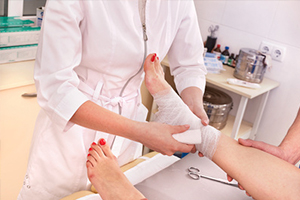
If the inflammation is mild, bursitis is treated at home and consists of rest and elastic bandages and bursa wraps with anti-inflammatory and anti-arthritic drugs.
Before visiting an orthopedist or rheumatologist an ice pack can be applied to reduce swelling and pain, or cooling ointments - as an emergency measure to relieve the symptoms of synovitis. For this purpose, ice is applied to the painful joint for 5-15 minutes every 4-6 hours.
Surgical treatment of synovitis bursitis
In cases where the lump over the joint is too large and taking too long to resolve, the doctor may perform a minimally invasive procedure—a puncture or drainage to drain the excess fluid. The puncture can be combined with arthroscopic lavage, a procedure in which disinfectant solutions are injected into the capsule, followed by drainage and the intra-articular administration of an anti-inflammatory steroid.
Surgical treatment of synovitis is also indicated with purulent inflammation in the periarticular capsule cavity and the formation of adhesions that interfere with the normal mobility of the joint. Severe purulent inflammation and large amounts of crystals deposited on the capsular lining may necessitate opening of the capsular cavity. This is done through a small incision (up to 1 cm) through which dead tissue, metabolic products, calcium deposits on the tendons and other foreign bodies are removed, providing fast and reliable relief even for severe acute and chronic bursitis.
Medicines for bursitis
The choice of medication for bursitis depends on the results of the synovial fluid analysis. If the composition is normal and free from white blood cells and infectious agents, anti-inflammatory therapy with nonsteroidal and steroidal drugs is carried out. If an infectious agent is detected during the examination, an individual antimicrobial treatment program is selected on the basis of the antibiogram.
anti-inflammatory drugs
Nonsteroidal anti-inflammatory drugs (NSAIDs) are used as the main treatment for bursitis. For mild inflammation, NSAIDs are prescribed topically, in other cases they are prescribed systemically (as tablets or injections) or in combination with others.
Severe inflammation that is associated with severe pain and cannot be treated with NSAIDs requires the use of hormonal drugs such as glucocorticoids (GCs). These are usually injected directly into the periarticular capsule after it has been flushed.
If the inflammatory process subsides before the drugs are administered, doctors predict a successful recovery with no further mobility impairments.
Both NSAIDs and HCs have side effects, so you should ask your doctor for an individual prescription before using them. Do not use NSAIDs if you have erosive or inflammatory lesions on the gastrointestinal mucosa.
The following NSAIDs are used in synovitis: naproxen, meloxicam, nimesil and others.
As well as GCs: dipropane, triamcinolone and others.
Antimicrobials and antibiotics
Before a serology or PCR, the doctor may prescribe broad-spectrum antibiotics. If the patient suffers from the effects of a particular infection, the doctor selects the synovitis drug that works best against this pathogen.
chondroprotectors
The chondroprotective agents glucosamine and chondroitin sulphate are recommended as a prophylactic, but also for injuries to the joint capsule. They help repair damaged cartilage, strengthen ligaments and tendons, and make the bursa more resilient to stress. This is the only group of drugs that improves the architecture of the joint and promotes healing of erosions without the formation of adhesions that impair mobility in a synovitis-damaged joint.
Heel Spur Symptoms
Symptoms of a heel spur The location of the spur in relation to the nerve fibers and nerve endings plays a crucial role - the closer the spur is, the more intense the symptoms. The location of the spur in relation to the nerve fibers and nerve endings plays a crucial role - the closer the spur is, the more intense the symptoms. In rare cases, the spur can be localized so 'well' that it can only be discovered after the symptoms of inflammation have subsided and during an X-ray examination.
The most noticeable symptom of heel spurs is pain. In the early stages of the disease, it may be subdued, presenting as discomfort after prolonged standing or walking. The nature of the pain is usually a tenderness, as if a small pebble had entered the shoe. Sometimes patients notice increased fatigue and a general decrease in foot strength.
Soon there are complaints of soft tissue injuries when suddenly getting out of bed or from a chair ('got up on the wrong foot' - that's heel spurs!), when climbing stairs, jumping or running and when spreading your feet.
The next stages are characterized by the clear symptoms of heel spur inflammation:
- Localized fever, warmth and tension in the heel area;
- Swelling that causes discomfort when putting on and taking off shoes (especially shoes that the patient previously found comfortable)
- reddening of the skin over the heel bulb;
- numbness of the foot that may extend above the ankle;
- Occurrence of thickening – thickening of the stratum corneum on the skin of the heel
- increased pain with walking and other activities that put pressure on the heel (eg, sitting on tiptoe);
- pronounced protrusions under the skin;
- formation of painful blisters;
- a tingling and burning sensation at the end of the day while resting.
If the pain is persistent, the patient's gait begins to change - a Limp.Limping, attempting to shift body weight onto the heel, or twisting the foot in place. In advanced cases, the characteristic symptom is heel spurs In advanced cases, the heel spur is associated with a transverse flat foot.This only aggravates the situation and contributes to the growth of heel spurs. Flat feet lengthen the patient's foot and the muscles in the lower leg stretch unnaturally. Pain can be felt not only in the knee or hip, but also in the back. An additional complication is. contortion of posture, intervertebral fractures.
Diagnosis of heel spurs
To diagnose a heel spur, the orthopedist or rheumatologist takes the medical history on the first visit, feels the affected area and examines the patient for inflammation, pain, abnormal bone deformities and osteophytes.
In order to make an accurate diagnosis, further examinations are required:
- ROENTGEN (helps determine the presence of a heel spur, its location and size);
- ULTRASONIC EXAMINATION (shows the condition of the soft tissues and tendons, as well as the outlines of the inflammation).
To the exclusion. ankylosing spondylitis, Rheumatoid arthritis, i other autoimmune diseasesas well as goutFor gout, specialists usually prescribe a general biochemical blood test (to determine inflammation, uric acid levels and a specific marker - rheumatoid factor).
In the presence of comorbidities and vascular complications, lower limb vascular Doppler ultrasonography and nerve conduction testing may be recommended. MRI may be recommended for patients with symptoms suggestive of severe nerve conduction disorders.
By combining these diagnostic procedures, heel spurs can be reliably distinguished from gout, osteoarthritis, bone tuberculosis, long-term trauma and other symptom-causing pathologies.
Causes of heel pain
Experts point out several factors that can cause discomfort in the foot area. They are often caused by accidental trauma to the foot or ill-fitting footwear. If the heel pain does not have a traumatic cause, the treatment should depend on the nature of the pathology. This symptom can be caused by:
- Arthritis.
- inflammation of the fascia.
- Achilles tendinitis.
- Infectious arthritis.
- Gout.
- Osteochondrosis of the spine.
- Osteoporosis.
diagnosis
When a patient experiences heel pain, the cause must be determined and treatment recommended. For this purpose, the doctor carries out an examination, after which he z. B. can cause laboratory and instrumental investigations:
| How is the diagnosis made? | Time |
|---|---|
| X-ray of the ankle | 10 mins |
| Ultrasound examination of the ankle | 30 minutes |
| morphology of the blood | 10 mins |
| MRI scan of the ankle | 30 minutes |
| electromyography | 300 minutes |
Causes and Symptoms of Gout
Gout refers to a group of arthritises. The term arthritis refers to any disease of the joints. The term 'gout' is used when referring to deposits of uric acid salts in various tissues of the body, most commonly in the joints and cartilage. How does this process come about?
Uric acid is a breakdown product of purines - special substances that are produced in our body and are also supplied to us with food. We get a lot of purines from eating fatty meat and fish (herring, sardines, cod), meat by-products (sausages, frankfurters), and fast food. Also when drinking alcohol (especially beer and grape wine), unnatural juices, sweet carbonated drinks and coffee. Then a large amount of uric acid is formed in the body, the elimination of which the kidneys cannot cope with. Another cause of gout is when the body produces a normal amount of this acid but the kidneys are unable to eliminate it due to an abnormality.
Uric acid salts (urates) build up in the joints, especially the smaller joints, and gradually destroy them. Damaged joints are most prone to salt build-up. The joints of the big toe (commonly referred to as the 'big toe') are often damaged by wearing uncomfortable, tight shoes. Gout can also lead to kidney stones, which in turn lead to kidney failure, which in some cases is fatal.
- Acute joint pain (especially after heavy eating or heavy alcohol consumption). Sometimes the feeling is so unbearable that even if you drape a sheet over an arm or leg, the pain can still be felt. The pain begins at night, subsides during the day, and then returns. This can be the case for several days or even months;
- redness and swelling of the joint;
- fever in the area of the joint up to 39-40 ° C;
- Fever;
- general weakness.
How do you treat gout on your feet?
Since the Middle Ages, gout has been referred to as the 'disease of kings' because it was the holy men who could indulge in hearty meals and were prone to gluttony. In order to fight gout, you must first change your diet. You should limit the consumption of meat and its by-products, give up fatty foods, legumes and fish caviar, beer, wine and, if possible, other alcoholic beverages. Gout patients are usually prescribed diet table 6. It is recommended to drink at least 2 liters of fluids per day. Mineral waters like 'Borjomi', 'Narzan', 'Essentuki' are helpful.
In acute attacks of gout, it is recommended to put ice on the inflamed areas and make poultices with dimethoxide. Pain relievers and anti-inflammatory drugs that do not contain steroids are indicated.
American researchers have found a connection between calcium and ascorbic acid deficiency and the development of gout. Therefore, taking these substances as part of the treatment and prevention of the disease is necessary in consultation with the doctor.
In the case of gout, physiotherapy improves well-being considerably. One of the most advanced and modern treatment methods for gout is shock wave therapy. When the affected tissue is exposed to shock waves of a certain frequency, it leads to loosening of salt microcrystals and thickening of connective tissue, tenfold increase in blood flow, which in turn leads to salt absorption and tissue regeneration.
In particularly severe cases, surgical methods are used to remove uric acid deposits in the joints. However, in order to avoid this, prevention of joint diseases such as gout in the feet should be carried out.
Prevention of gout in the feet
In order to maintain the mobility and function of the ankles into old age, the following preventive measures should be observed:
How to diagnose heel bursitis
An examination by a specialist is usually sufficient to diagnose Achilles tendon bursitis or heel bursitis. In addition to a visual examination, an X-ray is required to detect any abnormalities in the bone and joint tissue. An MRI scan helps make the soft tissues in the heel area clearly visible. To analyze the exudate, a puncture can be performed to determine the nature of the infectious process.
Both acute and chronic heel bursitis require good medical care. The treatment of this condition is based on the general principles of treating inflammation and has several main goals:
- pain control and elimination of the focus of inflammation;
The treatment of Achilles tendon bursitis and carpal bursitis begins with immobilization of the affected limb by applying an immobilization splint. During treatment, attempts should be made to spare the limbs as much as possible. Treatment tactics then depend on the severity of clinical symptoms and the presence of infection.
Treatment for acute bursitis includes medication, physiotherapy, and if the inflammation is suppressed, surgery or puncture can be done.
treatment at home
Medical science does not dispute the effectiveness of some home remedies for heel bursitis. However, it is important to understand that they can have a positive effect only if they are used in addition to the comprehensive treatment prescribed by the doctor. Remember that home treatment does not replace or limit the treatment prescribed by the doctor.
With the correct use of the methods of home medicine, it is quite possible to relieve discomfort. Below are some easy recipes:
- Flaxseed Compress. Flaxseeds heated in a dry pan are poured into a burlap sack or wrapped in a handkerchief. This remedy is applied to the inflamed area for 2-3 hours every day for 2 weeks, preferably before bedtime.
Bursitis can cause severe pain and affect full mobility and normal living. If treatment for bursitis is not started quickly, the symptoms and possible complications of the disease can lead to the need for surgical removal of the bursa.
Do not delay your visit to the doctor, because with modern treatment you can maintain full mobility of the joint and lead a normal life again.
The unique techniques and innovative equipment of the 'Halo!' clinics enable a quick and accurate diagnosis and, most importantly, the correct prescription of the necessary effective therapy.
Read more:- cysts in the ankle.
- Why does the femur hurt?.
- Why the shin hurts.
- The tarsal bone hurts from above - what to do?.
- Your child's leg hurts and which doctor to see.
- Why does the galley muscle hurt?.
- The long fibula muscle hurts.
- Heel bone tendon sac in Latin.