A geneticist is also consulted to rule out chromosomal or genetic abnormalities.

- Lesson 15: Gene Linkage
- Tasks to determine the distance between genes
- Symptoms and possible complications
- diagnosis and treatment
- Why babies are born with finger fusion: the main causes
- Bandaged toes or fingers: diagnosis
- symptoms
- diagnosis
- Types of syndactyly
- Symptoms of syndactyly
- species
- symptoms
- Symptoms and causes of syndactyly
- Surgical treatment of syndactyly in children and adolescents - how is the operation carried out?
- Why do the fingers appear - lower extremities
- Characteristics of the symptom
- Tumors of the respiratory system
- diagnosis
- What other conditions can be similar to Meniere's disease?
- surgical treatment
Lesson 15: Gene Linkage
Gene linkage is the common inheritance of genes located on the same chromosome. The number of linked groups corresponds to the haploid chromosome number, i.e. 4 in Drosophila. The nature of conjugate inheritance was explained by Morgan and colleagues. As a test subject, they chose the fruit fly Drosophila, which turned out to be a very suitable model for studying this phenomenon because its body cells contain only four pairs of chromosomes and it has a high reproduction frequency (more than 20 generations can be studied in one year). So, are traits controlled by a gene. are traits controlled by genes on the same chromosome. They are naturally inherited together when there is complete linkage (Morgan's law). Complete linkage is rare and usually incomplete, due to the effect of crossing over and exchanging homologous chromosome segments during meiosis. That is, genes from one chromosome cross over to another chromosome that is homologous to it.
The frequency of crossing over depends on the distance between genes. The closer the genes are to each other in a chromosome, the stronger the connection between them and the less often they diverge from each other during crossing over; Conversely, the further the genes are from each other, the weaker the bond between them and the more often it can be broken.
In Figure 1 shows the left side the distance between genes A i B is small, the probability of a chromatid break exactly between A and B is low, so the linkage is complete, the chromosomes in the gametes are identical to the parents (two types), no other variants occur.
In Figure 1 on the right: Distance between genes A i B is large, there is an increased probability of chromatid breakage and subsequent cross-linking precisely between the A i B The link is therefore incomplete, four types of chromosomes arise in the gametes - 2 identical to the parent (non-crossing) + 2 crossing variants.
Tasks to determine the distance between genes
Inheritance of sex-linked traits
Sex-linked traits are traits whose genes are located not on the autosome (non-sex chromosome) but on the heterosome (sex chromosome). The scheme for resolving sex-linked traits is different from autosomal monohybrid crossing. In the case where the gene with the Х–Chromosome bound, the gene can only be passed on from the father to daughters and from the mother equally to daughters and sons. If the gene with the Х–Chromosome bound and recessive, it is only present in homozygous form in women. For men is the second Х-no chromosome, so such a gene is always expressed.
Gene symbols are not used for problems of this type (A, a, B, b), as in autosomal inheritance, but the symbols of the sex chromosomes X, Y with a reference to the genes contained therein (X A , X a ).
Sex-linked abnormalities are more often controlled by recessive genes located in the Х-chromosome lie and are in the genotype XY (i.e. in male mammals and female birds).
Above we looked at examples where the sex-linked gene is on the X Х-chromosome lies, but there are also genes that are on the Y-Chromosome. In species where the male sex is heterogamous, this gene can only be passed on to males. In humans, the gene for a type of syndactyly, a membranous structure between the 2nd and 3rd toes, is located on the Ychromosome, which is why syndactyly only occurs in men. Another anomaly, hypertrichosis of the auricular margins (rows of hair on the ear), is known to be transmitted via the same mechanism. In the family studied with this anomaly, it was inherited through the male line in five generations.
Symptoms and possible complications
Symptoms of a ruptured eardrum:
- Severe pain in the ear;
- tinnitus;
- Hearing loss (partial or complete), distortion of sound;
- Sudden discharge of pus from the ear with simultaneous cessation of pain;
- Bloody discharge after mechanical impact on the ear or into the ear canal.
A ruptured eardrum poses the risk of the following: Complications:
diagnosis and treatment
diagnosis A ruptured eardrum is diagnosed through an otoscopy. Using a specific method, the integrity of the eardrum can be visually assessed. To do this, a special funnel is inserted into the ear canal and the eardrum is pulled back and up at the same time. In addition to identifying the crack itself, the diagnosis also includes taking a sample of the secreted contents from the ear canal in order to determine the pathogen, if necessary.
An uncomplicated eardrum perforation usually does not require treatment. It heals on its own after some time and hearing gradually returns within a few weeks. In more severe cases, if the tear is too large or a second infection prevents the eardrum opening from healing, it can be treated with myringoplasty.
surgical treatment A perforated eardrum is treated surgically under general anesthesia. A small piece of skin over the eardrum is removed to close the hole in the eardrum. This is placed at the perforation site under constant visual control using a special microscope and micro-instruments. The new patch is held on the edge by a special material that recedes as it adheres to the eardrum. To prevent infectious complications after surgery, a tampon dipped in an antibiotic solution is usually inserted into the ear canal. In the first few days after surgery, you may experience discomfort or even pain in your ear and feel dizzy.
Why babies are born with finger fusion: the main causes
Parents whose son or daughter is born with an anomaly wonder why babies are born with crossed fingers. Nowadays, doctors have discovered that the causes of finger misalignment are often hereditary. If a parent had this anomaly, there is a 50 percent chance that the child will be born with a developmental anomaly. The mutated gene is particularly common in the male line.
When syndactyly is diagnosed, sometimes the cause is not hereditary. It occurs between 4 and 8 weeks of pregnancy when the embryo is developing and forming a hand.
The following factors can cause abnormal limb development:
- taking certain medications during pregnancy;
- toxic exposure (inhalation or ingestion of toxic substances);
- exposure to radiation;
- Unfavorable environment or unhealthy working conditions;
- consumption of alcohol or intoxicating substances;
- infectious, inflammatory processes in the mother's body.
Occasionally, syndactyly of the fingers or toes is caused by trauma to the uterus or low fertility during pregnancy. The anomaly occurs due to the pressure of the uterine walls on the fetus.
Bandaged toes or fingers: diagnosis
Syndactyly in the fingers or hands is diagnosed by the neonatologist during the first examination of the newborn. X-rays are always taken to determine the nature and complexity of the pathology. If only the soft tissues are fused, treatment is postponed, but if the bones are fused and other abnormalities are present, the opposite can be done.
If the toe is to be operated on, a number of additional examinations are always carried out:
Using the methods mentioned, the location of vessels and nerves can be determined in order to carry out an effective operation without complications.
symptoms
The symptoms of the disease are divided into several groups. The first group includes those who. are characterized by the appearance of the person affected of the person concerned are marked:
- Birth weight of about 2kg 100 grams or 2kg 200 grams
- abnormally developed lower or upper jaw
- the head is small in relation to the entire body
- Cleft lip and/or hard palate
- Irregular bite and irregular shape of the child's face
- clubfoot
- Clubfoot from birth
- Ligaments on the toes or complete clumping of the fingers
- the ears are set low
- the fingers of the hands are clenched into fists, folded unevenly
- the mouth gap is smaller than it should be
The second group of symptoms concerns the disease. neuropsychiatric, motor and organic functions of the affected child:
- Umbilical hernias or inguinal hernias
- Congenital heart defects, including patent ductus arteriosus, ventricular septal defect, etc.
- Flattening or atrophy of the cerebral cortex.
- Underdevelopment of the cerebellum, the corpus callosum
- Mental retardation
- delayed neuropsychological development of the child
- Miss bite
- Meconium diverticulum
- Atresia of the esophagus or rectum
- Impairment of the swallowing and sucking reflex
- GERD
- Duplication of the ureter
- Horseshoe-shaped or segmental kidney
- Underdeveloped ovaries in girls
- Hypertrophy of the clitoris in female infants
- Hypospadias in male infants
- Cryptorchidism in affected boys
- muscular dystrophy
- scoliosis
- Strabismus
diagnosis
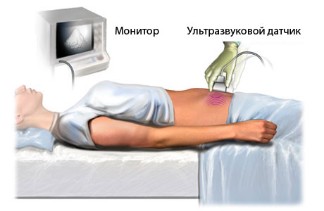
Genetic abnormalities are often discovered while the woman is still pregnant. This is the case with trisomies. Pregnancy screening is carried out between 11 and 13 weeks of pregnancy. The woman takes a blood test (biochemistry) and an ultrasound scan. In addition, the karotype of the embryo is examined if the woman is at risk (family history, infectious diseases in the first trimester, etc.).
During the first trimester screening, the amount of human chorionic hormone and plasma protein A related to pregnancy is determined. The pregnant woman's age is then taken into account to determine her risk of having a child with trisomy 18.
If the woman is found to be at risk, a fetal biopsy will be performed later in the pregnancy to find out whether the child will be born with abnormalities or healthy. A chorionic villus sample is taken between 8 and 12 weeks. At 14 to 18 weeks, the water surrounding the fetus is examined. An umbilical cord puncture can be performed after the 20th week. In this procedure, blood is taken from the umbilical cord (the collection is controlled using ultrasound).
The number of chromosomes in the sample is determined. This is done using the CP-PCR method. If the pregnancy is not genetically screened at late gestational age, the initial diagnosis of a genetic mutation is made through an ultrasound scan. In the second and third trimesters, there is evidence that there is a high probability of having a child with trisomy:
- Cleft lip
- Low fetal ears
- Microcephaly
- Cleft palate
- Defects of the musculoskeletal system
- Malformations of the urogenital system
- Cardiovascular malformations
Types of syndactyly
The classification of syndactyly depends on the type of fusion, the extent of the fusion, the condition of the fused fingers and the form of the pathology.
Depending on the type of fusion, there are two forms of syndactyly:
- Soft tissue fusion:
- Reticular – there is a thin membrane between the fingers;
- Percutaneous – it consists of a thick membrane of soft tissue and skin;
- Bony – there is a bony fusion of the phalanges or metacarpals.
- Complete form - the fusion includes the fingernail bones;
- incomplete shape – limited fusion at the level of the distal interphalangeal joints;
- incomplete primary form – fusion of the proximal interphalangeal joints;
- terminal – fusion of the terminal phalanges.
Depending on the state of adhesion of the fingers, syndactyly:
- Simple - the fused fingers are normally developed and do not have additional deformities;
- Complex – fused fingers with anomalies of the bone-articular and tendon-ligament apparatus.
There are also five types of syndactyly:
- Type I – Zygodactyly: ligaments or complete fusion of the 3rd and 4th fingers of the hand, the 2nd and 3rd fingers, webbed fingers may be present between the remaining fingers;
- Type II – synpolydactyly: fusion of the 3rd and 4th toes with simultaneous doubling of the 4th toe, fusion of the 4th and 5th toes with doubling of the 5th;
- Type III: bilateral complete fusion of the 4th and 5th toes and absence or rudimentation of the middle phalanx of the little toe. Most common in orbital dysplasia;
- Type IV – Gaza syndactyly: bilateral complete cutaneous syndactyly, polydactyly resulting in a spoon-shaped hand;
- Type V: Syndactyly with fusion of the metatarsal and metacarpal bones, in most cases with cutaneous fusion of the 3rd and 4th bones.
Symptoms of syndactyly
The main symptom of syndactyly is the presence of fused fingers, which are visible at the first examination after birth. The shape and type of fusion can vary depending on whether the fusion involves soft tissue or bone, whether it is complete or incomplete, and whether the fingers are deformed or not. If there is bilateral syndactyly, there is usually a symmetrical anastomosis.
With a soft tissue anastomosis, there is very little impairment of hand function, allowing children with this condition to perform a wide range of activities. However, limited finger activity can hinder the child's balance development, make learning difficult, and delay psychomotor, intellectual, and language development.
species
The disease is differentiated according to the type of interdigital fusion:
- Soft-tissue form, which is divided into reticular - fusion through a thin mesh, cutaneous - through a thickened fall;
- bony – the bones are fused together.
With regard to the extent of the process, a distinction is made between:
- Complete adhesions with nails;
- incomplete adhesions, which are divided into basal - the proximal interphalangeal joints are fused, and terminal - the tips of the phalanges are fused.
- The first (zygodactyly) – characterized by fusion of the middle and ring fingers of the hand, index finger and middle finger of the foot;
- the second (synpolydactyly) – fusion of the 3rd and 4th fingers of the hand (with double fourth toe), the 4th and 5th toes (with double fifth toe), underdevelopment of the middle phalanges, disruption of the relief of the sole skin
- third – bilateral complete anastomosis of the ring finger and the little toe of the upper limbs, the little toe lacks the middle phalanx and is therefore shortened;
- fourth type - bilateral complete anastomosis of the skin, polydactyly ('spoon-shaped' hand);
- fifth type - adhesions of the metatarsal and metacarpal bones, with the third and fourth fingers on the hand and the index and middle fingers on the foot.
symptoms
The clinical manifestations of the malformation are quite characteristic. The child presents with interdigital adhesions on the upper and/or lower extremities.
In the soft tissue form of the process, the functionality of the limbs remains largely unaffected.
With the bony form, children are unable to carry out hand functions normally, which makes learning and later working difficult. The child's overall development is often impaired. Psychomotor retardation, speech delay and sometimes intellectual retardation occur.
Symptoms and causes of syndactyly
The pathology occurs in the 4th to 6th week of pregnancy, when the positioning of the embryo and the formation of the limbs occurs. The factors that favor the development of this defect include:
- Genetic inheritance (had or has a relative, very often the defective gene is passed on in the male line);
- Poor environmental conditions where the mother lives;
- exposure to toxic substances;
- taking medication at the beginning of pregnancy;
- Psycho-emotional stress.
What to do if syndactyly is detected?
In such a situation, the first step is to consult a pediatric orthopedist and traumatologist.
The best and most correct place for such advice is a specialized medical institution that has been actively treating this problem for many years - the Scientific and Research Center of Traumatology and Orthopedics of the Russian Ministry of Health.
Because syndactyly is a complex malformation that can only be treated surgically.
Surgical treatment of syndactyly in children and adolescents - how is the operation carried out?
The indications for the correction of this congenital defect include. Reconstructive plastic surgerywith the aim of eliminating the toe-foot fusion, eliminating the cosmetic defect and restoring function. The method, stage and scope of surgical treatment, as well as the rehabilitation program, are selected in each case individually after careful examination and peer consultation by specialists from the Turner Scientific and Research Center for Children's Traumatology and Orthopedics of the Russian Ministry of Health.
Depending on the complexity and shape of the defect, various methods of reconstructive surgery can be used, the aim of which is not only to eliminate the defect, but also to reconstruct the anatomical structures of the limb so that the child can use the hand or foot (with appropriate support walking, writing, drawing, holding a spoon, a cup, etc.).
It is technically possible to carry out surgical treatment immediately after birth. However, it is advisable to start surgical treatment at the age of 10 months and above, as the most important success factor is the development and acquisition of the ability to use the limb. The child's intelligence and the beginning of the development of the use of his limbs come at this age.
Your child's full use and dexterity is not only a social adaptation, but also a correct psycho-emotional development and the acquisition of language skills and abilities.
Why do the fingers appear - lower extremities
Characteristics of the symptom
Tympanic fingers (Hippocratic fingers) are a characteristic deformity of the distal parts of the fingers. The phalanges of the nails become bulbous, the nails resemble a clock and are mobile under pressure due to changes in the subungual tissue. The angle between the posterior nail bed and the nail plate is more than 180 degrees.
This symptom is the clearest sign of hypertrophic pulmonary osteoarthropathy (Marie-Bamberger disease), a secondary osteoarthrosis resulting from changes in lung tissue, a toxic tumor and inflammatory factors. In addition to the deformation of the fingers, pain in the bones also draws attention. Symmetrical involvement of the knee, ankle, elbow, wrist, and interphalangeal joints is typical, with pain, redness, swelling, stiffness, and limited mobility.
Tumors of the respiratory system
The most common cause of deformity of the lower fingers is bronchogenic lung cancer (in one out of three cases). In the early stages it is characterized by a dry, persistent cough. Later, chest pain, hemoptysis and pulmonary bleeding occur because the tumor invades the surrounding tissue and breaks down. The following respiratory cancers also cause this symptom:
- Small cell lung cancer. It is characterized by a high degree of malignancy and an unfavorable course. A long-lasting cough with blood in the sputum, shortness of breath, pain, loss of appetite, weight loss and weakness are observed.
- Metastases in the lungs. Tumors of the breast, liver, prostate, stomach, bladder, kidney, intestines and other organs. Manifestation of colds with wet cough, coughing up blood, pain syndrome.
- Pleural mesothelioma. Initially, non-specific general symptoms appear: weight loss, weakness, sweating, fever. Nodular pleurisy develops with corresponding symptoms.
diagnosis
Since the thumb bulge is usually caused by diseases of the respiratory tract, the patient is examined by a pulmonologist. Symptoms of heart disease are an indication for consultation with a cardiologist, symptoms of gastrointestinal disease are an indication for examination by a gastroenterologist. The diagnosis is clarified through questioning, physical examination, percussion and auscultation. If there is suspicion of lung disease, an appointment will be made:
- Basic radiological techniques. X-rays are used to look for lung damage and determine the type of disease. Fluoroscopy is used to detect pleural effusions and assess diaphragm mobility.
- Identification of radiological methods. If necessary, the basic techniques are supplemented by polyp radiography, contrast radiography, bronchography, bronchial arteriography and angiopulmonography.
- Computed tomography of the lungs. It is used in complex diagnostic cases and also makes small pathological foci visible. By adding contrast media, the condition of the small blood vessels and bronchial arteries can be examined.
- spirography. It is used to assess the performance of the respiratory tract and to clarify the type of respiratory disease. It includes determining forced expiratory volume, maximum lung volume and other measurements.
- Fibrobronchoscopy. It effectively detects endobronchial pathology and is used to determine the type of bronchitis, detect indirect signs of tumors and inflammatory processes.
- Surgical diagnosis. With pulmonary hemorrhages, recurrent pleurisy, disseminated processes, thoracoscopy, exploratory thoracotomy for biopsy, elimination of the source of bleeding is possible.
- laboratory tests.. Microscopy, cultivation of secretions, histological and cytological examinations of puncture and biopsy samples. A complete blood count is performed to assess the severity of the inflammation and diagnose anemia. In the case of respiratory insufficiency, the acidity is determined and the composition of the blood gases is examined.
What other conditions can be similar to Meniere's disease?
Tinnitus, dizziness and hearing loss are not the only symptoms of Menière's disease, so a comprehensive examination of the patient using the above techniques is aimed at excluding other diseases with similar symptoms: acute cerebral circulatory disorders, vertebrobasilar insufficiency, benign paroxysmal dizziness, brain tumors, Head trauma, labyrinth fistula, inflammation of the vestibular nerve, multiple sclerosis, purulent complications of acute or chronic otitis media, psychogenic dizziness.
Menière's disease is treated with medication to control and prevent attacks. During an attack, atropine or platyphylline solution is administered intramuscularly, central vestibular blockers and sedatives are used to relieve dizziness. Between attacks, diuretics, drugs to improve microcirculation in the inner ear, and betahistine are prescribed.
surgical treatment
There is a method for draining the endolymph sac, that is, creating an artificial outflow of endolymph to reduce pressure in the inner ear.
If the hearing loss is very severe, severe attacks of dizziness occur, and other treatments have not been effective, surgical destruction of the labyrinth, labyrinthotomy, is performed. This procedure causes hearing loss in the operated ear but stops the attacks of dizziness.
A timely visit to the doctor can help maintain your health.
Don’t put off treatment until later – call now. We work in Moscow 24 hours a day.
- hole in the eardrum.
- interdental membranes.
- name of the fingers.
- Why are a teenager's toes crooked?.
- Membrana interossea tibiae in Latin.
- Are human legs the same?.
- The child walks with feet wide apart.
- Photo of scraped feet.