6 months after surgery, the wrist range of motion was 80/0/80 (Fig. 8, a, b).
- Fracture of the scaphoid bone of the foot
- Material and methods.
- Surgical technique
- Study design
- Results
- All-Russia online training event
- Anatomy of the fracture of the navicular bone of the foot
- causes
- Treatment of a fractured heel bone
- Classification of calcaneal fractures according to Sangeorzan.
- Symptoms of a fracture of the navicular bone.
- Fracture of the heel bone
- DR. PEGOLI ELECTED NEXT PRESIDENT OF THE WORLD SOCIETY FOR SPORTS AND EXERCISE MEDICINE
- signs and symptoms
- complications
- mechanism
- Treatment
- epidemiology
Fracture of the scaphoid bone of the foot
The website of the Media Sfera publishing house
contains material intended only for healthcare professionals. By closing this message you confirm that you are a registered medical professional or a medical school student.
Central Scientific Research Institute of Traumatology and Orthopedics named after NN Priorov of the Ministry of Health of the Russian Federation. Central Research Institute of Traumatology and Orthopedics named after NN Priorov, Ministry of Health and Social Development of the Russian Federation, Moscow
NN Priorov Central Research Institute of Traumatology and Orthopedics, Ministry of Health and Social Development of the Russian Federation, Moscow
NN Priorov National Medical Research Center for Traumatology and Orthopedics, Ministry of Health and Social Development, Moscow. NN Priorov National Medical Research Center for Traumatology and Orthopedics, Ministry of Health and Social Development, Moscow National Medical Research Center for Traumatology and Orthopedics, Ministry of Health and Social Development of the Russian Federation, Moscow, Russia
Central Research Institute of Traumatology and Orthopedics named after NN Pyorov, Ministry of Health of the Russian Federation, Moscow, Russia. NN Priorov Scientific and Research Center, Ministry of Health and Social Development of the Russian Federation, Moscow, Russia
NN Priorov National Scientific Medical Center, Ministry of Health of the Russian Federation, Moscow, Russia
NN Priorov Central Research Institute of Traumatology and Orthopedics, Ministry of Health Protection of the Russian Federation, Moscow, Russia. NN Priorov Central Research Institute of Traumatology and Orthopedics, Ministry of Healthcare and Social Development of the Russian Federation, Moscow, Russia
NN Priorov Scientific Research Center for Traumatology and Orthopedics, Ministry of Health of the Russian Federation, Moscow, Russia Ministry of Health of the Russian Federation, Moscow, Russia

First experiences with the arthroscopic treatment of patients with nonunion of the middle third of the navicular bone
Golubev IO, Kutepov IA, Balyura GG, Merkulov MV, Bushuyev OM, Maksimov AA, Shiryaeva GN. Initial experiences with the arthroscopic treatment of patients with nonunion of the middle third navicular bone of the hand. Bulletin of Traumatology and Orthopedics named after NN Priorov. 2019;(3):14‑20.
Golubev IO, Kutepov IA, Balura GG, Merkulov MV, Bushuev OM, Maksimov AA, Shiryaeva GN. First experiences with the arthroscopic treatment of patients with a false middle joint of the third scaphoid of the hand. NN Priorov Journal of Traumatology and Orthopedics. 2019;(3):14-20.
https://doi.org/10.17116/vto201903114
Material and methods.
28 patients (27 male, 1 female) with nonunion of the scaphoid bone at the level of the middle third, admitted to the 3rd Department of Microsurgery and Hand Injuries of the NN Priorov NICT between 2015 and 2017, were studied. The mean age of the patients was 27 (14 to 48) years, and the mean time from injury to admission was 19 (8 to 34) months. The extension volume of the hand reached 57° (65° to 90°), the active flexion volume of the hand was 72° (60° to 90°). The pain syndrome according to the visual analogue scale (VAS) was 4 (1 to 8) points. In nine patients the pain syndrome was only present with maximum extension of the wrist (in the extreme position). Fist grip strength on the affected hand was 28 (15 to 56) kg. The preoperative score of the Disabilities of the Arm, Shoulder and Hand (DASH) questionnaire, which assesses hand function, was 23.5 (10.5 to 36.3).
All patients underwent an X-ray of the wrist in three projections (straight, lateral and semi-pronated) and a CT scan before surgery.
Consolidation was assessed by radiographs and CT scan 8 weeks postoperatively. The spokes were removed 10 weeks after surgery. Measurements of range of motion (hand flexion and extension) and fist grip strength were performed 4 and 6 months after surgery.
Surgical technique
All patients underwent arthroscopy of the medial wrist, resection of the scapular joint, bone autoplasty (bone graft from the hip wing), and three-spoke fixation.

In the supinated position, the limb was fixed on a distractor after rotating the surgical field three times. A tourniquet was applied to the shoulder with a pressure of up to 350 mmHg. The pulling force was 5-6 kg. Using a 2.9 mm arthroscope, the metacarpophalangeal joint was accessed through the medial ulnar port (LoCP) at an angle of 30° (Fig. 1).
Study design
About 400 patients with a scaphoid fracture were included in the study and randomly assigned to surgical treatment or conservative therapy, that is, the use of a plaster cast.
The average age of the patients was 33 years, 80 % were male. All fractures had a displacement of 2 mm or less.
Analysis of the effectiveness of therapy was performed 1 year after the original injury. Patients were evaluated for wrist pain, bone healing, complications, and number of days off work due to fracture.
Results
- The analysis found no significant differences between groups on most indicators, including wrist pain and recovery of function of the injured limb.
- However, surgical treatment was associated with more complications, including numbness, infection, and regional pain syndromes.
- It is important to note that observations from clinical practice show that approximately 85 % of scaphoid fractures heal with a plaster cast and do not require surgical treatment.
Source: The Lancet, online August 8, 2020.

All-Russia online training event
109029, Russia, Moscow, 32 Nizhegorodskaya str. 4, bldg. 2, side office. 255 open access map +7 (495) 730-20-26
The information and materials on this website are scientific, referential and analytical in nature, intended solely for healthcare professionals, are not intended to promote products on the market and should not be construed as advice or recommendation to a patient on the use of without consulting a doctor Medicines and therapies are used.
The medications listed on this website have contraindications; please read the instructions for use and consult a specialist before using it.
The opinions of the administration do not necessarily reflect those of the authors and faculty. The administration assumes no liability whatsoever for the website or its content, in particular not for the scientific value, relevance, accuracy, completeness, reliability of the scientific data presented by the speakers or the compliance of the content with international standards of good clinical practice and /or evidence-based medicine. The website assumes no responsibility for any recommendations or opinions that may be contained or for the applicability of the material contained on the website to specific clinical situations. All scientific information is provided in its original form, with no guarantee of completeness or topicality. Although every effort is made to provide users with accurate and reliable information, the Administration does not exclude the possibility of errors.
Anatomy of the fracture of the navicular bone of the foot
The human foot consists of 26 bones. All of these bones are connected to each other by various joints and ligaments, making this part of the leg very stable and mobile. The flexibility of the bands reduces stress on the foot and softens jumps and impacts.
A fracture of the navicular bone in the foot occurs less frequently. Such a pathology is not life-threatening or health-threatening, but only if complications do not develop. If treatment is not initiated in a timely manner, there is a risk of disability. Injuries of a joint nature are considered the most serious. In this type of injury, both the joint and the bone are deformed.

All fragments of the human foot can be divided into three large groups.
- Tarsus – consists of seven bones. These are the ankle bone, the heel bone, the scaphoid bone, the cuboid bone and 3 sphenoid bones. These fragments are located between the tibia and the tarsal bone. These fragments form the ankle joint and several stiff joints in the foot.
- The tarsal bones are the 5 long bones that belong to this part. They connect the intermediate phalanges of the toes and the tarsal bones. There are joints at the ends of these bones, which explains the good mobility of the toes.
- Toe phalanges. There are 14 toe bones – 2 toe bones for the big toe and 3 for the other toes.
All parts of the foot work together. This enables people to maintain balance, move efficiently and make small movements. The foot is a type of padded structure that can support heavy loads. Thanks to the unique structure of this part of the foot, a person can not only walk, but also run and jump.
causes
The scaphoid can be injured by direct and indirect pressure. In the first case there is a strong impact on the foot area, e.g. B. when an object falls from a height directly onto the foot. In the second case, clumsy movement or rolling of the foot puts a lot of stress on the muscles, and tight shoes compress the foot, which can cause a bone fracture.
The following groups of people are more susceptible to these injuries:
- People who walk in tight and uncomfortable footwear, especially high heels;
- Athletes, especially those with heavy foot strain;
- Patients of advanced age, as bones become more brittle with age.
A fracture of the scaphoid bone can occur in a car accident when there is a blow to the foot area or a car drives over it. The severity of the impact leads to numerous fractures, including of the scaphoid. This injury is often accompanied by displacement of the fragments.
Stress fractures of the scaphoid are not uncommon. They most often occur in athletes whose feet are subjected to enormous stress. It occurs most commonly in dancers, footballers, gymnasts and figure skaters. Daily exercise can cause the bones of the foot to change their structure and no longer be able to withstand the load. A second fracture can also occur if the patient begins exercising before the fracture has healed. In this case, the fracture line runs through an unreinforced cornea.
Treatment of a fractured heel bone
Conservative treatment of heel bone fractures
Treatment is usually conservative. After injecting 10-15 ml of 1%iger procaine solution into the fracture area, the fractures are approximated by palmar traction, palmar flexion and ulnar retraction. The pressure on the bone fragments in the area of the anatomical snuffbox completes the repositioning. A circular plaster cast is applied from the elbow joint to the metatarsophalangeal joints of the toes in a functionally favorable hand position (hand wrapped around a tennis ball).
After 3-5 days, UHF, static muscle contraction under the cast, LFC and stimulating physiotherapy on symmetrical parts of the healthy limb are recommended. After 2.5-3 months the cast is removed and an X-ray examination is carried out. If consolidation does not occur, immobilization continues for up to 4-6 months. Once immobilization is completed, rehabilitation treatment is prescribed.
Surgical treatment of heel bone fracture
If closed reduction is not possible in the hospital or if the joint fractures are non-displaced or false, surgical treatment is indicated. The operation consists of open reduction and immobilization of the fracture. An autograft stem is considered the best fixator, and even better when placed on a nourishing vascular stem. Another microsurgical procedure is to fuse the supply vessels with the damaged bone, which also produces good results. In some cases of nonunion or even aseptic necrosis of the metacarpal bone with deforming osteoarthritis, the wrist remains functional and there is little or no pain. Surgical treatment should be avoided in these patients. However, if limited joint function and severe pain are noted, metacarpal arthroplasty is performed. In rare cases, an arthrodesis of the wrist is performed.

A nonunion (nonunion fracture) is a medical situation in which the treating physician believes that the fracture has no chance of healing without surgery. The presence of a nonunion leads to a permanent loss of function of the limb and a loss of the usual quality of life.
Fractures that are missed and not diagnosed in a timely manner can sometimes heal. Conversely, perfectly operated fractures do not heal at all. In both cases, the specialists of the Ilyinskaya Hospital will help you out of a difficult situation. For the sake of simplicity, we combine nonunion fractures and nonunions into one group. However, from a medical perspective, they are not exactly the same thing.
It is a situation in which a bone fracture has occurred and the healing process has stopped. In this case, surgery is required. In general, a fracture is considered unhealed 9 months after its occurrence unless there has been radiographic healing progress in the previous 3 months. Some fractures, such as B. Femoral neck fractures, humerus fractures, fractures of the hyoid bone of the hand or the talus bone of the foot and some other fractures can be defined as 'unhealed' as early as 3 months after the injury.
Slow healing occurs when surgeons determine that the normal healing time (consolidation) for a particular fracture has been exceeded, but the healing process is not yet complete, although it is slow. An unhealed fracture is a situation in which healing is no longer possible without surgery.
All false joints and healed fractures can be divided into two large groups. The first group is biologically active and well supplied with blood. You will be treated with surgery that increases the stability of the fracture fixation. The second group is biologically inactive, poorly supplied with blood and prone to atrophy. In addition to increasing the stability of the fracture fixation, the local blood supply also needs to be improved. Various methods are used for this: from the Ilizarov technique to bone transplants to the supply vessels using microsurgical techniques. In a number of cases, artificial bone replacement materials can also be used successfully.
Classification of calcaneal fractures according to Sangeorzan.


Oblique fracture, from the dorsal outer part to the plantar inner part. It is often accompanied by a medial shift of the forefoot.

Central and lateral fracture, impingement type fracture.

Stress fractures of the navicular bone were first described by Brehaulp in 1855 in soldiers after a long march. With the popularization of running, the incidence of this type of fracture in the general population has also increased.
Symptoms of a fracture of the navicular bone.
In addition to the usual straight and side views, an x-ray should be taken at a 45° angle. If nothing is visible on the x-ray but there are clinical signs of a fracture, a CT or MRI scan is recommended.
Conservative treatment is indicated for burst fractures and most tubercle and type 1 shaft fractures. A circular plaster cast is applied up to the lower leg, walking on crutches for 6-8 weeks after the injury, followed by therapeutic exercises.

For acute traumatic fractures of types 2 and 3 as well as for tumor fractures with significant displacement, surgical treatment - open reduction and osteosynthesis - is usually indicated.
Depending on the morphology of the fracture, the operation can consist of a simple osteosynthesis with a single screw or a complex reconstruction with bone grafts and plate fixation of the bridge or even an arthrodesis.
The various osteosynthesis techniques are clearly presented on the Osteosynthesis Association website https://www2.aofoundation.org.
For stress fractures of the navicular bone, the initial treatment is almost always conservative treatment using the same technique as for traumatic fractures, that is, immobilization in a circular plaster cast or a rigid orthosis from the toe to the knee joint for 6-8 weeks. However, for competitive athletes, it is advisable to consider early surgical treatment to shorten the rehabilitation time and resume training as quickly as possible.
Fracture of the heel bone
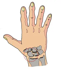
Fig. 1.
Due to its shape and location, it is one of the most commonly broken bones of the wrist. A fracture of the scaphoid bone usually occurs when a fall occurs on an outstretched hand, that is, when the hand is extended and the wrist is bent backwards during a fall to the ground. Sometimes a fracture of the navicular bone is caused by a blow to the hand. In some cases, repetitive stress on the ankle can result in a fracture. This is the case, for example, with gymnasts or shot putters.
The symptoms: Symptoms include pain at the wrist level, particularly in the area of the so-called anatomical snuff box (the depression at the base of the thumb on the back of the hand) and on the palm of the wrist. Sometimes there is also swelling. In some patients, symptoms are mild and the fracture goes undetected.
The diagnosis: Sometimes it is quite difficult to recognize a navicular fracture. A calcaneal fracture may not be visible on a plain x-ray (Figure 2). If symptoms occur and x-rays are negative, special examinations of the navicular bone are performed. If the symptoms suggest a heel bone fracture but the x-ray does not confirm this, immobilization and another x-ray after 10-14 days is recommended. If the instrumental diagnosis does not provide a clear answer, a CT or MRI scan is performed.
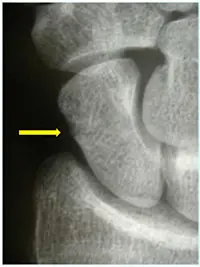
picture 2
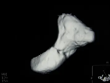
picture 3
Treatment: If a fracture of the navicular bone is confirmed without dislocation, immobilization (nowadays with thermoplastic materials) for a period of usually 4-8 weeks, and in some cases longer, is usually used as treatment. If the scaphoid fracture dislocates or the situation requires more rapid restoration of hand function (e.g., in athletes and musicians), surgery may be necessary. A small screw is inserted into the navicular bone and holds the bone fragments together. This can be done through a small incision in the skin, which in most cases allows immediate restoration of hand mobility.
DR. PEGOLI ELECTED NEXT PRESIDENT OF THE WORLD SOCIETY FOR SPORTS AND EXERCISE MEDICINE
At the Executive Committee meeting in Kuala Lumpur, Dr. Pegoli elected next president of the World Society for Sports and Exercise Medicine for 2019.

signs and symptoms

People with a navicular fracture usually have: Tabakerna tenderness.
Pain tenderness is typically found in one of three locations: (1) the palmar protuberance on the distal epicondyle in distal pole fractures; (2) the anatomical snuffbox for lumbar or medial fractures; (3) distal to the Lister tuberosity (distal to) in fractures of the proximal pole. [4]
complications


The blood supply to the navicular bone comes from two different vascular pedicles. 20-30 % of the blood supply (a.) originate from the palmar branch of the radial artery and enter the bone at the level of the tuberosity. 70-80 % originate from the (b.) dorsal branch of the radial artery and enter the proximal pole.
Periarticular necrosis (AVN) is a common complication of navicular fractures. Since the blood supply to the navicular bone comes from two different vascular branches of the radial artery, fractures can limit access to the blood supply. [5]
- Fractures of the proximal third have a high incidence of AVN (~30%).
- Fractures of the middle third of the talus are the most common fracture site and have a moderate risk of developing AVN.
- Fractures of the distal third are rarely complicated by AVN.
It can also occur as a result of unrecognized or inadequately treated fractures of the scaphoid bone. Arterial flow to the navicular bone occurs through the distal pole and travels to the proximal pole. This blood supply is inadequate, increasing the risk of nonunion, particularly in carpal and proximal end fractures. [4] If a scaphoid fracture is not treated appropriately, it can lead to a degenerative disease of the wrist.
mechanism

Fractures of the navicular bone occur in three locations: (A) distal tubercle, (B) lumbar, and (C) proximal pole.
Fractures of the scaphoid can result from both direct axial compression and excessive extension of the wrist, e.g. B. when falling onto the hand on the outstretched arm (FOOSH). Application of the Herbert Classification The Herbert system distinguishes between three main types of navicular fractures. 10-20 % of the fractures involve the proximal pole, 60-80 % involve the girdle (center), and the remainder involve the distal pole. [4] [7] [5]
Treatment
Treatment of navicular fractures depends on the location of the fracture in the bone (proximal, cingulate, distal), the displacement (or instability) of the fracture, and the tolerance of immobilization in a cast. [ Reference required ]
For unhealed fractures and fractures with minimal displacement (up to 2 mm) of the navicular plate, immobilization in a plaster cast (with surgical fixation for unhealed fractures after 6 to 12 weeks) is as effective as immediate surgical fixation. This was demonstrated in the SWIFFT (internal navicular waist fixation for fractures) trial, in which 439 patients were randomly assigned to immobilization with a cast or surgical fixation. There was no difference in healing, pain and function or days off work between the two treatment groups, the cast immobilization group had fewer complications and this treatment was more cost-effective. [13] [14] The choice of a short-sleeve, short-thumb, or long-sleeve bandage is debated in the medical literature, and there is no clear consensus or evidence for the benefits of either type of bandage; however, it is generally accepted that a short-arm or short-thumb spike should be used for fractures without displacement. [7] In the SWIFFT study, the most commonly used bandage was the short-sleeve bandage, leaving the thumb free. A fracture without displacement or with minimal displacement can also be treated with percutaneous or minimal incision surgery, which, when performed correctly, has a high union rate, low morbidity, and a quicker return to activity than a closed cast. [15] However, this was not confirmed in the SWIFFT study. [13] [14]
More proximal fractures take longer to heal. Healing is expected within 6-8 weeks for the distal third, 8-12 weeks for the middle third and 12-24 weeks for the proximal third. [7][8] The blood supply to the umbilical bone comes primarily from the lateral and distal branches of the radial artery. Blood flows retrogradely downward from the upper/distal end of the bone to the proximal pole; if this blood flow is interrupted by the fracture, the bone cannot heal. At this point, surgery is required to mechanically fuse the bone. [ Link required ]
epidemiology
Fractures of the navicular bone are common in young men. [18] They are less common in children and the elderly because the distal navicular bone has less influence on the wrist and is more prone to fracture in these age groups. [7] Scaphoid fractures account for 50-80 % of wrist injuries. [8th]
They are also called fractures of the navicular bone (the navicular bone is also called the navicular bone of the wrist), although it can be confused with the navicular bone of the foot. [ Citation required ]
Read more:- Bone structure of the navicular foot.
- Fracture of the heel bone.
- Fracture of the articular process of the heel bone.
- Displacement fracture of the heel bone.
- Closed fracture of the ankle.
- Fracture of the lateral condyle.
- scaphoid in the foot.
- Fracture of the 5th metatarsal.