If you wear calf-high boots, you don't have to try to take them off. Simply loosen or untie the laces. Only when the shoe puts pressure on the ankle can you try to pull it slightly, but it's better not to do that. The limb should be elevated to reduce swelling. Painkillers can be taken.

Broken ankle

Each ankle joint consists of an external, lateral malleolus and an internal, medial malleolus; these are different parts of the fibula and tibia. From the outside we see them as two thickenings on the inside and outside of the ankle joint.
The two ankles form the 'fork' of the ankle joint, through which the weight of the human body is transferred to the foot. A fracture is a break in the integrity of the bone and can affect various anatomical structures. It is possible to break just the medial ankle, just the medial ankle, or both.
If the bones are dislocated or broken, the fracture is considered complicated. Subluxation of the joint can further aggravate the situation. Sometimes the fragments break through the skin and tissue - in this case it is an open fracture.
Treatment of an ankle fracture
If the bones are not displaced and there are no other complications, bone fragments or torn ligaments, a plaster cast is usually sufficient for about 4 to 8 weeks. In the case of an uncomplicated fracture, modern orthoses and bandages can also be used to immobilize the leg. They are made of durable plastic or metal, covered with fabric and attached with Velcro. The bandage can be adjusted to the size of the leg and can be removed and the skin rubbed off if necessary, but this must not be done without the doctor's approval.
In the case of a closed fracture and dislocation, a reduction, that is, a reduction of the joint, is carried out before applying the bandage. This is done in the hospital under local anesthesia. A plaster cast is then applied.

Surgical treatment is necessary if the bone cannot be repaired without surgery or if there is a subluxation and other complications. Surgery is considered very effective, the ankle heals faster and complications are rare. The surgery is usually performed a few days after the injury.
Regardless of the method used to return the bones to their correct position, rest must be maintained. There is no walking on the foot and walking with crutches is required.
diagnosis
Ankle fractures must be differentiated from ligament injuries. For an accurate diagnosis, an x-ray of the ankle joint is taken. On the x-ray, the traumatologist can see exactly how the fractures have moved and how extensive the injury is.
The x-ray shows mainly the dense tissue, while the condition of the soft tissue is difficult to assess. In cases of doubt, additional CT and MRI examinations are ordered.
Current treatment methods
After the ankle joint is restored to the correct position, it takes a long time for all the damaged tissue to recover. Physiotherapy can help speed up the healing process. It improves blood circulation, facilitates lymphatic drainage and reduces swelling.
Definition and type of technology
Radiography is the main method of radiological examination, which involves taking an x-ray: a shaded image of the organs on x-ray film.
The examination is carried out using medical x-ray machines. The X-rays they produce penetrate the human body and are recorded by the system. The analog devices then create an image on X-ray film, which needs to be developed. More modern digital X-ray systems have a sensitive detector that immediately transmits the X-ray image to a computer screen.
What do the x-rays show?
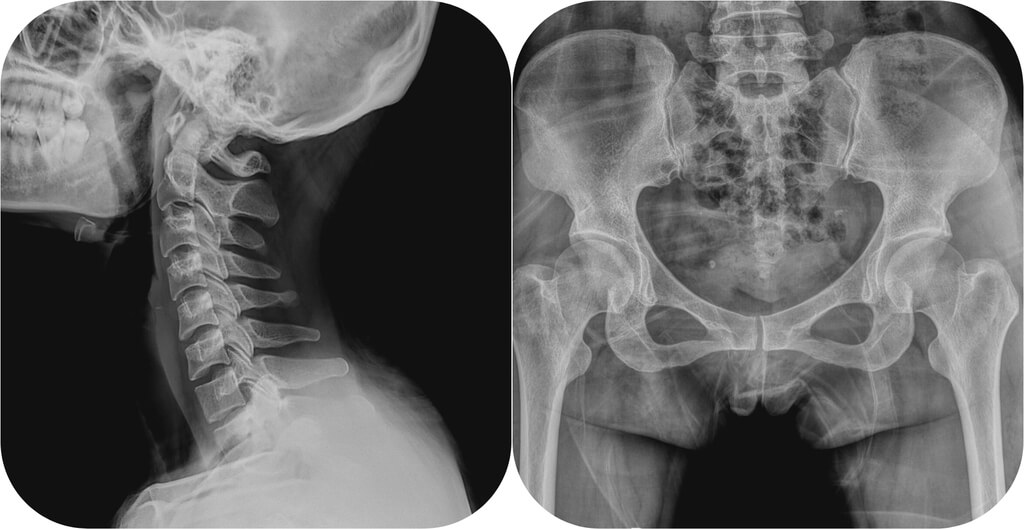
The doctor sees shadows of varying intensity on the x-ray: the areas where bones are replaced are white, the soft tissues are gray; the lungs look black on the x-ray. X-ray images are high-contrast because different tissues receive different X-rays: the denser the tissue, the brighter it appears on the X-ray image.

X-ray images are inherently negatives, so the lighter areas on them are called opacities. For example, a dense and bright area of pneumonia against the background of a 'dark' lung surrounded by air is referred to by the doctor as a shadow. A bone fracture will be visible as a darker 'break' on a light 'field' of bone.
The shadow images created by X-rays provide the doctor with information about the condition of various organs (lungs, heart, stomach, lymph nodes, bones, spine, etc.) and allow the detection of various pathologies: foci of inflammation, destruction (destruction), dystrophy, tumor nodes, abnormal organ development.
Read more:- fracture of the elbow in the foot.
- Structure of the human ankle.
- X-ray anatomy of the ankle.
- dislocation of the ankle.
- Treated subluxation of the ankle.
- The ankle bruise is where the picture is taken.
- contusion of the ankle.
- pelvic subluxation.
