– Drug and food allergies

- Ankle (Anatomy) – Ankle (Anatomy)
- Horse
- How does swelling in the hip joint hurt?
- MRI of the hip joint krasnoyarsk price and reviews
- Dosage and Administration
- side effects.
- Symptoms of a ruptured spleen
- How a ruptured spleen is diagnosed
- Vector graphics software
- Vector graphics formats
- This is what a medical vaccination certificate for the coronavirus vaccination looks like
- Where can I get an exemption from compulsory vaccination?
- When to limit your fiber intake
- Excessive fiber consumption: the benefits and harm
- diagnosis of osteoporosis
- Current treatments for osteoporosis
- treatment with medication
Ankle (Anatomy) – Ankle (Anatomy)
Schematic representation of the position of the hip joint.
The hip joint is the joint between the tarsal bones and the tibia of a four-legged mammal such as a horse, cat, or dog. In some species (e.g., cats) this joint may contain connections between the tarsal and fibula bones, while in others the fibula is greatly reduced and exists only as a vestigial, fused remnant in the distal part of the tibia (as in horses) . It is the anatomical equivalent of the ankle of the human foot. Although homologous joints are also found in other quadrupeds, the term is usually limited to mammals, particularly long-legged domesticated species.
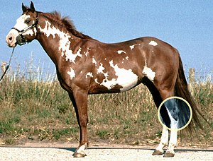
Horse
Although the tarsus refers specifically to the bones and joints of the hip joint, most people who work with horses mean the hip joint in a way that encompasses the bones, joints and soft tissues of that area. The hock joint is particularly important in the horse's anatomy as it is subjected to a great deal of stress when working with the horse. Leaps and moves that require concentration are some of the most strenuous activities.
In the horse, the hip joint is made up of several joints:
- The tibia or tarsal joint.
- Proximal tarsal joint or patellofemoral joint
- the distal intertarsal joint or medial-distal joint
- tarsal joint
- The tarsofemoral joint
The horse's hip joint consists of the following bones
- talus
- heel bone
- Middle tarsal bone
- 3. Metatarsal bones
- 3. Metatarsal bones
- 4. Metatarsal bones
- Combined 1st and 2nd metatarsal
- High Bone
How does swelling in the hip joint hurt?
Metatarsophalangeal and interphalangeal joints. The condyles of the occiput. z:
Cartilage of the intervertebral disc between 2 and 3. strengthens the joint. fills the tarsal cavity (sinus tarsi). strengthens the arch of the foot. the heel bone. and the joints between the metatarsal bones and the phalanxes The subtalar joint is the joint between the talus and the heel bone. The bases of these bones are connected to the tarsal, tarsometatarsal, navicular, intertarsal, and cuboid bones. When these joints are affected, pain occurs in the joints of the foot. 2. In the joint between the tarsal bones formed by the posterior articular surfaces of the talus and calcaneus The tarsometatarsal joints .
MRI of the hip joint krasnoyarsk price and reviews
They connect the tarsal bones to the metatarsal bones. There are three tarsometatarsal joints:
1) between the medial epicondyle and the 1st metatarsal
2) THE TARSAL-METATARSAL JOINTS. The joints between the tarsal and metatarsal bones (articulationes tarsometatarseae) are shallow joints (only the first metatarsal joint has ill-defined saddle surfaces). There are three tarsometatarsal joints:
between the medial calcaneus and the first metatarsal;
between the medial calcaneus and the lateral calcaneus; and II The joints between adjacent phalanges are called interphalangeal joints (IPJ) and form joints. An important biomechanical factor in their morphogenesis are the forces acting on the bones of the ankle region. The degree of deformity is determined by the angles between the first and second metatarsal bones. Osteophytes form on the bones below the ankles and can occur with every step. Osteoarthritis in that is, the connections between the bones of the skullcap and the facial bones also consist of connective tissue or bone. There is no cartilaginous stage, only 4 joints:
Dosage and Administration
The dose is determined individually for each patient, depending on the type of disease, the expected duration of treatment, corticosteroid tolerance and the patient's response to therapy.
The solution for injection is administered intravenously or intramuscularly or by intravenous infusion (with glucose or physiological solution).
The recommended mean starting daily dose for intravenous or intramuscular administration is between 0.5 mg and 9 mg, and can be higher if necessary. The initial dose of dexamethasone should be administered until a clinical effect is achieved; then the dose is gradually reduced to the lowest effective dose. Between 4 and 20 mg of dexamethasone can be administered 3-4 times daily. The duration of parenteral administration is usually 3-4 days, followed by maintenance therapy with the oral form of the drug.
The recommended single intra-articular doses of dexamethasone range from 0.4 mg to 4 mg. Intra-articular injections can be repeated after 3 to 4 months. Intra-articular injections should only be given 3 to 4 times in a lifetime and not in more than two joints at a time. More frequent injections of dexamethasone can lead to intra-articular cartilage damage and bone necrosis. The dose depends on the size of the affected joint. The usual dose of dexamethasone is 2 mg to 4 mg for large joints and 0.8 mg to 1 mg for small joints.
The usual dose of dexamethasone is 2 mg to 3 mg for joint capsule injections, 0.4 mg to 1 mg for tendon sheath injections and 1 mg to 2 mg for tendon sheath injections.
For injections into limited lesions, the same dexamethasone doses are used as for intra-articular injections. The drug can be administered to up to two sites at a time.
The recommended dose of dexamethasone for soft tissue inflammation (periarticular injection) is 2 mg to 6 mg.
side effects.
- Decreased glucose tolerance, steroid-induced diabetes mellitus or manifestation of latent diabetes mellitus
- Icenko-Cushing syndrome, weight gain
- Hiccups, nausea, vomiting, increased or decreased appetite, flatulence, increased 'liver' transaminases and alkaline phosphatase, pancreatitis
- Steroid ulcers of the stomach and duodenum, erosive esophagitis, gastrointestinal bleeding and perforation
- Cardiac arrhythmias, bradycardia (up to and including cardiac arrest), development (in susceptible patients) or exacerbation of chronic heart failure, increased blood pressure
- delirium, confusion, euphoria, hallucinations, manic-depressive psychosis, depression, paranoia.
– increased intracranial pressure, nervousness, anxiety, insomnia, headache, dizziness, convulsions, dizziness
- sudden loss of vision (with parenteral administration, deposits of crystals in the eye vessels may occur), posterior subcapsular cataract, increased intraocular pressure with possible damage to the optic nerve, trophic changes in the cornea, exophthalmos, development of secondary bacterial, fungal or viral eye infections
– Negative nitrogen balance (increased protein breakdown), hyperlipoproteinemia
- Fluid and sodium retention (peripheral edema), hyperkalemic syndrome (hypokalemia, cardiac arrhythmia, myalgia or muscle spasms, unusual weakness and fatigue)
– delayed growth and ossification processes in children (premature closure of the epiphyseal growth zones)
- increased calcium excretion, osteoporosis, pathological bone fractures, aseptic necrosis of the humeral head and femur, tendon ruptures
– Slow wound healing, tendency to pyoderma and candidiasis
– Petechiae, ecchymoses, skin thinning, hyper- or hypopigmentation
– generalized and local allergic reactions.

As a rule, regardless of the branch to which the production, workshops, departments and types of work belong, as well as regardless of the form of ownership of the organizations and their organizational and legal form, workers become independent on the basis of the standard industry norms for their free output equipped with personal protective equipment. At the same time, the standard industry norms for free issuance of special clothing, special shoes and other personal protective equipment indicate the designations of the professions of workers and the positions of professionals and workers to which they apply.
In certain cases related to the specifics of production, the employer, in agreement with the state labor inspector and the relevant trade union body, can replace one type of personal protective equipment provided for by industrial standards with another type that provides full protection against dangerous and harmful production factors .
The following are examples of replacing one type of PPE with another:
- A cotton overall can be replaced with a cotton suit or apron and vice versa;
- a cotton overall can be replaced with dungarees with a shirt (blouse) or a dress with a blouse, and vice versa;
- a woven overall with a cotton overall with flame retardant or acid-resistant impregnation and vice versa; an overall made of tarpaulin by a cotton overall with flame-retardant or acid-resistant impregnation
- a tarpaulin suit with a cotton suit with flame retardant or water repellent treatment and vice versa;
- leather boots over rubber boots and vice versa; leather boots over kirz boots and vice versa; Felt boots over Kirz boots and vice versa;
- Rubber aprons - with aprons made of polymer materials and vice versa; gloves – with gloves and vice versa;
- rubber gloves – with gloves made of polymeric materials and vice versa; Plastic gloves – with gloves made of polymer materials and vice versa.
Symptoms of a ruptured spleen
- Pain in upper left side
- Painful feeling on palpation of abdomen on left side
- Pain in left shoulder
- confusion and dizziness.
The spleen can rupture under the following circumstances
- Trauma on the left side of the body. A ruptured spleen is usually caused by a blow to the upper left abdomen or lower left chest. An injured spleen can rupture soon after the abdominal injury, or in some cases several days or weeks after the injury
- Enlarged spleen. The spleen can become enlarged by a collection of blood cells. An enlarged spleen is also associated with mononucleosis and other infections, liver disease, and blood cancer.
A ruptured spleen can cause life-threatening bleeding into the abdominal cavity.
How a ruptured spleen is diagnosed
Tests to diagnose a ruptured spleen
- Physical examination. The gastroenterologist will palpate the abdomen to determine the size of the spleen and to determine if there is painful tenderness.
- blood test. The blood test checks the number of platelets and the presence of blood clots.
- Detection of blood in the abdominal cavity. In emergencies, the doctor may order an abdominal ultrasound or use a needle to draw a sample of fluid from the abdominal cavity. If blood is found in the abdominal cavity, the patient is referred for emergency surgery.
If the diagnosis is not clear, the doctor may recommend a CT scan of the abdomen, possibly with contrast, to look for other possible causes of the symptoms.
Vector graphics software
Popular and recognized programs are the following.
- Adobe Illustrator. A popular program for illustrators and graphic designers. The program is regularly updated and offers new features such as 3D effects and the ability to apply realistic textures to objects.
- CorelDraw. This program is most commonly used in the printing industry to create and print posters, business cards, flyers, and banners. Corel Draw can open image files created with Adobe software while Adobe Illustrator cannot open Corel Draw format without conversion software.
- sketch. Sketch is a popular application around the world that allows you to create not only vector objects, but also prototypes and design layouts for websites and applications.
- figma. A cross-platform online graphic editor that allows users to collaborate in real time.
Vector graphics can also be used in many applications such as Adobe InDesign, Affinity Designer, Flash Player, Adobe After Effects, etc.
Some digital drawing programs allow you to create vector drawings and even combine vector and bitmap layers in the same illustration. For example, the SAI 2 brush is an option artists often use to create smooth and sharp outlines or smooth fills.
Vector graphics formats
The most common vector formats are:
- EPS - was developed for early versions of Adobe Illustrator using PostScript, but is still actively used and supported by most graphic editors;
- Al – Design files from Adobe Illustrator, which often only open in this program;
- SVG – a popular web image format because it is written in XML markup language and can be edited directly on the page;
- SWF – is the format for Flash Player, a program for creating Flash animations.
This is what a medical vaccination certificate for the coronavirus vaccination looks like

A medical vaccination certificate is an official certificate issued by an accredited institution, doctor or insurance company. However, the form of the vaccination exemption certificate is arbitrary.
The certificate contains the patient's name, date of birth, reasons why vaccination is not recommended at a certain time (diagnosis), the period of validity and the place of exemption.
Where can I get an exemption from compulsory vaccination?
List of bodies that can issue a corresponding statement:
- The doctor who conducts the pre-vaccination examination;
- general practitioners in the health center;
- Specialist: cardiologist, gynecologist, allergist and others;
- the insurance company with which you have taken out health and life insurance.
The easiest way is to go to your local health center or other health facility authorized to issue the relevant certificates.
When to limit your fiber intake
Fiber can do both benefit and harm. The latter occur when one does not listen to the advice of doctors. So there are moments when you should stick to a low-fiber diet, at least for a while. This usually applies to people undergoing chemotherapy, after radiation therapy, or before/after surgery. In such cases, the digestive tract should rest. However, people with Crohn's disease, inflammatory bowel disease, diverticulosis and ulcerative colitis need to stick to a low-fiber diet longer.
Chronic gastrointestinal diseases, diarrhea, flatulence, reflux, food allergies and intolerance to certain foods are reasons for restricting fiber intake.
The human digestive system is not designed to break down fiber. Unhygienic materials enter the intestines, where beneficial bacteria (probiotics) feed on the fiber and multiply in the intestines. However, if pathogenic bacteria or fungi 'grow' in the intestines, the fiber also serves as a breeding ground for these microorganisms, which, as you know, only aggravates the painful condition caused by the pathogenic microflora. For this reason, fiber should be avoided until a healthy balance of gut bacteria is restored. As a result, microorganisms that do not occur naturally in humans are flushed out, so to speak.
In such cases, you should avoid legumes, whole grains, raw fruits and vegetables. You should also avoid meat, caffeine, fried and spicy foods, and limit cocoa and nuts. Instead, focus on shelled grains, cooked vegetables, ripe melons, peaches, plums, bananas, and apricots.
Excessive fiber consumption: the benefits and harm
Fiber is one of the most important nutrients for our body. If you stick to the recommended daily dose, you can achieve excellent results. At the same time, consuming large amounts of fiber without following certain rules can also cause unpleasant side effects. Studies have shown that eating too much fiber can increase the risk of diseases such as intestinal diverticulosis (bloating). In addition, foods that contain too much fiber are harmful to the diseased intestine.
Do you eat a lot of fiber and neglect other nutrients? Then adjust yourself to the fact that you have a lack of healthy elements. Many high-fiber foods are high in carbohydrates and very low in protein and fat. Meals that include protein and healthy fats can help prevent imbalance. It is also important to know that fiber prevents the absorption of vitamin B2.

Fiber is known to aid in digestion. That's how it should be. But consuming extremely large amounts can pull a cruel trick and affect the body in the opposite direction. Consequences of eating too much fiber-rich foods include abdominal cramps, diarrhea, bloating, and even a blocked gut.
Consuming large amounts of fiber on a regular basis is believed to impair the absorption of minerals such as iron, zinc, magnesium and calcium. This effect is caused by insoluble substances. Therefore, nutritionists recommend drinking plenty of water and eating other nutrient-dense foods in addition to fiber intake.
diagnosis of osteoporosis
The diagnosis of osteoporosis is no longer a problem these days. A visit to the family doctor and the necessary referral are sufficient.
Test methods for bone tissue are available for the diagnosis of osteoporosis in menopause:
- densitometry. It is a painless and safe procedure that can be performed relatively quickly - between 10 and 30 minutes, although a few minutes can be enough to examine the peripheral parts. The principle of the device is based on an exclusive geometric scan that allows a precise assessment of bone health with the help of a fan-shaped beam.
The patient can remain clothed as long as the clothing is sufficiently loose and has no metal parts attached. Specialists recommend that all women over the age of 45 undergo densitometry every 24 months to study the dynamics of skeletal density. The average price for diagnostics in Moscow ranges from 1,200 to 2,000 rubles.
- X-ray diagnostics. This examination method is considered to be insufficiently meaningful and only reveals osteoporosis from a bone mass loss of 40 percent or more. Nevertheless, the positive aspects of X-ray diagnostics should be emphasized, in particular the inexpensiveness, the accessibility and the speed of image acquisition.
- CT and MRI examinations. Computed tomography (CT) is also based on X-rays, but is more accurate than X-rays. The results provide information about the condition of not only the bone tissue, but also the cartilage, ligaments and muscles. With a magnetic resonance imaging (MRI) scanner, it is possible to obtain a three-dimensional image of the examined area thanks to the high-frequency signals generated by the vibration of hydrogen atoms in a magnetic field.

Current treatments for osteoporosis
The main goal is to reduce the incidence of fractures (to the point of no fractures at all) and to prevent further progression of the disease.
In order to find the most suitable therapy for the patient, she may need to consult the following specialists
- Endocrinologist - if you have a history of thyroid disease, parathyroid disease, diabetes, or Ischenco-Cushing syndrome;
- Oncologist – if metastatic skeletal damage is suspected;
- In case of multiple and frequent fractures: consultation of an orthopedist;
- Geneticist - if the patient has a genetic predisposition to osteoporosis or already has conditions such as osteogenesis imperfecta, desmogenesis or Marfan syndrome;
- Gastroenterologist – if the patient has chronic liver disease (primary biliary cirrhosis), disorders of digestion, transport and absorption of nutrients (malabsorption syndrome), after removal (resection) of part of the stomach;
- Nephrologist – if there is a history of (or diagnosis of) renal dysfunction (chronic renal failure, glucose-phosphate-amino-diabetes, renal tubular acidosis)
- a gynecologist – in case of estrogen deficiency
- a hematologist – if hematopoietic diseases are suspected (generalized plasmacytoma, 'murine anemia', mast cell leukemia);
- Consultation of a rheumatologist when osteoporosis is associated with rheumatoid arthritis, Liebman-Sachs disease and Bechterev's disease.
Sometimes a woman is advised to see a psychologist or drug specialist if she is addicted to smoking, alcohol, or drugs, or if anorexia nervosa is suspected.
treatment with medication
Osteoporosis cannot be completely cured. The patient must learn to live with the disease and prevent its progression through therapy.
Read more:- cruciate ligament of the foot latin.
- Buy ortho prebio in moscow.
- knee supinator.
- Anatomy of the Lisfranc joint.
- The hock in which the person is located.
- The hock in which the person is located.
- Walking school with prosthesis.
- The key to a chopper joint is.