Congenital right-sided severe clubfoot

- Clubfoot: causes, symptoms, treatment
- Etiology and pathogenesis
- Hip dysplasia: types and symptoms
- symptoms of dysplasia
- Congenital clubfoot
- Symptoms of Clubfoot
- Treatment of clubfoot in children
- Treatment of clubfoot
- Technique for carrying out redression
- Diagnosis of clubfoot
- Treatment of clubfoot
- ambulance
- This is no fun: How to prevent the development of a club foot in a child - parental measures
- A visit to the chiropodist – first things first
- Massage for clubfoot in children: correction of pathology at an early stage
- Massage techniques in the treatment of clubfoot
- Physiotherapy for children is recommended for the following diseases:
- Differences in exercise therapy for children at different ages
- For children under one year old
- Physiotherapy in preschool age
- Additionally
- When should you see a trauma surgeon?
- Diagnosis of flat feet in children
- Different types of treatment for flat feet in children
Clubfoot: causes, symptoms, treatment
Clubfoot is a foot deformity characterized by an unnatural position and deviation from the longitudinal axis of the lower limbs. It is usually congenital, affects both feet and is most commonly diagnosed in boys. Without proper treatment, this condition can become disabling. With modern treatment methods for clubfoot, the defect can be completely eliminated.
Depending on the characteristics of the anomaly and the defects associated with it, the following forms of clubfoot are distinguished:
- idiopathic - the talus is shortened, its neck is misaligned, and it is characterized by equinus (horse foot);
- positional – the talus and heel bones are unchanged, the articular surface is normally developed, but in a state of subluxation;
- Congenital – an anomaly of a secondary nature caused by pathological development of other parts of the muscular and skeletal system;
- Syndromic – a combination of congenital clubfoot and extrafascial abnormalities.
Depending on the type of disease, the following types of clubfoot are distinguished:
- Varus – the heel is turned inward;
- Equinovarus – the foot is turned inward and downward;
- Valgus – the heel is turned outward;
- Supination – the heel is turned forward and the forefoot is bean-shaped.
A distinction is also made between unilateral and bilateral deformities.
Etiology and pathogenesis
Acquired clubfoot occurs less frequently than congenital clubfoot. It can be caused by:
- Bruises, trauma, burns;
- Unsuitable footwear;
- Inflammatory diseases in the body;
- Rickets;
- Inherited factors;
- abnormalities in the CNS;
- Slow development of muscle tissue and tendons;
- paralysis, etc.
Other diseases of the musculoskeletal system also cause a foot defect.
Doctors are not always able to determine the cause of congenital clubfoot, only in 20 cases out of 100. The most common pathological factors include.
- toxicosis during pregnancy;
- bad habits of the expectant mother;
- viral infections;
- nodules in the amniotic fluid;
- heredity;
- avitaminosis;
- Pressure of the amniotic sac, uterus, umbilical cord on the lower limbs of the fetus;
- taking certain medications or drugs;
- lack of water, etc.
Poor ecological conditions and unhealthy working conditions are not insignificant. Any negative impact on the fetus during the first trimester can lead to clubfoot.
The ligaments and muscles of the ankle and foot undergo a pathological process. The ligaments and flexor tendons of the foot are shortened and underdeveloped. The muscles are misaligned and the ankle bone is bulging forward and outward. If left untreated, the foot subluxates and prevents normal walking.
Hip dysplasia: types and symptoms
Hip dysplasia is a congenital disorder. It can manifest itself in one or both hips at the same time and is classified into different types depending on the severity (and risk of consequences). The DHHS includes:
- Congenital hip dislocations;
- predislocations;
- subluxations;
- Immaturity of the hip joint (only detected by x-rays).
Groups at risk for developing dyspepsia include premature infants, children with an abnormal (non-cephalic) fetal presentation at birth, and children from multiple pregnancies.

Due to the hormonal-related vulnerability of the joint tissue and pelvic structure, girls are much more likely to be affected by hip dysplasia than boys. In addition, this pathology often runs in families, which means that a hereditary predisposition may be 'responsible' for the development of dysplasia.
Treatment for dysplasia begins as soon as the diagnosis is made. And it must be recognized in the first few days after birth. From a therapeutic point of view and with regard to the prognosis for recovery, the first three months of the child's life are crucial; after that, treatment is considered delayed and the prognosis worsens.
symptoms of dysplasia
The symptoms of hip dysplasia in children can also be recognized independently. A doctor should be consulted if a child exhibits the following symptoms
- Lack of lateral extension of the legs: If it lies on its back and the legs are bent at the knees, they should easily touch the surface;
- A clicking sound can be heard when the legs are spread;
- When he/she lies on the stomach with the legs extended in close contact, asymmetry of the buttock folds is noticeable;
- One leg appears to be facing outward compared to the other leg.
Congenital clubfoot
In contrast to hip dysplasia, congenital clubfoot occurs mainly in boys. Causes of this condition include:
- Irregularly shaped ankle muscles;
- abnormalities in the bone structure of the foot;
- Innervation abnormalities (nerve conduction abnormalities) of the limb.
Symptoms of Clubfoot
The symptom of this pathology is clubfoot - a violation of the normal alignment of the foot in relation to the ankle joint, as a result of which the foot is turned outward. The foot may become deformable when trying to change position, or it may become stuck in one position. Both unilateral and bilateral clubfoot can be observed. At the first suspicion, a doctor should be consulted: As with dysplasia, the earlier treatment is started, the faster the child will heal.
Treatment of clubfoot in children
Treatment begins as soon as the diagnosis is made. If the disease was diagnosed in the maternity ward, a pediatric orthopedist should be consulted, who will determine what is necessary depending on the stage of the disease and show techniques for therapeutic massage and foot exercises.
The child will then receive a massage at the community health center, where the doctor will assess whether medical splints are required and how long they need to be worn.
In severe cases where the pathology is neglected, a plaster cast may be required, followed by adapted orthopedic shoes.
Treatment of clubfoot
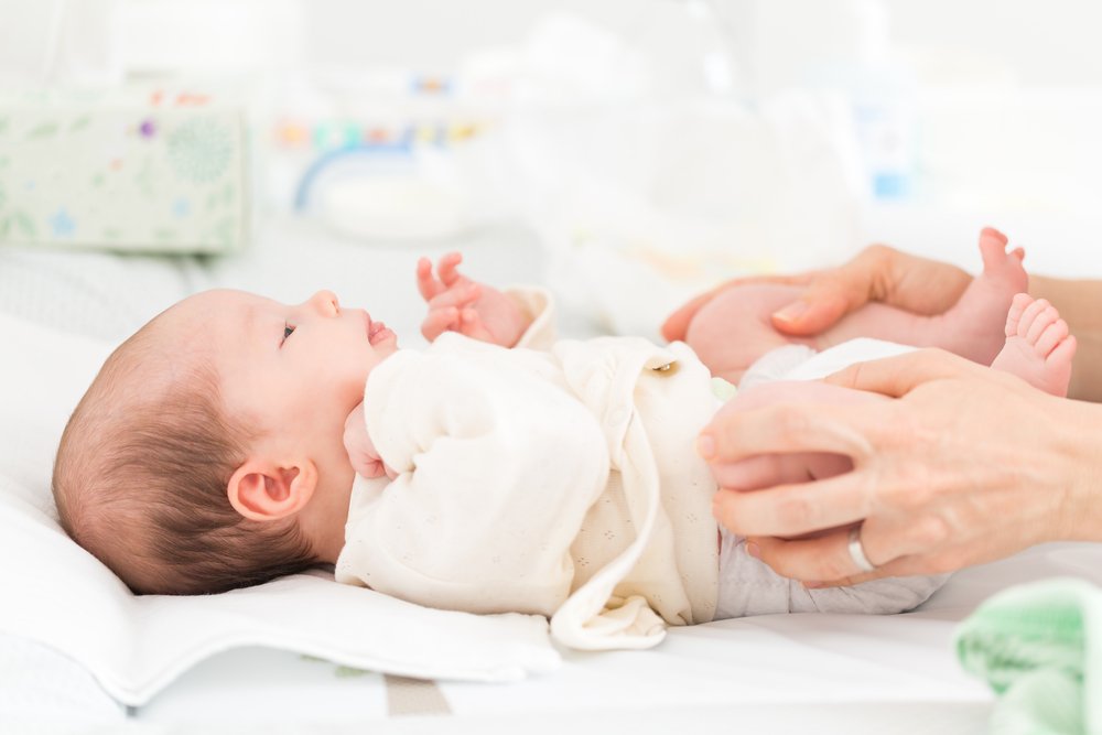
Treatment of patients with congenital clubfoot should begin as early as possible, that is, from birth, when the tissue can still be easily stretched and compressed. The main goal of treatment is to eliminate all elements of clubfoot. This is done manually by gradually repositioning (repeated, monotonous movements) of the foot over 5-10 minutes, which is carried out by the child's mother each time the newborn changes position.
After discharge from the hospital, when the infant's skin is less susceptible to damage, the resulting foot correction is fixed with a flannel or gauze bandage, bending the leg at the knee after each adjustment. To prevent the flannel bandage from falling off, Ettingen put it on the Kleol, Kuslik attached it to the bra, etc. However, only 20 percent of the children recovered from the bandage treatment.
According to Frumin AE, the redressed foot is fixed with a plaster bandage from the age of 2-3 weeks.

Technique for carrying out redression
The hand with the clubfoot and the other hand hold the heel and correct the distal part of the foot - eliminating supination by rotating the foot and moving the distal part of the foot outward.
The flexion of the foot is not corrected by applying pressure with the sole to the distal part of the foot in order not to create clubfoot, which is another type of complex foot deformity with dire consequences. Therefore, in addition to lowering the heel, the sole should be compressed at the projection of the Lisfranc and Chopar joints. The reduction is carried out in such a way that the soft tissues are not damaged, but only stretched.
If the child's skin is normal and firm, the doctor performs resurfacing and fixes the achieved foot correction with a plaster cast. The doctor holds the foot in the corrected position (with the lower leg bent at the knee) until the cast has hardened. To prevent the cast from slipping during movement, it is sometimes advisable to place the cast in the middle third of the thigh, with the lower leg flexed to 90°.
Diagnosis of clubfoot
It is important to determine whether the clubfoot is a true clubfoot (caused by a malformation of the foot bones) or a positional clubfoot.
With positional clubfoot, the patient's foot is more mobile and is actively or passively brought into a normal position. The equinus is only weakly pronounced. Transverse folds are visible on the back of the foot, which indicate sufficient mobility. Postural valgus foot usually resolves spontaneously within the first few weeks of life, but conservative treatment is indicated if this form of valgus foot is identified.
X-rays are not meaningful in children under 3 months of age because the bones at this age are usually still cartilaginous and are not visible on X-rays.
For children older than 3 months, radiographs are taken in two projections: anteroposterior and lateral. The X-rays are taken with the sole and dorsiflexion of the foot at its maximum.
An ultrasound scan is carried out to examine children under 3 months old. This method is completely harmless, but less informative because it only shows one of two levels (side or top view).
Treatment of clubfoot
The orthopedist selects the treatment of clubfoot depending on the severity of the disease. Treatment should be as early, consistent and continuous as possible.
The result depends on the degree of clubfoot. In mild clubfoot forms, 90 % cases can be corrected without surgery. Severe club feet can only be corrected conservatively in 10% of cases.
The conservative treatment of clubfoot begins in the first few weeks of life, because the bony structures of the child's foot are very soft during this time and the foot can be corrected without surgery.
Physiotherapy and foot massage are recommended. The foot is gently immobilized with flannel bandages. As soon as the shape of the foot is corrected, a special splint is placed on the child. If you have a severe clubfoot, a plaster cast is applied to gradually bring the foot into the correct position.
Children with clubfoot are then treated with physiotherapy, massage, therapeutic exercises and orthopedic shoes. The feet are covered with special polyethylene splints overnight.
If conservative correction of clubfoot fails, surgery is performed. Surgical treatment is performed when the child is 1 to 2 years old and includes tendon, ligament and aponeuroseplasty of the foot. In the postoperative period, plaster casts are prescribed for up to 6 months.
ambulance
The symptoms of clubfoot are very specific, making it difficult to confuse this condition with other foot abnormalities. The congenital form is characterized by the following features, among others.
- The heel is displaced inwards;
- The patient has a transverse bend of the foot;
- The ankle joint has little or no mobility;
- The size of the foot is small.
Acquired foot disease has a slightly different clinical picture:
- The characteristic gait changes for no apparent reason;
- The position of the knees is changed;
- The mobility of the feet is impaired;
- The feet turn against each other;
- The thumb is 'pointed inwards'.
It is important to see an orthopedist immediately at the first signs of this disease. Treatment of clubfoot in children and adults is more effective in the early stages of the disease.
This is no fun: How to prevent the development of a club foot in a child - parental measures
Clubfoot is an anatomical abnormality of the ankle joints. It looks like this with the foot turned inwards and slightly downwards. In this position it is not possible to fully step on the foot.
It is important to know that at some point clubfoot can no longer be corrected. Then it is no longer possible to help the child. Therefore, treatment should be initiated as soon as this disorder is detected.
You can read how parents should deal with a club foot in a child in our article.
A visit to the chiropodist – first things first
To reduce fears and worries, an appointment with a podiatrist should be made.
The doctor will assess the condition of your child's feet and make the necessary treatment recommendations to stop the development of the disease.
Orthopedic surgeons typically recommend the following measures when treating and preventing the development of clubfoot.
- a massage course;
- some sports suitable for people with this anatomical defect, such as: B. Dancing or gymnastics;
- a set of therapeutic physical exercises to eliminate clubfoot;
- the use of massage mats at home;
- the use of orthoses and/or orthopedic shoes.
All of these recommendations are aimed, on the one hand, at strengthening and stretching the muscles and, on the other hand, at activating or relaxing them, depending on the orthopedist's recommendation. It is important to remember that the course of the massage - its duration and features - is prescribed exclusively by the doctor. As far as sports are concerned, any sport can be chosen, but the injury rate of the sport in question must be taken into account. Now it is important to prepare the child's body for proper development', so doctors recommend dancing, gymnastics (not Olympic sports), swimming, etc.
Learn more about these methods of clubfoot treatment and correction that you can do yourself at home.
Massage for clubfoot in children: correction of pathology at an early stage
Clubfoot massage in children is an almost unbeatable option for treating congenital anomalies. The most important thing is timely detection of the anomaly. Massage can completely relieve the child of a condition that becomes incurable in adulthood. The misalignment, which persists throughout life, brings complications for its wearer.
Clubfoot is more common on both sides, with boys representing the main contingent. In addition to the external appearance of the foot - the edge of the sole is turned inwards - there is a significant restriction of mobility in the ankle joint. The deformed foot affects foot function and shape as well as gait and posture. Clubfoot massage in children helps to relax the muscles of the lower limbs - internal and external groups - and reduce their increased tone. This is done using the usual massage techniques:
- Cancel;
– shaking;
- Strain; Shake; Stretch and vibration.
To strengthen the stretched and weakened muscles of the lower leg - anterior/external groups - more intensive manipulations are used:
– kneading;
– rubbing;
- Beat.
Massage techniques in the treatment of clubfoot
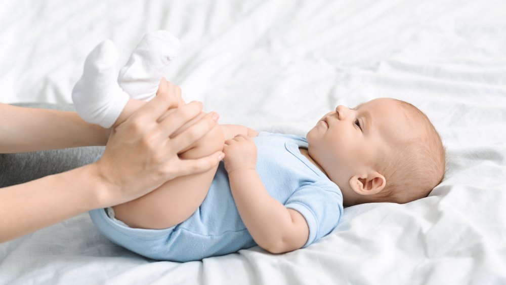
Massage in the treatment of clubfoot in children is recommended after general strengthening treatment. The sequence of the massage is as follows:
– First the feet are rubbed all around, then the shins, thighs, Achilles tendon and soles of the feet are massaged one after the other.
– Stroke the back of your thigh from the back of the knee to the buttock crease. Stroke your thigh vigorously and shake it gently to relax the muscles.
– Massage the back of the lower leg in a differentiated manner.
– The stroke is carried out from the heel to the back of the knee, with the hand being guided along the calf muscle.
– The clubfoot massage in children continues with stroking the Achilles tendon. The Achilles tendon is also stretched, supplemented by vibration. During treatment, the foot should be in slight pronation so that the outer edge of the foot is raised. During the massage, the inner edge of the foot is stretched and the outer edge is tightened.
The next step in massaging children with clubfoot is to massage the front surface of the foot:
– The foot is stroked lengthwise, then you move on to the back of the foot – during the massage the foot is held at a 90 degree angle to the lower extremity.
– Different types of rubbing are used in foot massage: pinching, stroking, pressing, stroking, scissoring and light stroking.
– We rub the hock and ankle with a precise circular movement. The foot should be properly supported (slight pronation position).
– Massage the forefoot by rubbing firmly and applying pressure to the ankle joint and using gentle tapping with shear.
– When massaging children with clubfoot, the knee joint is rubbed and stroked in circular movements.
– The foot is blocked so that the hand rests on the sole of the foot, which is bent backwards while pressure is applied to the outer edge of the foot. The foot is then turned slightly outwards.
– The foot is turned outwards. The movement occurs along the longitudinal axis, very gently lowering the inside of the foot and raising the outside.
Physiotherapy for children is recommended for the following diseases:
- Lung infection;
- Congenital torticollis;
- hip dysplasia;
- Rheumatism;
- bronchial asthma;
- postural defects;
- clubfoot;
- clubfoot;
- Rickets;
- CEREBRAL PALSY.
It is important to start the exercise complex at a young age. If the disease remains untreated, the child will lag behind in its development and functional impairments may occur. Carrying out LFC treatment improves metabolism, prevents deformities of the musculoskeletal system and delays in growth and development. Proper technique and timely action can improve the prognosis and reverse the diagnosis.
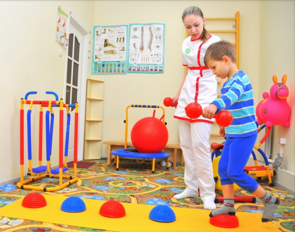
Physiotherapy for children has its own specifics. The most common of these is a large number of game-based exercises. The programs are based on age and diagnosis, but also take into account the child's level of development as well as their mental and motor skills.
Differences in exercise therapy for children at different ages
For children under one year old
An infant's first year of life requires more physical exercise than that of an adult. Physical exercise has a positive effect on children's physical and mental well-being. With regular exercise, children develop their motor and language skills in a timely manner and develop properly.
Exercise is important for premature babies, but also for lethargic, anorexic, nervously excitable and sick children. Physiotherapeutic gymnastics is particularly suitable for children with physical or mental disabilities.
- Physiotherapy for children in the first year of life consists of reflexive, passive and active exercises.These are movements that the child carries out in response to external stimuli. These movements are not carried out consciously. When the instructor performs the exercises for the child, we call them passive exercises. If, on the other hand, the child performs them independently, they are referred to as active exercises. LFC is often supplemented by massage therapy.
- At the age of 1.5 to 3 months, babies are characterized by increased muscle tone and innate reflexes. Therefore, only reflex exercises are recommended at this age.
- At the age of 3 to 4 months, the tension of the flexor and extensor muscles in the shoulders is coordinated. It is recommended to emphasize passive exercises for infants.
- Between 4 and 5 months, the baby's leg and neck muscles are strengthened. The exercises are different for this age.
- At 6 to 9 months of age, the baby develops voluntary movements. Therefore, the exercise program includes active exercises that promote crawling, sitting and standing.
- At 9 to 12 months of age, coordinated movements and the desire to walk are developed. The LFC aims to develop and strengthen these skills.
Physiotherapy in preschool age
Additionally
A very common injury in children is subluxation of the radial head at the elbow joint. Three bones meet in the elbow joint: the humerus, the ulna and the radius. These bones are held together by ligaments. In young children, the ligaments are very flexible and loose and can slide easily around the bone. As we age, the ligaments become stronger and slipping is no longer as easy.
This injury occurs when the child is violently pulled by the hand: the father twists, lifts the child strongly by the wrists (the child must be lifted by the armpit) or even when the parents hold the child by the hand the toddler slips and gets stuck on the hand - and a subluxation occurs.
At the time of injury you may hear the joint cracking. Normally, when an injury occurs, the child feels a short, stabbing pain that subsides almost immediately. The main symptom of the injury is that the child stops bending the arm at the elbow - children keep the injured arm fully extended.
As soon as possible after the injury, the child should be seen by a trauma surgeon who will repair the subluxation and put the ligament back in place.
When should you see a trauma surgeon?
Children often fall, bump, or injure themselves in one way or another. How do you know when a bandage and iodine is enough and when to go to the emergency room?
- Any cut or puncture wound should be examined by a doctor. Do not put greens or iodine in the wound! This leads to additional chemical burns of the wound. Do not apply cotton wool to an open wound - its fibers are difficult to remove from the wound. If the wound is very dirty, rinse it with clean water. Then cover the wound with a clean cloth (sterile gauze, tissue, etc.), apply a pressure bandage, and go to the emergency room as soon as possible. The doctor will carry out initial surgical treatment of the wound, clean it thoroughly (you probably won't be able to do this yourself very well), restore the integrity of all structures and apply a bandage.
- If there is significant swelling at the injury site. This may indicate that it is not just a bruise, but also a fracture, sprain, or torn ligament.
- If the child loses consciousness, even for a short time. This may be an indication of a head injury, which can have serious consequences.
- If the child vomits after the injury. Vomiting, nausea, and paleness also indicate a possible brain injury.
- If the child hit his head. A head injury may not be immediately noticeable, but it can have very serious consequences.
- If the child was hit in the abdomen. A blow to the abdomen can cause damage to internal organs and internal bleeding.
- If the child fell from a height (chair, table, etc.), fell from a bicycle, etc. Sometimes there are no external symptoms, but the internal organs are damaged.
- If the child is restless and behaves unusually.
In general, if you have any doubts, it is better to calm down and consult a doctor. Injuries in children – this is a topic where it is better to exaggerate than to trivialize. You don't have to shy away because you're worried about distracting the doctors in the emergency room or trauma center for nothing. Your child's health comes first!
Diagnosis of flat feet in children
Diagnosis and treatment of flat feet is performed by a podiatrist. During the first consultation the following steps will be carried out:
- Anamnese. The specialist will have a conversation with the child, parents or carer. The symptoms, the time of occurrence and the preceding factors are asked.
- Investigation. The doctor carefully looks at the child's lower limbs and footwear. Visual inspection may detect lateral deviation of the foot and abnormal changes in length. The podiatrist will also ask the patient to walk around the office to assess gait:
Diagnosis and treatment of flat feet in children. Eranov Nurali Fayzievich Eranov Sherzod Nuralievich. Re-health journal, 2020.
At the age of 5-6 years, it is difficult to diagnose flat feet. This is due to the intensive growth and development of the musculoskeletal system. The following diagnostic methods are used to confirm the diagnosis:
- Plantography is the examination of the foot based on its impression. This procedure is used for both diagnosis and prevention. In children's centers it is actively used in preventive examinations. Essentially, a dye is applied to the foot, which the patient then places on a white sheet of paper. The doctor uses the dye to assess the condition of the foot.
- Computer-aided plantography. This is a more modern version of this investigation. A special surface is used that the patient steps on. The examination is performed while sitting and standing to examine the foot without lifting body weight. The information obtained is processed by computer.
- Podometry. This procedure makes it possible to assess the distribution of body weight on the feet.
- X-ray in two projections. In most cases, it is used for differential diagnosis and to assess the condition of the bones of the foot.
- Electromyography. It is used to assess the bioelectric activity of the muscle fibers of the lower leg and foot.
Different types of treatment for flat feet in children
Many parents ask themselves the question: 'Can flat feet be cured? Effective treatment of pathology is possible even in childhood. The earlier the disease is detected, the greater the chance of a complete cure. This is because children's bones, muscles and ligaments are not yet fully developed and can therefore be easily corrected.
Self-treatment is strongly discouraged. Only a qualified podiatrist can explain how to correct flat feet and stop the foot deformity. For this purpose, the child is prescribed comprehensive treatment, which includes:
- Massage. The correct massage technique normalizes blood circulation in the lower limbs, strengthens ligaments and muscles, and eliminates leg fatigue. The massage can be carried out both with the help of a professional and at home.
- Therapeutic exercises. Walking on heels and toes, making circular foot movements, and lifting objects off the floor with your feet are most effective. LFK should be carried out daily to achieve the desired effect.
- Physiotherapy. Phonophoresis and electrophoresis are recommended to improve prognosis.
- Medication. To relieve pain, non-narcotic analgesics in tablet or ointment form can be prescribed. In some cases, nonsteroidal anti-inflammatory drugs and muscle relaxants may be prescribed to relieve muscle spasms.
- Orthopedic shoes. They help distribute the load on the feet and relieve the clinical symptoms of the disease. Wearing orthopedic shoes is also an effective prevention of flat feet.
If conservative therapy does not have the desired effect, surgical treatment is considered. The optimal age for surgery is 8-12 years. The type and tactics of the operation depend on the clinical case and are selected individually by the surgeon.
Read more:- Clubfoot in 7-year-old children.
- What is clubfoot?.
- Clubfoot in children therapeutic exercises 7 years old.
- Why does a child develop clubfoot?.
- Congenital clubfoot.
- 1 year old child with clubfoot.
- clubfoot.
- Clubfoot Treatment.