Photo, video: Depositphotos / pixelstock; 5-tv.ru
- Foot deformities and their treatment
- Causes of foot deformities
- Egyptian foot: lover of solitude
- Roman foot: a social animal
- Type 2 – Leader.
- Guy #4 – Listener
- The Egyptian Foot: Characteristics, Personality and Shoe Type
- The German foot: characteristics, personality and shoe type
- Foot treatment at the Osteopathic Center Neonatus Sanus
- diagnostic methods
- In this case, attention should be paid to the foot and posture.
- The shape of the baby's feet.
- Description of the operation
- Correction of the true curvature of the legs
- Indications for the operation
- Treatment of tinea pedis
- Guidelines
Foot deformities and their treatment
The human foot carries an enormous load and allows people to move freely in space. Fluctuations in body weight always lead to changes in the arch area of the foot.
All deformities are characterized by a permanent change in the natural appearance. These changes can affect the length of individual bones as well as the strength of tendons and ligaments. When a person suffers from a limb deformity, their gait is impaired and they experience discomfort and pain when exercising.
The cause of these changes is the abnormal distribution of body weight on the foot. Deformity changes occur equally frequently in men and women, regardless of age. People with chronic joint and bone diseases, athletes and people who work with excessive strain on their feet are at risk.
Causes of foot deformities
The most common causes of foot deformities include:
- Trauma;
- Connective tissue diseases
- genetic predisposition;
- prolonged exposure to cold temperatures;
- congenital malformations of bones and joints.
Even minor deformations of the foot can lead to diseases such as arthrosis or osteochondrosis. In addition, people with deformities can suffer from altered posture, including scoliosis.
Egyptian foot: lover of solitude
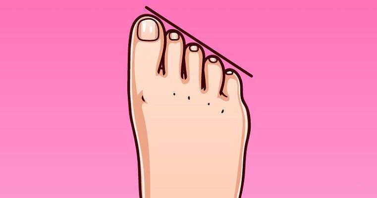
People with an Egyptian foot shape have a longer first toe and the others are proportional but shorter.
Character traits: These people are quite secretive and do not like to open their souls to everyone they meet. They are often viewed as impulsive and rebellious. 'Egyptians' love to dream; many of them want to escape reality.
Egyptian footwearers like to live in their own world; Emotions often take precedence over rationality. They can be quite fickle in their desires, many want to be pampered.
Roman foot: a social animal
In people with a Roman foot shape, the first three toes are longer than the other two.
Character traits: While the owner of the Egyptian foot seeks solitude, the 'Roman' wants to be in company. You enjoy meeting new people and discovering new cultures. Many travelers have a truly Roman foot shape. They also tend to have a well-proportioned, 'balanced' body.
Negative personality traits include arrogance and self-confidence, which often border on risk-taking.
Type 2 – Leader.
Shape: The second toe is larger than the others and the foot has a triangular shape. This foot is also known as the 'Greek foot'.
'Leaders' are motivated to achieve a specific goal, want to reach the top in both their personal and professional lives and are willing to do anything and break through walls. With this approach, they actively develop and are richly rewarded.
However, the rigidity of behavior does not allow everyone to be with him: the 'leader' cannot convert them and they must either leave or endure.
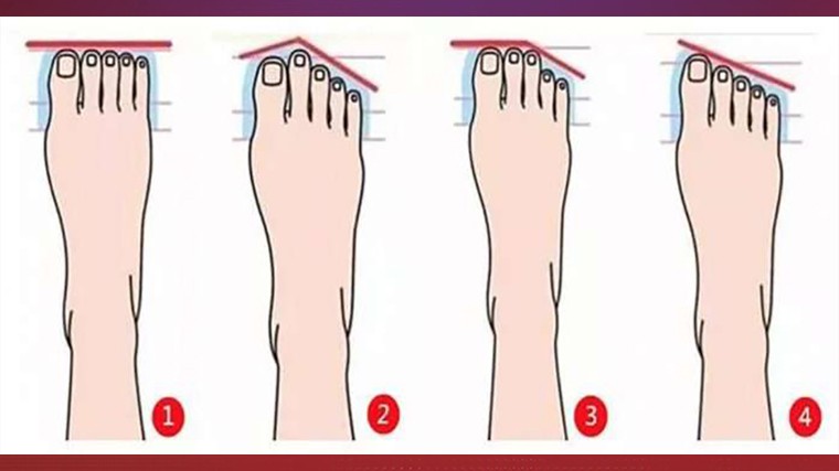
Guy #4 – Listener
Egyptian' foot shape: each toe is lower than the previous one, its length is evenly reduced.
Gentleness and sensitivity are their characteristics, they have a soft, calm character.
One weakness is their lack of initiative: even if they have an interesting development idea, they prefer to keep it to themselves and not make it public. Not to mention the ability to make decisions, the 'listener' will be relieved to be able to place this responsibility on the shoulders of someone with a different foot structure.
Previously, 5-tv.ru suggested looking up and looking into a person's eyes to recognize their character by the color of their eyes.
The Egyptian Foot: Characteristics, Personality and Shoe Type
Il The Egyptian foot is not difficult to see since they is the most common form in the world population. It is a plantar structure in which the length of the big toewhile pull the other fingers together until they reach the scales, which creates a 45° angle. While the Roman foot is considered a squatting position, the Egyptian foot is generally longer and narrower.
- personalityA person with the Egyptian foot is characterized by, among other things. enigmatic character and a reserved personality.That's why he or she doesn't like to open up to others and hides some secrets. Thanks to these properties, it is not surprising that The Egyptian foot is also known as the solitaire foot.
- The shoe type: The toe of the Egyptian foot that should be protected is definitely the big toe, which is the longest. If you recognize this shape on your feet, Avoid wearing closed-toe shoesThey are tight This can cause the bones of the big toe and second toe to repeatedly hit, making walking very painful. Choose instead Shoes with rounded or open toes, They ensure good blood circulation in the toes and the necessary freedom of movement.
- A star athlete known for his Egyptian foot? The unique and inimitable Julia Roberts!
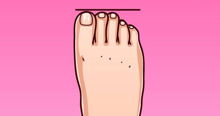
The German foot: characteristics, personality and shoe type
An observation about the The German foot.If you look at the German foot you will recognize it. that the big toe is longer than the others, who has one instead of the other is almost the same length and has a more rounded shape.
- If you have one Germanic foot certainly stands out for yours Person particularly rational and methodical. You don't like to dream, but rather face reality as it is. So, no frills on the head, just but keep your feet firmly on the ground.. For these qualities of yours, They hate changes and unexpected events.
- They have a certain regularity, German feet do not need any special footwear. As always, it is important to prefer shoes that you feel comfortable in and in which you can move completely freely and comfortably.
- As you can see in the photos below, Jennifer Aniston clearly has a Germanic foot.
Foot treatment at the Osteopathic Center Neonatus Sanus
Our osteopathy and neurology clinic on Vasiliev Island, Neonatus Sanus, has extensive practical experience in the treatment of foot and musculoskeletal dysfunction.
The comprehensive and qualitative approach to dental treatment is based on 3 principles: dental health, spinal health and foot health as a whole.
In our center you can receive a good examination, treatment and advice from leading specialists in St. Petersburg.
diagnostic methods
This method is the simplest and most informative.
Using a special device (plantograph), an impression of the foot is made and the degree of deformity is determined:
- Grade I – the cavity covers less than half of the foot;
- Grade II – the depression covers less than a third of the foot;
- Grade III – the depression covers less than a third of the foot; Grade III – no deepening.

In this case, attention should be paid to the foot and posture.
What is the relationship between these seemingly distant body parts: the foot and the spine? Very easy!!! Our body, from feet to head, is an evolutionary structure in which all limbs are connected. The feet are the 'foundation' of the body. Defects in the foundation inevitably lead to distortion of the entire structure, impair posture and gait, lead to pain in the spine and premature 'wear and tear' of the joints. According to information in the literature, at least 80 % of the population have musculoskeletal problems that are directly or indirectly related to foot deformities.
The first deformations and dysfunctions of the feet and posture appear in childhood and accompany people into adulthood. All children are born with flat feet, and the arches of the feet and the curvatures of the spine begin to actively form when they assume an upright posture.
Your children's feet will support them throughout their lives. It is believed that from the first steps of a child to old age, a person circles the earth four times. The foot is a complex structure consisting of 26 bones that are connected to each other in a special way by ligaments, joints, muscles and tendons.
At birth, children's feet are not yet formed, and the future bone structures are represented by cartilage. The first phase of arch formation is completed by the age of 8-9, when the child's foot begins to take the shape of an adult foot. During hormonal maturation, further correction takes place to functionally refine the structures of the foot.
The shape of the baby's feet.
The insecure child aged 6 to 18 months usually has an O-shaped foot shape (varus). When the child begins to walk upright, he tries to maintain balance by spreading his legs wide apart. This pulls the knees inward toward the midline and an X-shaped (varus) leg shape may gradually develop by 2.5 to 3 years of age. In this context, one of the patriarchs of Russian orthopedics, MO Friedland, wrote that a child who begins to walk 'must go through a difficult school of balance'. With a very small footprint and a high center of gravity, children must first learn to maintain their balance while standing and moving. The body's musculoskeletal corset is then strengthened and the legs are typically aligned: the foot, shin, knee and hip are centered and aligned along a single line - the vertical axis of the lower limbs.
Description of the operation
Leg shape correction – correction of the real or false curvature of the legs using methods of plastic surgery and orthopedic cosmetology. The correction strategy and the choice of surgical methods and techniques depend on the cause of the aesthetic problem. A true curvature requires orthopedic correction. False curvatures are corrected by realigning the volume of soft tissues and restoring the inner contour of the leg with implants or lipofilling.
How is orthopedic leg correction performed?
Both true and false curvatures are successfully treated by plastic surgery and orthopedic cosmetology. If the curvature is incorrect, soft tissue surgery is sufficient: implants or lipofilling are used to correct the curvature. With a true curvature, surgery on the bony structures is required to correct the shape of the legs.
Correction of the true curvature of the legs
Correcting a true leg curvature requires reshaping of the bony structures. Orthopedic leg correction is performed on the right and left shin bones at the same time and consists of several steps:
- The operation is performed on the shinbone. The surgeon inserts a special orthopedic brace through small incisions in the skin. As a rule, Ilizarov rods or spokes are used. The advantage of a spoke bandage is that the diameter of the spoke is smaller than the diameter of the rod and the scars on the skin are less visible after the bandage is removed. After the brace is inserted, an osteotomy is performed, that is, part of the bone is cut out to reposition the bone, correct the curvature of the legs and reshape their shape. The osteotomy is performed through a short surgical approach, the scar on the skin is inconspicuous, fades over time and is visually almost invisible.
- In the next step, the shape of the legs is corrected by gradually changing the tension of the spokes of the orthopedic brace. During regular medical check-ups, the bones are straightened by correcting the braces until the curvature of the leg is completely corrected.
- immobilization – The final phase of leg correction, which begins once the bones are straightened. At the same time, the actual rehabilitation begins. The rehabilitation period requires disciplined behavior towards all the surgeon's recommendations. It is important to have regular follow-up visits to ensure the splint is fitting securely and to strictly control physical activity.
- Final stage of leg correction – Removal of the appliance after complete restoration of bone integrity and correction of congenital or acquired defects.
Indications for the operation
The inner arch of the leg is usually fusiform. The anatomically normal shape of the leg is characterized by a curved inner contour with contact points at the hip, shin, knee and ankle joints.
With leg curvature, the inner contour is missing in the middle or lower third. In the O deformity, often referred to as varus, there is no inner contour contact at the knee joints but there is closure at the ankle joints. Due to the nature of the deformity, the inner contour of the leg takes the shape of an O.
With an X-shaped deformity, there is still contact at the knee joints, but there is no foot contact at the ankles. Due to this feature of the deformity, the internal contour is X-shaped. This type of deformity is called a valgus deformity.
A varus or valgus deformity can be caused by either a curvature of the tibia or atrophy/hypotrophy of the muscles and soft tissues in the lower leg. A curvature caused by a defect in the bony structures is called a true curvature. If the cosmetic defect is due to abnormal development or regression of the soft tissues, the curvature of the leg is called false curvature.
The leg shape defects under consideration can be congenital or acquired. Congenital defects result from abnormal development of the bones or muscles of the lower limbs. Acquired defects can arise from illness, fractures, accidents or trauma. A false curvature can also result from a fracture of the femur or tibia; in this case, the defect in the inner contour of the leg is caused by atrophy of the tibial muscles after long-term immobilization of the lower limbs.
Indications for correcting the shape of the foot:
Treatment of tinea pedis
The safest treatment option for tinea pedis is the use of topical antifungals, but relapses are common and require continued therapy. Alternative, longer-term treatment includes itraconazole 200 mg orally once daily for 1 month (or as pulse therapy: 200 mg twice daily for 1 week per month for 1-2 months) and terbinafine 250 mg orally once daily for 2-6 weeks. Concomitant use of external antifungal therapy may reduce the relapse rate.
To prevent relapses, it is necessary to reduce the moisture content of the skin on the feet, including when wearing footwear. It is very important to wear permeable or open-toed shoes and change socks, especially in the warm season. The spaces between the toes should be wiped by hand after washing. It is also recommended to use desiccants; antifungal powders (e.g. miconazole), Gencian violet (not approved in the Russian Federation), baths with Burov solution (5%iger aluminum subacetate) and 20-25%iger aluminum chloride solution overnight for 1 week, then 1-2 times/week after Requirement.
Guidelines
The diagnosis should be suspected when patients present with interdigital, ulcerative, hyperkeratotic, or vesicular lesions on the fingers and/or toes.
Dyshidrotic eczema, plantar psoriasis, and allergic contact dermatitis should also be considered.
Treatment consists of topical and sometimes oral antifungals and drying agents.
Copyright © 2023 Merck & Co, Inc, Rahway, NJ, USA and its subsidiaries. All rights reserved.
Read more:- Egyptian foot in men.
- X-shaped legs photo.
- What to do if your teen has crooked legs.
- flat feet.
- types of toes.
- How much does crooked leg surgery cost?.
- Why are the fingers curved?.
- flatfoot μb.