Administering a lethal dose will result in serious effects on the body. The effects on the CNS consist of inhibiting the respiratory center, slowing the heartbeat, lowering blood pressure and putting the patient in a coma. If left untreated, these conditions can lead to death.
Lozartan
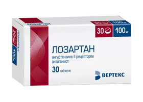
Illustration of the drug
- Latin name: losartan
- ATX code: C09CA01
- Active ingredient: losartan
- Manufacturer: VERTEX AO, R-Pharm Novosyolki Ltd, Gedeon Richter-RUS AO, Pranapharm, Ozon Ltd, KRKA-Rus, Tatkhimpharmpreparaty AO, Biocom ZAO (Russia), Teva Pharmaceutical Plant AO (Hungary), Gedeon Richter Poland Ltd (Poland), KRKA (Slovenia)
form of release
- Coated oval tablets 25 mg white tablets with markings;
- 50 mg white oval film-coated tablets marked '50';
- 100 mg white oval film-coated tablets marked '100'.
Losartan is a selective, competitive antagonist that blocks the AT1 subtype receptors in various tissues such as the brain, adrenal cortex, liver, kidney, heart and vascular smooth muscle, thereby reducing the effects of angiotensin II.
Administration of the active substance of the drug leads to a reduction in the total peripheral resistance (post-loading) and venous return of the heart (pre-loading). All physiological effects of angiotensin II, including stimulation of the release of aldosteroneare blocked by the action of Lozartan. The reduction in blood pressure is independent of Renin-angiotensin system.. Taking this drug increases plasma renin activity by clearing angiotensin II.
The effect of this drug was in the study Life (Losartan Intervention For Endpoint reduction in hypertension study)at the 9193 participants with essential hypertension. The subjects were 55-80 years old and had a blood pressure of 160-200 mmHg. After treatment with Lozartan, blood pressure was reduced by 13 % and the mortality rate in these patients was reduced by 25 %.
Treatment prices:
| service | Price (RUB) |
|---|---|
| types of therapies | |
| Standard detox therapy | 3 500 ₽ |
| Double detox therapy | 6 000 ₽ |
| Enhanced detoxification therapy | 7 500 ₽ |
| Maximum detox therapy | 9 500 ₽ |
| Fast sobering up at home | 7 500 ₽ |
| Inpatient stay at home for 1 day | 22 000 ₽ |
| Highly specialized hospitalization | 15 000 ₽ |
| Inpatient treatment | |
| Accommodation | |
| Economy room (6 beds) | 2 000 ₽ |
| Standard room (4 beds) | 3 000 ₽ |
| Superior comfort room (2 beds) | 5 500 ₽ |
| VIP room (1 bed) | 12 500 ₽ |
| Individual 24-hour stand | 5 000 ₽ |
| Medical and social rehabilitation 21 days | 140 000 ₽ |
| service | Price (rubles) |
|---|---|
| Initial consultation with a narcologist | for free |
| Consultation with a psychologist | 3 000 ₽ |
| Psychiatric Counseling | 5 000 ₽ |
| Home coding torpedo | 7 500 ₽ |
| Fast deprivation and encoding (duplex) |
tramadol
Drug manufacturers report an alarming increase in drug addiction among the population. Increasingly, drugs are being used not for relief but for a fleeting intoxication. Systematic drug intoxication leads to permanent damage to the liver, kidneys, heart and other body systems.
The most common form of drug addiction occurs in adolescents and young adults. The young body is often not up to the enormous strain and 'gives up' in the formative phase. Young people taking tramadol can be disabled at best, die at worst.

What is tramadol?
Tramadol is a powerful drug used in severe pain syndromes. The painkiller is used for severe postoperative pain, biliary colic, myocardial infarction and for certain diagnostic procedures. Taking it as prescribed by a doctor does not have a strong toxic effect, while systematic uncontrolled intake of tramadol leads to irreversible cachexia.
The drug was made by a pharmaceutical company made famous by a substance like Contergan. This drug caused mutations in the patients' fetuses: as a result of its intake, children were born with deformed limbs. Tramadol does not have a similar composition to Thalidamide, but it is also highly toxic and dangerous for the body if taken continuously.
Composition of tramadol
The effect of the analgesic is 10 % comparable to that of morphine. The substance blocks mu-opioid receptors in the brain, which are responsible for transmitting pain impulses. This is the reason for the analgesic properties of the drug. The drug has sedative and antidepressant effects. The molecule O-desmethyltramadol is released in the brain, which has a narcotic effect and is highly addictive. Thus, tramadol is a precursor to a drug like O-desmethyltramadol.
Individual risk of arterial vascular disease
- Presence of a known pathology of atherosclerotic etiology in the coronary, carotid or renal arteries;
- Abnormal pulse changes in the vessels of the lower limbs;
- Ischemic pain in the lower limbs;
- Presence of one of the risk factors for atherosclerosis - hypertension, smoking, dyslipidemia, hyperhomocysteinemia;
- Age over 50 years and presence of diabetes.
In patients with vascular pathology, an accurate anatomical diagnosis can be made using modern research methods, such as: B.
- duplex scan,
- Ankle Brachial Index or BPI,
- Finger Bar Index or FBI,
- segmental pressure measurement,
- pulse wave recording,
- dopplerometry,
- stress tests.
If necessary, these data can be supplemented by MRI, CT, aortoarteriography. These examinations provide information for the creation of a treatment plan.
In patients at risk of arterial disease of the lower limbs, it is advisable to check the pulsation of the arteries of the lower limbs and the condition of the skin of the feet.
A simple test to measure blood pressure in the legs called the 'Ankle Brachial Index' is also available. Ankle Brachial Index (ABI). It is recommended to take the LPI measurements on both legs. Evaluation of the Doppler waveform can provide even more valuable information for assessing blood flow to the limbs, thereby providing the physician with additional information for diagnosis.
In some cases of non-compressed arteries (inability to lower blood pressure with a cuff), the LPI is ineffective. This is the case with diabetes or elderly patients.
Classification of the forms of the diabetic foot
- 0 – high-risk diabetic foot with deformities, calluses, hyperkeratosis, without ulcerative defects;
- 1 – stage of superficial skin ulceration;
- 2 – stage of deep cutaneous ulceration involving subcutaneous adipose tissue, muscle tissue, tendons and without bone lesions;
- 3 - stage of deep ulceration with bone involvement;
- 4 - stage of limited gangrene
- 5 - stage of extensive gangrene.
Depending on whether one of the pathological components predominates, a distinction is made between ischemic (5-10 %), neuropathic (60-75 %) and mixed neuro-ischemic (20-30 %) forms of the diabetic foot:
The ischemic (neuronal ischemic) form of the diabetic foot develops against the background of diabetic angiopathy and is characterized by impaired blood supply to the limbs due to changes in the large and small vessels as a result of atherosclerosis, which develops much faster in diabetics than in other people . In the course of the ischemic form, persistent edema, intermittent claudication, constant pain and fatigue in the legs, and a change in the color of the skin of the limbs to blue are intensified. The feet and lower legs become cold, and as the disease progresses, ulcers or necrosis develop on the edge of the heel or on the toes.
The neuropathic form of the diabetic foot is divided into three forms:
The neuropathic form is caused by damage to the nervous system of the distal limbs as a result of diabetic polyneuropathy. The first symptoms of the disease appear in those places where the foot is most stressed during physical exertion. Over the long term, neuropathy causes the foot to deform and shift the support to specific areas where the skin thickens and later forms an ulcer.
Under these circumstances, the patient experiences partial numbness, burning and 'tingling' in the limb. After some time, the sensitivity of the leg decreases to such an extent that the ulcer is no longer noticed. Because of this insensitivity, the sufferer is unable to respond to thermal or mechanical stimuli, which can further aggravate the damage and easily lead to infection.
Prognosis and prevention of diabetic foot disease
Prophylactic measures to prevent the development of diabetic foot
- Meticulous foot care and daily foot examination to detect changes;
- regular control of blood sugar levels at home;
- continuous monitoring by a diabetologist and a podiatrist;
- Compliance with the diet and drug regime;
- Avoiding tight shoes in favor of orthoses and orthopedic shoes;
- exercises for the feet;
- Fear of foot injuries.
A number of factors must be considered when determining prognosis, including appropriateness of treatment, duration of diabetes, and comorbidities.
Causes of diabetic foot
The most important elements in the pathogenesis of diabetic foot ulcers are angiopathy, neuropathy and infection.
Uncompensated hyperglycemia over a long period of time in diabetes leads to specific changes in the blood vessels (diabetic macroangiopathy and microangiopathy) and peripheral nerves (diabetic neuropathy).
Angiopathies lead to a decrease in the elasticity and permeability of blood vessels, an increase in blood viscosity, which impairs innervation and normal tissue trophicity, and leads to a loss of sensitivity of nerve endings in the feet.
Increased peptide glycosylation reduces joint mobility, which is accompanied by deformation of the limb bones and disruption of the normal biomechanical loading of the foot (diabetic osteoarthropathy or Charcot's foot). Due to the changed blood circulation, reduced sensitivity and reduced tissue defenses, any small trauma to the foot (bruise, abrasion, fracture, microcut) turns into long-term, non-healing trophic ulcers, which are often infected by staphylococci, colibacilli, streptococci and anaerobic microflora.
The various bacterial enzyme groups loosen the surrounding tissues, spreading infection and necrosis to the subcutaneous fat, muscle tissue, bones and ligaments, increasing the risk of abscesses, mucus formation and limb gangrene.
complications
If the body is not properly treated, cut or fissured, infection can occur. Microorganisms that have got into the bladder cause inflammation, which is manifested by redness, swelling and severe pain in the bladder. If left untreated, the blister will soften and pus will ooze out under pressure. Inflammation originating from the cornea can spread to the surrounding tissues and lead to the formation of a corneal abscess or phlegmon, the bones of the foot (osteomyelitis), the synovial membranes and the joints of the foot.
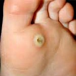
Identifying callus formation by its characteristic appearance is not difficult. Callus should be distinguished from ridges, inflammatory lesions in the joints of the metatarsal bones, Morton's disease, and genetically caused increased keratoses of the skin. Some warts, especially foot warts, can resemble a callus. A characteristic symptom is that the wart is very sensitive and hurts when rolled over, while a callus hurts when pressed.
When the patient visits a dermatologist, the doctor needs to find out what the wart is related to. To do this, he will ask the patient about his job, interests, sports and other hobbies, and the type of footwear he usually wears. If the callus is on the foot, the doctor will examine the patient for deformities and diseases of the foot and, if necessary, refer him to a podiatrist, rheumatologist or orthopedist. Important is the presence of diseases such as diabetes, neuritis, obliterative arteritis, varicose veins with chronic venous insufficiency. If these diseases are detected in the anamnesis, an endocrinologist, neurologist, vascular surgeon or phlebologist should be consulted to determine the most appropriate treatment tactics for corns.
treatment of corns
A corn that is not painful usually does not need treatment. If pain does occur, the triggering factor must be eliminated, e.g. E.g. tight shoes, tool handle friction, etc. Special bladder pads can help reduce friction and pressure on the cornea when walking. These can be made by hand. A circle with a hole in the center is cut out of a soft, sufficiently thick fabric. The circle is placed so that the callus is in the center of the hole. If the callus is under the toes, special toe caps made of felt, rubber or soft plastic are used. If the callus is on the toes, spacers, bushings and toe caps are used. Toe spacers prevent toes from rubbing together. Sleeves and toe caps protect the sides and tips of the fingers.
In the case of dry calluses, cornification is treated in the beauty salon as a separate treatment or as part of a pedicure or manicure. For this, special softening masses, salicylic acid or a pedicure are used. Soft calluses and blisters should be treated with antiseptics and a protective bandage fixed with a plaster.
If possible, orthopedic treatment is carried out for foot deformities. If there is an accompanying pathology that leads to impaired innervation or blood supply in the area of the callus, the treatment should be carried out in collaboration with the appropriate specialist. If conservative treatment of the cornea is unsuccessful, it should be removed. The callus can be removed by cryoablation, laser ablation, electrocoagulation, radiofrequency ablation, or surgical excision.
MRI SCAN
A series of Tl, T2, T2 tinn, DWI and SWI imaging studies in the Ipex planes visualized subcostal structures. The midline structures were not relocated.
Multiple T2, T2-tinn foci without perifocal infiltration ranging in size from 0.3 cm to 0.6 cm (gliosis) were noted in the white matter in the predominantly frontal and medial lobes.
The lateral cerebral ventricles are asymmetrical, D
No additional masses were found in the area of the periaqueductal horn. The internal auditory canals are not dilated. Eye sockets, optic nerves, pituitary gland - no abnormalities. The basal cisterns are not enlarged or deformed.
The convex subarachnoid spaces and sulci are not enlarged. The lateral cerebral fissures are symmetrical, not widened. The cerebellar amygdala is located at the level of the greater occipital foramen. The transition between skull and spine is unremarkable.
There is a small irregular thickening of the maxillary sinus membrane that shows no fluid level. Pneumatization of the other facial cavities is not significantly affected.
conclusion: MR image showing multiple supratentorial foci of vascular origin in the frontal and parietal lobes. Small parietal inflammatory lesions in the maxillary sinus membrane (no fluid level).
MRI showed vascular changes and age-related hypertension. The presence of tinnitus can be due to atherosclerotic changes in the blood vessels of the head and neck and can also be an early sign of sensorineural hearing loss. These are small foci that arise from chronic insufficient blood flow to the brain; they occur in hypertension and atherosclerosis.
These foci are not threatening, they are vascular foci that indicate pressure fluctuations.
These foci are not dangerous, they can be congenital or caused by fluctuations in blood pressure. Almost everyone your age has this herd.
Read more:- Why amputate legs for diabetes?.
- Diabetic foot ulcers.
- X-ray of Charcot's foot.
- hole in the heel.
- Heel bone tendon sac in Latin.
- What can I do if my feet are irritated by new shoes?.
- foot bone in Latin.
- Photo: Diabetic heels.