Here you will find out what flat feet are, what causes them and what consequences they can have if they are not treated early.
- Clubfoot in a child
- types of disease
- Causes of valgus arches deformity
- diagnosis
- clinical picture.
- diagnosis
- Clinical method
- Flat foot in a child
- Symptoms of illness
- causes
- Plantar examination of the foot children of primary school age.
- symptoms of the disease
- causes
- Flat foot exercises for children aged 1.5 and over. Set for advanced users.
- Balancing cushion exercises for flat feet
- Why do foot deformities occur?
- Transverse flatfoot
- Causes of flat feet
- How can an osteopath treat this problem?
Clubfoot in a child
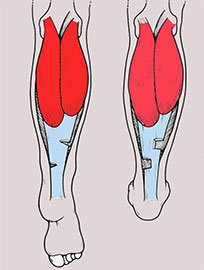
Clubfoot in children has a lower arch and an X-shaped curvature of the foot: the toes and heel point outwards, the metatarsus points inwards.
The arch of the foot begins to form when the child learns to walk. Humans are initially born with flat feet because their feet have never been loaded and supported. With the first steps, the feet begin to acquire a normal shape and structure under the influence of external factors. At around 1.5 years old, an arch of the foot is already visible. This will gradually build and strengthen over the course of several years. This process is completed by the age of 4-5 years. Depending on the individual characteristics of your child's body and the developmental anomalies of the musculoskeletal system, endocrine system and nervous system, this period can vary up or down.
Your child is at risk if an orthopedist or neurologist diagnosed in the first year of life
- Insufficient or excessive muscle tone;
- delayed motor or psychomotor development;
- Underweight or overweight.
A valgus condition in children can be be easy or obvious. When the diagnosis is made, the specialist measures the abnormality in degrees. Abnormal arch formation can also be caused by the inability of the child's ligaments and muscles to fix the ankle joint in the correct position due to overstretching. Improper weight distribution in the foot and excessive pressure on the longitudinal arch are also common.
Important!!! In the early stages of the disease, special footwear can help correct the alignment of the feet and prevent the disease from worsening. If you notice that your child's feet protrude slightly while training to walk, you should urgently consult an orthopedist.
types of disease
There are two types of valgus deformity: it can be congenital or acquired.
Congenital form The congenital form develops in utero as a result of a misalignment of the bony structure of the foot. In this case, the diagnosis is made in the first months after birth.
Acquired deformity It is caused by weakening of the ligaments and tendons and abnormalities in motor development. It occurs up to 1 year of age or earlier. Sometimes the causes are trauma, a long time in a cast, premature foot position, dysplasia, etc.
- Orthopedic footwear;
- prevention of rickets;
- Minimizing stress on immature feet up to 7-8 months of age;
- Regular check-ups with an orthopedist.
Causes of valgus arches deformity
- Congenital factors (genetic predisposition to foot deformity, weakness of connective tissue and tendon-muscular apparatus);
- Traumatic (fractures of the hind and metatarsal bones, ankles);
- Consequences of poliomyelitis, nerve and muscle diseases;
- Rachitic bone deformities, osteoporosis;
- Excessive load on the lower limbs, standing work, heavy physical work, long walking;
- overweight, obesity;
- wearing uncomfortable, ill-fitting footwear;
- Developmental disorders: fatty bones, ossification disorders (osteochondropathies);
- Endocrine disorders.
- Pain in the back of the foot;
- Local pain when putting on shoes;
- Diffuse, aching pain at the end of the day;
- Difficulty choosing and wearing shoes;
- swelling of the ankles;
- Sinking of the longitudinal arch of the foot, significant sinking and protrusion of the scaphoid bone;
- Additional sesamoid bones around the ankle joint.
- Medial rotation of the tibia;
- Bellaire's sign (pain on the inside of the knee joint);
- Development of gonarthrosis, arthrosis of the ankle and foot joints.
Internal displacement of the kneecap with rotation of the lower limbs
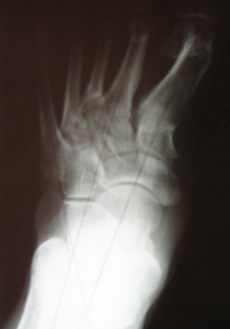
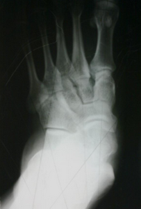
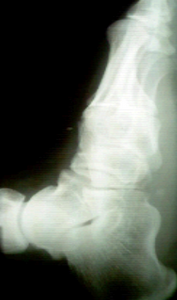
diagnosis
- Clinical examination, complaints, anamnesis – examination by the orthopedist.
- Radiological examination.
- Plantography – scraping of the foot to detect flat feet, zone of greatest stress.
- Other instrumental diagnostic procedures – magnetic resonance imaging, computed tomography.
clinical picture.
The children complain of fatigue, pain in the feet and lower legs (the degree of pain depends on the severity of the deformity). The parents point out a stilted gait, frequent falls on flat surfaces and that the children are not always able to fully participate in play with their peers.
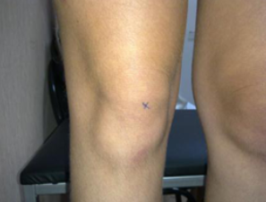
Changes in the load and support of the foot affect the wear of the shoe: the heel wears more on the inside, the sole of the shoe wears more on the outside. As you get older, you may experience problems in your knees (especially on the inside), hips and spine.
Since all children with flat feet have a more or less disturbed spatial relationship in the upper musculoskeletal system, hyperlordosis phenomena occur in the lumbar spine, often with compensatory kyphosis in the thoracic spine and forward tilt of the pelvis. The biomechanical movement sequences are impaired - the gait is disturbed. Internal twisting of the lower limbs is the most common cause ofClaw formation,Corridor'.
diagnosis
Clinical method
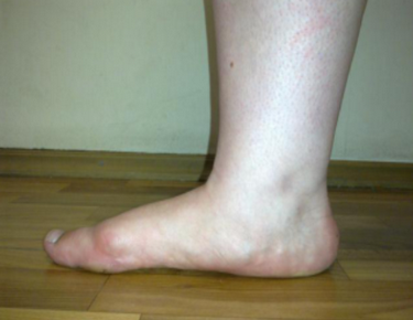
The foot is examined in a freely dangling state and under stress while standing and walking. During the posterior examination, the alignment of the hindfoot is determined by drawing a line through the midpoint of the Achilles tendon and the midpoint of the calcaneal tuberosity, which represents the axis of the hindfoot. A deviation of the axis in the vertical plane or outwards, in the valgus direction, up to an angle of 6° is considered normal.
An outward deviation of more than 6° is pathological (pes valgus). The relative length of the toes varies from person to person. Depending on the length of the toes, a distinction is made between the Greek foot -I III > IV > V, the Egyptian foot -I > II > III > IV > V and the rectangular foot in between -I = II > = III > = IV > = V
The peculiarities of the foot structure favor the development of certain static changes. In the Greek form of the foot, in which the 1st metatarsal is shorter than the 2nd and sometimes the 3rd, transverse flatfoot is most common, as is overloading of the head of the 2nd metatarsal; in the Egyptian foot shape the development of a static valgus.
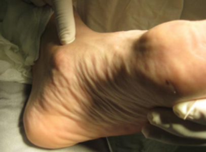
W firm, rigid flat feet A number of typical changes can be identified during the examination:
- 1. The longitudinal part of the arch of the foot is not present and the foot is supported by the entire surface of the sole of the foot. The forefoot is in dorsiflexion relative to the hindfoot, which in turn is in sole flexion relative to the forefoot. Below the medial malleolus there is a protrusion formed by the head of the talus bone, which is displaced inward, forward and downward. Sometimes there is a second bulge below the head of the talus - the inner horn of the heel bone.
- 2 The forefoot is in abduction relative to the rear foot. The angular curvature of the long axis of the foot becomes particularly clear when one considers the line along which the inner edge of the foot rests on the ground; the apex of the external abduction angle of the forefoot lies in the metatarsal region.
- 3 The heel is tilted outwards (pronation). When looking at the foot from behind, the inner edge of the heel rests on the surface. The stirrup, lowered from the middle of the calf muscle, runs to the inside of the heel. The inner malleolus protrudes, the outer malleolus is flattened; A depression can be seen in the area of the sinus tarsi.
- 4 The forefoot is supinated in a compensatory manner compared to the hindfoot. Heel pronation and forefoot supination cause arch atrophy.
- 5 In some cases, the metatarsals fan out and the big toe is brought inward, resulting in flattening of the longitudinal arch and flattening of the transverse arch.
Flat foot in a child
Children over 3 years old can be diagnosed with flat feet. Before this point, all children have flat feet. However, after the age of 3, the feet should align and assume a normal position. The abnormal shape of the foot can be detected during this period. If it is too loose or too wide, problems may arise in the future. In children, experts differentiate between these types of flat feet:
In children, the most common cause of this condition is heredity. Very often the child is born with ligament problems and muscle weakness. There are also abnormalities in the structure of the talus, its abnormal formation even in the mother's womb. Acquired flat feet are not uncommon even in adolescence. This is a consequence of the factors mentioned above:
The latter is rare, but it still happens. When choosing children's shoes, you should always keep in mind that the body is 'soft' during the growth phase and can easily be influenced by external factors.
Symptoms of illness
When a child begins to walk, he places his foot in one way or another; Over time, the child grows, the foot takes on its normal shape and the child moves as it should. If this does not happen, these are the first signs of valgus flatfoot. The development of this condition can have serious consequences that affect the child's entire life. To recognize the disease, parents should pay attention to the following symptoms
- pain when wearing shoes;
- problems choosing shoes;
- changes in the toes;
- Blow;
- pain in feet at the end of the day;
- Development of osteoarthritis.
Over time, the foot develops an X-shape. The child has an awkward gait. He gets tired quickly and often complains of pain in his feet. It is important not to overlook these symptoms and seek medical attention in a timely manner. Valgus deformity often leads to the development of such complications:
There is no need to panic if such symptoms occur. It takes many years for serious complications to develop. However, if you take care of your child in a timely manner, he can quickly recover and forget about possible flat feet.
causes
The child's disease can be congenital or acquired. In the first case, possible developmental anomalies of the fetus in the womb are the decisive factor. In the second case, the appearance of this pathology is due to abnormal development of the child's tendon apparatus after birth. If the causes are not classified, they are generally as follows
- congenital bone weakness;
- genetic predisposition;
- obesity at a young age;
- trauma to the feet;
- thyroid disease;
- wearing unsuitable footwear.
In some cases, the child develops valgus foot after injuries to the ligaments, muscles and bones of the lower limbs. Sometimes a child's lower limbs need to be worn in a cast due to problems at birth. This is also a common cause of developing a valgus foot. In addition, neuromuscular diseases can occur as a result of this pathology:
Plantar Examination of the foot Children Children of primary school age.
Sports Physical education, correction flat feet, child, exercises, Foot, toes toes, Musculoskeletal system–Musculoskeletal system apparatus, kindergarten Old, motorcycle Task, kindergarten kindergarten.
postural defects, Musculoskeletal system–Motor skills Device, childConnective tissue, posture formation, diseases, correct size, kindergarten Old, childhood body, chest.
symptoms of the disease
The disease develops gradually. Obvious symptoms include pain upon exertion, especially when walking, wearing tight and short shoes. They may also include:
- pain, muscle spasms in the lower limbs;
- impairment of the gait pattern;
- metatarsal hypertrophy;
- large deviation of the first toe;
- ingrown toenails;
- corns and calluses on the soles of the feet;
- flat feet.
Synovitis, an inflammatory process of the periosteum capsule, and soft tissue inflammation can also occur. If the pain is left untreated, it becomes permanent. When you see a doctor, you will notice the following external symptoms, which vary depending on the affected joint:
When the patient's feet are brought together, the heels are splayed, the lateral malleolus is soft, and the medial malleolus protrudes. In addition, palpation causes noticeable pain due to the constant tension in the following areas:
Without treatment, the valgus deformity of the foot persists throughout life. Until recurrence, which can occur at any age, patients rarely seek medical attention and typically live with the condition throughout their lives.
causes
The main cause of valgus curvature is congenital connective tissue pathology - a disorder of collagen synthesis, which is part of the structure and provides strength to bones, muscles and joints. Other causes are just 'ankle':
In addition, certain endocrine diseases such as diabetes or thyroid disease can be the cause of the deformity. In children, the curvature is usually visible immediately after birth, but sometimes it can also be caused by external circumstances:
Valgus curvature of the foot in adults occurs in the following cases with already acquired flat feet:
The problem is that it puts strain on the spine, knee and hip joints and causes additional health problems.
Flat foot exercises for children aged 1.5 and over. Set for advanced users.

The exercise device is supplemented by a compensating cushion. This is a rubber disc that is partially filled with air.
For children aged 2 and over, some of the exercises can be replaced with balance exercises on the pillow. This trains the foot and lower leg muscles as well as the balance system, which in turn creates a stable and balanced muscle tone in the legs. The surface of the balancing cushion has a ridged surface that additionally stimulates the soles of the feet and improves microcirculation in the tissue.
Balancing cushion exercises for flat feet
Place your child in a high chair and place a balancing cushion under his feet. Give him your hand and ask him to get up from the chair. Initially, help the child maintain balance. Later, teach the child to stand up and balance without your help. Repeat the exercise 5-7 times.
Ask your child to stand on the balance cushion and stay on it for up to a minute, first with their eyes open, then with their eyes closed for a few seconds.
Once your child has learned to stand securely on the pillow, play a ball game with him. First it throws the ball to you and you catch it. Then you throw the ball to the child and he has to catch it, keeping it on the pillow. Repeat the exercise 8-10 times.
'Lifting on one level. Place a balance cushion on the step. The step can be made from a sturdy cardboard box. The child should climb onto the step where the balance cushion is located and then step back down. Repeat the exercise 5 times for each foot.
Why do foot deformities occur?
The foot is a very complex structure consisting of more than 20 bones, many tendons and ligaments. It is often compared to a clock where all parts are connected. A misalignment of one wheel does not immediately cause the entire mechanism to fail, but over time the problems accumulate - and sooner or later the system fails. It's the same with the foot: congenital or acquired anatomical defects affect the entire biomechanics and lead to foot deformities.
If we look at the foot, we notice that it is not flat, but has two arches - a longitudinal and a transverse arch. These arches absorb the impact on the ground and distribute the load accordingly. However, in some people the foot flattens and develops flat feet. There are two types of flat feet: transverse and longitudinal.
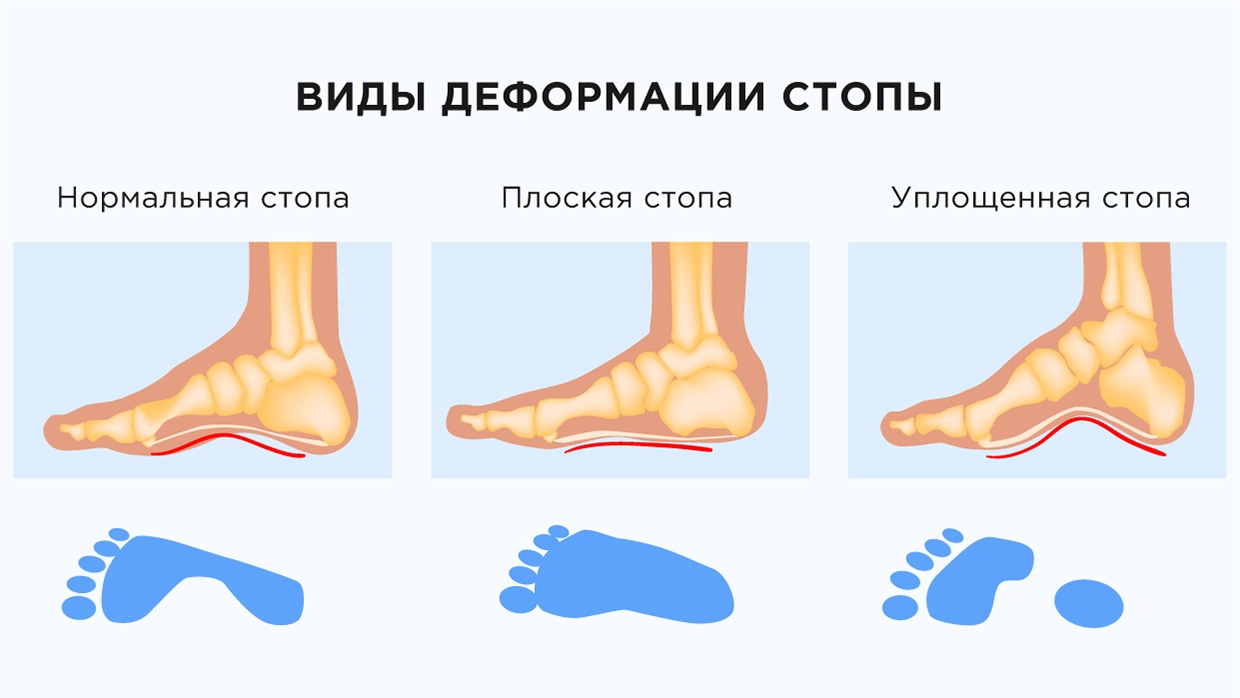
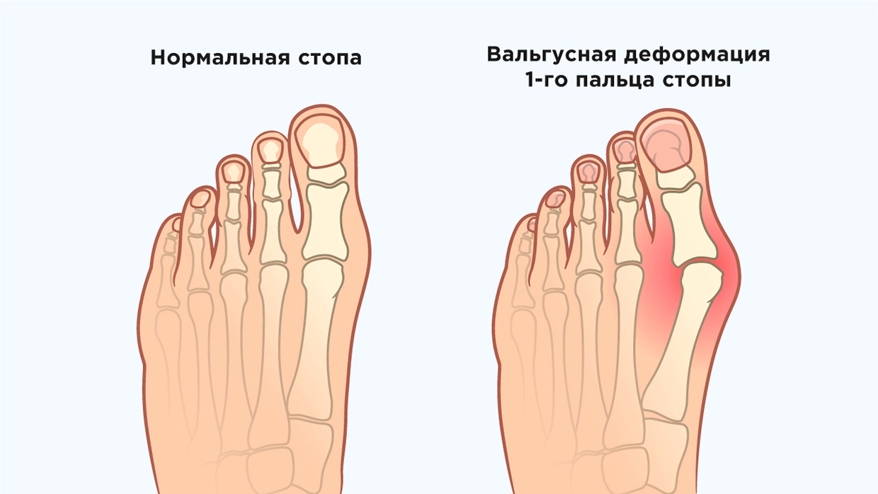
Transverse flatfoot
Excess weight, weight bearing and pregnancy put strain on the foot and cause it to flatten. Most patients with transverse flatfoot are women in their 30s and 40s. Sometimes the problem also occurs in children. A visible consequence of this type of flat foot is, among others, the following hallux valgus – This is a thickening on the big toe. Sometimes a callus forms on it, which can become inflamed. The little toes begin to deform and take the shape of a hammer toe. Women with a foot deformity may have difficulty putting on shoes, and the toes, metatarsals, and back of the foot may begin to ache and swell.
Causes of flat feet
- vitamin deficiency;
- Diseases of the musculoskeletal system and metabolic disorders;
- sedentary lifestyle;
- injuries, including congenital injuries;
- Rickets;
- hereditary factors;
- rapid growth of the child;
- unsuitable footwear (that cannot support the foot).
Premature babies and children who have had an intrauterine infection are most commonly affected. Children with hip dysplasia are also at risk.
How can an osteopath treat this problem?
Osteopathy treats the body as a whole, which is an undeniable advantage of treatment over other methods. The osteopath treats the cause, the essence of the problem, and not the effect. He works on the leg to relieve stiffness and muscle tension, improve blood circulation and give the foot the correct physiological alignment, and he also works on the spine, as vertebral misalignments that are not visible to the eye have a negative impact on functioning affect the entire body. After the session, the osteopath shows the practitioner a series of exercises to consolidate the effect. The number of sessions depends on the individual, but is usually around three, depending on the severity of the deformity.
The pathology is successfully corrected at an early age. If your child is struggling with this, don't hesitate to see an osteopath. Our center will help you forget about this problem.
Read more:- flat feet (valgus foot).
- Massage for flat feet.
- Which doctor treats flat feet?.
- flat feet.
- Shoes for flat feet.
- flat feet and valgus.
- Orthotics for flat feet.
- Supinators for flat feet.