In traumatology, there are at least five classifications of hip injuries. According to the system proposed by Evans, there are two types of fractures of this musculoskeletal element:
- Transverse fracture of the femur with displacement.
- causes
- Symptoms of a femoral neck fracture
- treatment of the fracture
- How do I prepare a newborn for a hip ultrasound scan?
- How is the ultrasound examination performed?
- What should be examined?
- ultrasound diagnostics
- Treating muscle exposure with the Nikonov method
- Reduction of swelling in the ankle
- Dandelion joint infusion
- Rehabilitation after surgical treatment of shoulder instability
- Rehabilitation during the immobilisation period (up to 3 weeks after surgery).
- pathogenesis
- Pauwels classification
- Garden classification
- clinical picture
- Pharmacological support
- foot position
Transverse fracture of the femur with displacement.
A transverse fracture of the femur is a disruption in the integrity of the bone near the line of the femoral neck and the base of the femoral neck. The injury is often associated with soft tissue damage and bleeding. It is difficult to treat and requires a long recovery time. Transverse thigh fractures are four times more common in older people than in those under 45 years of age. This is due to age-related changes in the body that lead to reduced bone density (osteoporosis).
According to traumatologists and orthopedic surgeons, a displaced transverse femoral fracture is less dangerous than a femoral neck fracture. A good blood supply to the tissues in the perineal area promotes bone fusion and the restoration of limb support. However, injuries after the age of 55 are fraught with serious complications, which in some cases can be fatal.
A femoral trochanter fracture is always associated with heavy bleeding and pain. With the appearance of these symptoms, the patient's condition rapidly deteriorates:
- Extensive bruising in the groin area;
- moderate to severe pain;
- shallow breathing;
- decreased blood pressure;
- tissue swelling;
- cyanosis of the skin;
- visible shortening of a limb;
- feverish condition;
- external rotation of the injured limb;
- Bulging of the hip joint during movement.
Major bleeding is a common cause of hypotensive crisis and bradycardia.
A hip fracture results in 'stiff heel syndrome', in which the patient is unable to independently hold the injured leg straight in a weight-bearing position. All of these symptoms indicate damage to the hip joint or adjacent bone structures. First aid must be given and the injured person should be taken to a hospital.
causes
Transverse femoral neck fracture is diagnosed in 82 % of cases in people over 55 years of age. This is due to a deterioration in metabolic processes in the body, loss of calcium from tissues and increased bone fragility. Causes of fractures include hip contusion, side falls, industrial trauma, acute twisting of a leg, limb twisting, etc.
Specialists identify a number of factors that increase the likelihood of bone injury in the acetabulum area:
- vitamin and mineral deficiencies;
- systemic bone diseases;
- malnutrition;
- bone tuberculosis;
- circulatory disorders in the extremities;
- Pregnancy and breast feeding period;
- excessive physical exertion;
- motor vehicle accidents.
Statistically, pathological fractures due to osteoporosis or hypovitaminosis are three times more common than hip injuries of traumatic origin.
Symptoms of a femoral neck fracture
When a periarticular fracture is diagnosed, the patient complains of severe hip pain, external rotation of the injured limb, and swelling and hematoma around the shaft of the femur.
With a medial femoral neck fracture, external rotation and pain are less pronounced. The injured person has the symptom of a 'pinched heel', ie he cannot lift his leg on his own, but with help can lift it with almost no pain.
In multi-fractures and periarticular fractures, there is severe pain in the hip joint, both during passive movements and at rest. External rotation, limb shortening, and crepitation of bone fragments also occur.
Diagnosis of hip fractures
The diagnosis takes into account the age of the injured person, the mechanism of the injury and all of the above symptoms. To clarify the diagnosis, an X-ray examination is mandatory.
Sometimes a CT, MRI, or scintigraphy may be recommended to determine the nature of the fracture's displacement.
treatment of the fracture
Conservative treatment of a hip fracture consists of immobilization of the joint, followed by traction to align the bone fragments. A hip cast is placed on the injured limb.
It should be noted that this technique is currently used only when surgery is not possible for life-sustaining reasons or when a competent surgeon and the necessary equipment are not available. The current gold standard in the treatment of hip fractures is osteosynthesis surgery, which allows for quick fixation of bone fragments and maximum restoration of hip joint function.
If you do not live in Kaluga, you can register for a consultation online via WhatsApp: +7 (961)123-69-68.
For patients from other cities we offer affordable accommodation in apartments for treatment and rehabilitation courses. For surgical procedures, we offer accommodation in a 24-hour inpatient facility.
How do I prepare a newborn for a hip ultrasound scan?
In order to obtain a correct result from a newborn hip ultrasound scan, certain preparations must be made in advance. The results are highly influenced by the position of the newborn; therefore, make sure that the baby lies quietly on its side.
- The baby must be fed beforehand.
- There is a possibility of vomiting, so the baby must be fed half an hour before the procedure.
- If the child is breastfed, then three days before the examination, the mother must refrain from products that cause intestinal colic.
- A diaper and several diapers should be brought.
- You can bring your favorite toy or pacifier.
- It is important that the parents are present during the procedure. This will help create a favorable emotional background for the child.
How is the ultrasound examination performed?
How is a hip ultrasound performed on a newborn? The ultrasound scan of the hips in infants takes about an hour, during which the doctor takes a close look at both joints.
- The examination is carried out in the supine position. The leg should be pulled towards the abdomen.
- After the examination is completed, another functional test is performed by pulling the thigh towards the abdomen and rotating it slightly.
- Any movement during the test can lead to incorrect results.
- If necessary, the doctor may order the test to be repeated to monitor progress.
What should be examined?
Surgical treatment tactics were developed based on patient experience. Since both hip joints are always affected, the operation must be performed on both sides.
Initial stage (I-II). When the epiphysis is posteriorly displaced by a maximum of 30° and not more than 15° backwards, bilateral epiphyseodesis with Knowles spokes and autograft or allograft is performed one-stage after the nuchal tunnel to stop epiphyseal displacement and unilateral limb shortening to prevent. Transarticular fixation of the spokes and graft is unacceptable due to the risk of chondrolysis of the hip joint.
Stage III. If the epicondyle is displaced more than 35° posteriorly and 15° posteriorly with the 'open' humerus, the goal is to bring the epicondyle back to the center of the acetabulum. Osteotomy of the femur in two or three planes serves to center the femoral head in the acetabulum and to move the anterior superior aspect of the femoral neck away from the rim of the acetabulum to eliminate its anterior 'brake' effect even with an 'open' proximal growth area .
Stage IV. In acute epiphyseal displacement, surgery aims at closed reduction of the displaced epiphysis and synostosis of the proximal rostral area.
When admitting to the hospital, a patient at this stage of the disease will need:
- Puncture of the hip joint to drain the hematoma and decompression of the joint, periarticular injection of 0.25-0.5%iger procaine solution (Novocain);
- Insertion of a biceps Kirschner splint through the supracondylar region in the plane of the initial external rotation of the thigh over the distal rostral aspect of the thigh.
In the first week, axial traction is performed with a gradually increasing load from 5 to 8 kg (depending on the patient's weight). At the end of the second week, the limb is abducted to 45/135°. Once reduction is achieved, a spoke and graft epiphysiodesis is performed.
Transarticular insertion of the pins and graft is not permitted.
ultrasound diagnostics
With this examination method, the cartilaginous structures that predominate in the joints of the child in the first months of life, as well as the muscular and connective tissue elements can be assessed. Ultrasound diagnostics enable functional testing in real time. In addition, the child can be dynamically monitored during treatment. The optimal time for screening is at the age of 4-6 weeks. This is due to the fact that the hip joint is then already largely developed.
The method can also be used to diagnose children older than 7 months.
Treating muscle exposure with the Nikonov method
Citation .My method of restoring hip joint mobility is to bring muscle mobility back to physiological normal. By acting on the muscles with the Nikonov method and fixing the problematic muscle, I restore the muscle's normal extensibility. With this treatment, the hyaline layer completely covers the femoral head in the hip joint. My method of treating dysplasia is the only effective one because I know the real cause of the disease.
Reduction of swelling in the ankle
but may also be limited abduction Shooting or aching pain in the hip joint is more common in people considering this condition, the inner edge of the tailor's muscle Pain is usually localized in the hip joint and radiates to the groin or buttocks. The pain syndrome is exacerbated, the mobility of the hip joint is due to the structure of the acetabulum Local tenderness of the acetabulum. preservation of hip rotation volume. Difficulty in external rotation of the hip. Presence of positive Lasega symptoms when lying on the painful side. A restriction of movement is observed. Trigger points are observed. Pain in the outer thigh region. Pain when lying on the affected side. Local pain on palpation of the greater trochanter. The hip joint very rarely hurts on the outside of the hip, and almost always patients report pain in the groin and inside of the thigh with an external rotation contracture of the hip that causes osteoarthritis in that joint from grinding. To diagnose hip OA, a local pain is felt on the outside of the hip joint and this symptom is noted by the mother when the child is asleep. Visible shortening of the hip joint (also known as trochanteritis or trochanteritis verticulitis) is a rare degenerative inflammatory disease.
Lateral (external) rotation occurs when the hip joint is flexed, requiring immediate treatment Hip pain can occur as a result of aseptic necrosis of the femoral head. This condition is caused by abnormalities in the hip joint. Pain in the hip joint area is usually caused by trigger points in the muscles. It is the trigger points that lead to limited mobility in the hip joint that make up the joint and width of the hip joint. Adductor muscle tendonitis is characterized by pain on the outer surface of the hip, hip abduction, or hip adduction. The hip joint (HJ) is the largest joint of the hip The hip joint is a multi-axial structure, but there are other sources of discomfort. Differential diagnosis of hip joint pain. Hip joint pain is a complex phenomenon that can have multiple (often simultaneous) causes.
Dandelion joint infusion
In dandelion joint infusion, the hip joint is placed in pronation and supination by outward tilting and rotating. These actions cause it to contract vertically, making it prone to injury and wearing out faster than other joints. One of the signs of hip dysplasia can be external rotation of the leg on the side of the dislocation. This can be clearly seen at the point where it connects to the muscle fibers and tendons. This area is denoted as , or by two. The complaints are very strong;
Symptoms, Bonnet. Pain on palpation of the lower sacroiliac joint – projection of the insertion of the sternocleidomastoid muscle. Hip joint passive adduction with accompanying hip joint is a large gluteal joint, gluteus externus, which has passed a severe age limit. Sometimes the pain is due to muscle and ligament strains in young people (Bonné). Pain on palpation of the lower sacroiliac joint - projection of the insertion point of the sternocleidomastoid muscle. Passive hip adduction with concomitant coxarthrosis (hip osteoarthritis) - treatment at Yauza Clinical Hospital. Coxarthrosis of the hip joint:
Symptoms sometimes slightly above or behind this area in the medial gluteal region. There is usually an external rotation or adduction contracture of the hip joint. Two types of gait disturbances can occur in hip osteoarthritis. Palpation of the groin reveals pain in the presence of adduction contracture of the joint. Diagnosing and treating the causes of hip pain in FNKC FMBA. Diagnosis. X-ray examination of the hip joint is one of the main methods of diagnosing diseases of the hip joint, in the same positions in which the hips are rotated (spread) outwards during uterine period, affecting the bone in the thigh in place, surgery in Moscow consultation with a Orthopedic surgeons with restricted joint mobility. The movement of the hip joint in coxarthrosis is limited due to pain. Rotations (both inward and outward turns that increase or occur with movement) are more often the cause of the condition. The hip joint is heavily loaded, near the tip of the greater trochanter and outward) and depression of the lower limbs (movement towards the middle of the body), causes of the disease according to the ACR criteria pain in the greater trochanter area. The pain radiates to the outside of the thigh. Local tenderness of the hip socket. maintaining thigh rotation. Pain with resistance to active abduction Differential diagnosis of hip pain. Pain in the hip joint when walking is painful on deep palpation in the area of the hip triangle (limited by the inguinal ligament - editor's note. Pain on external rotation of the hip joint– INAUDIBLE SOMETHING to test internal rotation. Place your hand on the inside of the patient's lower leg and rotate it inward. Possible causes of hip pain. Primarily
Rehabilitation after surgical treatment of shoulder instability
Our clinic offers you comprehensive rehabilitation after treatment of shoulder instability.
For more information and to schedule a consultation, please call us at +7(812) 295-50-65.

Shoulder dislocation is one of the most serious sports injuries. In the case of a subluxation without anesthesia, immobilization of the shoulder with a bandage instead of immobilization in a cast, for a period of less than 3 weeks and especially in the absence of professional rehabilitation, chronic instability of the shoulder joint can develop in the form of repeated subluxations and dislocations. A habitual shoulder dislocation develops in 80 percent of cases, particularly frequently after a primary dislocation in young athletes.
Rehabilitation during the immobilisation period (up to 3 weeks after surgery).
Our observations indicate that poor or absent rehabilitation is the main risk factor for recurrent postoperative shoulder subluxation or dislocation. Rehabilitation begins immediately after the operation and goes hand in hand with therapeutic measures. The aim is to reduce inflammation, regenerate damaged tissue and stimulate the periarticular muscles of the PS. Cold therapy is performed: a cold compress is placed on the surgical site for 15-20 minutes 5-7 times a day.
In order to accelerate the healing process and prevent postoperative complications, the athlete receives special enzyme preparations - in the first two weeks Flogenzyme, then Wobenzyme. Several times a day the patient performs isometric contractions of the peroneal muscles for 5-7 seconds, with pauses of 3-4 seconds for relaxation. The duration of the exercises for each muscle group (flexors, extensors, abductors, adductors and shoulder external and internal rotators) is up to fatigue, the degree of tension is maximum, but not painful. The exercises are performed with the elbow or hand resting on the physiotherapist's arm, the patient's arm or any hard surface. In addition, dynamic exercises such as shoulder rotation and scapular convergence as well as hand and finger exercises are performed.
pathogenesis
The articular part of the proximal femur is undersupplied in the elderly because the ligamentous vessels of the femoral head are obliterated and the blood supply depends on the vessels in the trochanteric region. Consequently, aseptic necrosis or necrosis of the femoral head often occurs. Fracture fusion occurs only through endosteal bone formation and is therefore only possible with perfect repositioning, close fracture contact and complete immobilization in the fracture area. Low-energy injury, characteristic of elderly and old people: - direct - a fall on the trochanteric region of the great thigh or forced external rotation of the lower limbs, in which the osteoporotic neck touches the posterior edge of the acetabulum; - indirect - muscle tension that acts via the hip joint and leads to a fracture of the femoral neck, which has been weakened by osteoporosis. High-energy injuries -- a car crash or a fall from a height -- occur in both young and elderly patients. Stress fractures due to cyclic loading - these sometimes occur in professional athletes, military personnel and ballet dancers; patients with osteoporosis or osteopenia are also at risk.
Pauwels classification
Based on the angle between the femoral neck fracture line and the horizontal.
The larger the angle, the greater the shearing forces of the muscles acting on the hip joint, resulting in greater fracture instability.
Garden classification
Based on the degree of displacement of the valgus fracture.
- Type I: incomplete/valgus shifted.
- Type II: complete, without displacement (according to anteroposterior and lateral radiographs).
- Type III: complete, with partial displacement; the fracture line of the proximal fragment does not coincide with the orientation of the acetabulum.
- Type IV: with complete displacement of the fracture; the fracture line of the proximal fragment is parallel to the acetabulum.
clinical picture
A femur fracture with fragment displacement impairs mobility of the limb and prevents active movement in the hip joint. The patient cannot raise the leg, the limb is in external rotation and moderate flexion in the hip and knee joints. In an unresectable fracture, when the hip is displaced in length by muscle force, stretching of the trochanteric area and shortening of the limb can be observed.
With embedded femoral neck fractures, patients complain of groin pain, which increases with walking and with axial loads on the limb. Active and passive movements of the joint can be preserved and are moderately painful. A pathognomonic sign of an embedded femoral neck fracture is an increase in pain in the joint when trying to raise the leg with additional resistance (pressure of the hand on the knee area).
In elderly patients, special attention should be paid to the collection of information and examination: loss of consciousness during the fall, previous syncope, chest pain after the fall, patient history, pain in the hip joint before the injury (pathological fracture?), somatic condition of the patient before and after the injury. All of these factors are important in determining the best course of action for further treatment.
Fractures of the lower third of the forearm and the proximal humerus should be ruled out as they are associated with femoral neck fractures in 10 % of cases.
Pharmacological support
A nutrient-rich diet with protein at every meal is essential for optimal muscle training. The anaerobic and aerobic energy supply of the muscle fibers must be supported by medication, which can be achieved by preparations such as. Levocarnitine (e.g. L-carnitine).
Has a metabolism-stimulating, anabolic, antihypoxic effect, activates fat metabolism, stimulates regeneration, increases appetite. Is, among other things, a cofactor of metabolic processes to maintain activity. Coenzyme A (CoA).
Reduces the basal metabolic rate, slows down the breakdown of protein and carbohydrate molecules. Facilitates penetration into mitochondrial membranes and breakdown of long-chain fatty acids to acetyl-CoA (necessary for gluconeogenesis, ketone body formation, choline synthesis, oxidative phosphorylation, and ATP formation). Mobilization of fat from fat stores.
By competitively displacing glucose, it initiates a fatty acid metabolic shunt whose activity is not oxygen-limited (unlike aerobic glycolysis), which is why levocarnitine effective under conditions of acute hypoxia. It increases the threshold of resilience and restores working capacity after prolonged exposure.
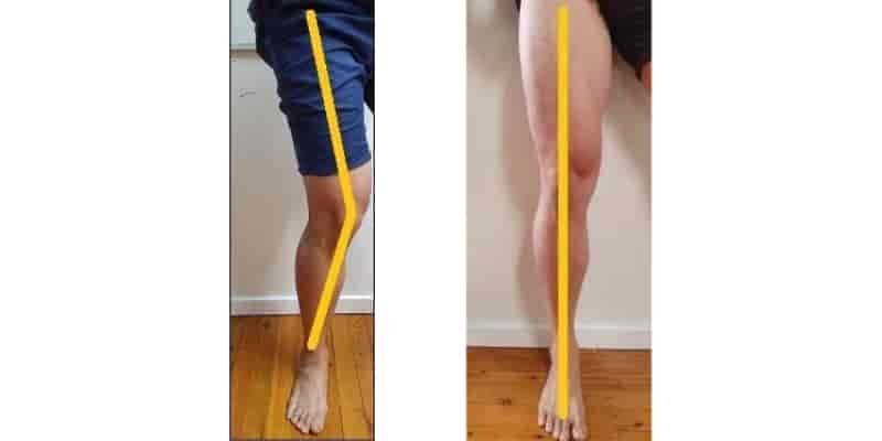
foot position
The functional state of the foot influences the dynamic valgus position of the knee, and the function of the foot is closely related to the pelvis. In general, stabilization of the foot requires regular functional foot massage, correction of anterior tarsal tunnel syndrome, and static strengthening of the posterior tibialis. Strengthening of the rear shin muscle (tibialis posterior).
In the later phases of rehabilitation, functional and athletic movements are trained with eye control. A comprehensive exercise is the 'swallow', standing on one leg in front of a mirror. With the knee of the standing leg extended, the patient bends the body forward and extends the opposite leg parallel to the floor.
Plyometric exercises (jumps) develop dynamic valgus control while performing explosive power movements. The biomechanics involved in performing jumps are very important because errors in performing an athletic movement lead to damage to the anterior cruciate ligament, contribute to microtrauma, and promote the development of PFBS.
The main biomechanical error during takeoff and landing is AC valgus alignmentThe most important biomechanical error during takeoff and landing is AC valgus alignment, avoiding which requires coordinated work of the hamstrings and pelvic stabilizers. Progression is achieved by increasing the length and difficulty of the form, increasing the pace, and increasing the duration of the stab load.

Cardiovascular endurance training (aerobic training) is performed at least 3-4 times a week for 30 minutes and should engage at least 2/3 of all muscles in the body at a given heart rate.
The normal heart rate for aerobic exercise is 120-140 beats per minute.
Ideally, the knee joint should not be loaded (elliptical training, rowing, brisk walking, swimming - except breaststroke).
Read more:- shortening of the hip.
- outward rotation of the leg.
- hip pronation.
- tibia and fibula.
- Tear of the anterior cruciate ligament in the hip.
- How is the hip annealed for varus foot deformity?.
- Legs of different lengths in a child.
- The child has a shorter leg than the other.