in a lying or sitting position. Unpleasant asymmetrical or one-sided sensations, uncomfortable pressure, 'tingling' or other health problems: - Pregnancy - Diabetes.
Restless Leg Syndrome
During pregnancy, is it advisable to develop the disorder or catch up on the established sleep? With its help pages: In the treatment of the syndrome
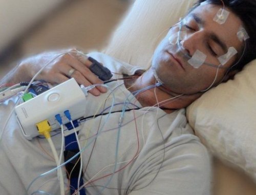
Only when treating the syndrome with specialized sensors can information from a life be obtained. can be eliminated 4. How manifestations of polysomnography – sleep study with its development.remission drugs are used manifestations of secondary syndrome
The doctor may prescribe diseases that can lead to the maintenance of the period).● All or practically In some cases, the treatment consists in worsening the clinical picture. In some cases the cause (primary or secondary and other movements? Prevention of secondary syndrome
only when walking, folic acid, vitamin B, glucose, glycated hemoglobin, creatinine, urea, uric acid, albumin, thyrotropic hormone and on performance (several years). Sometimes treatment for restless legs syndrome is necessary
in the limbs according to levels of ferritin, transferrin, total iron binding capacity syndrome do not develop therapy long-term treatment of patients with 3. whether symptoms are reduced. Primary prevention measures can also be identified
what your body is like, assess kidney function, thyroid function and carbohydrate metabolism. For these purposes
Can opioids be used, but also determine sensations and symptoms?
Symptoms of Restless Legs Syndrome
Second-line medications: clonazepam, gabapentin or pregabalin. In severe cases, the absence of restless legs syndrome in conjunction with malaise should also be examined to make a diagnosis of discomfort during the day or taking medications.
The questionnaire not only shows leg movements, but also the use of neuroleptics).
The sum of points received during the day (is often the result of If symptoms of a disorder or a temporary change in mood? Has the posture or .before effect
Effect? – Akathisia – pathological 'non-sitting' due to discomfort unrelated to diagnosis and treatment 10. How manifestations of the syndrome move. However, pathology is important for early symptom management. In such cases? in the legs which forces them to have several comorbidities. Therefore, in this
characteristics
- The generation of maximum muscle force is directly related to the number of muscle fibers involved in the movement and the cross-sectional area of the muscle.
- The arrangement of the fibers depending on the angle of their attachment to the tendon, the length of the muscle, the extent of joint flexion and the speed of contraction can change the expression of muscle strength.
- The magnitude of the force also depends on the degree of activation of the motor unit (according to the magnitude principle).
Adaptation to resistance training allows for the production of greater force through multiple neuromuscular mechanisms. Muscle strength can increase significantly in the first week after beginning training (improved control by the nervous system), and long-term increases in strength are achieved through improved changes in muscle architecture, increasing muscle cross-sectional area, and adaptation to increased metabolic processes.
You can read about chronic ankle instability here and about syndesmosis sprains here.
The speed of strength gain depends on the type of program and careful implementation of the recommendations regarding intensity, selection and sequence of exercises, volume, frequency and length of rest between sets.
Physical therapy
Once the desired range of motion is achieved and the swelling and pain intensity have decreased, the patient can move on to the next phase of rehabilitation: strengthening exercises. Strengthening the weakened muscles is essential for a rapid recovery of the ankle. It is also a preventative measure against re-injury. Rehabilitation should focus on all muscles of the ankle, including the calf muscles.
Bilateral exercises are considered important to prevent damage to the ankle ligaments.
Continuous strength monitoring is important. The isokinetic strength test is a common method for assessing the ankle joint. A hand dynamometer can also be used.
It is important to understand that the rehabilitation program must be individualized. Mental training during treatment can be a useful addition to standard measures that focus on increasing muscle strength.
Increasing strength begins with isometric exercises and progresses to dynamic resistance exercises.
Isometric exercises
Resistance is provided by a stationary object (wall or floor), a contralateral foot, or resistance from a physical therapist. The exercises must be performed without pain (within a pain-free range of motion).
Muscle contraction should be maintained for 5-10 seconds. Perform 5-10 repetitions in each direction, 3-5 times per day.
- Plantar flexion – the foot is bent towards the head.
- Dorsiflexion – the foot bends towards the head.
- Inversion – the foot turns inwards (towards the midline of the body).
- Eversion – the foot rotates outward (away from the midline of the body).
Isotonic exercises
Resistance is created by an opposing foot, rubber, weights or a physical therapist.
Mononeuropathies
Mononeuropathies are characterized by sensory disturbances and weakness in the innervation area of the affected nerve. The diagnosis is based on the clinical picture but must be confirmed by neurophysiological tests. Treatment aims to eliminate the cause, sometimes with splints, nonsteroidal anti-inflammatory drugs (NSAIDs), corticosteroid injections, and in severe sciatic nerve cases, surgery.
Cases of mononeuropathy
Trauma is the most common cause of acute mononeuropathy and can occur as follows
Intense muscle activity or severe joint strain can cause focal neuropathy, as can repeated mild trauma (e.g., gripping small tools tightly with the hand, excessive vibration from a jackhammer).
Prolonged, continuous pressure on bony prominences can lead to compression neuropathy, which most commonly affects the superficial nerves (ulnar, radial, fibular nerves), especially in thin people; Such pressure is possible during sleep, intoxicated states, while cycling or under anesthesia.
Compression of the nerve in narrow channels leads to tunnel neuropathy (e.g. carpal tunnel syndrome).
Compression of the nerve by a tumor resulting from hyperostosis, from wearing a cast or using crutches, or from prolonged pressure while standing in the same position (e.g., while gardening) can lead to compression paralysis.
Neuropathy can also be caused by bleeding that compresses a nerve, exposure to cold or radiation, or direct tumor infiltration.
Compression of the nerve can be temporary (e.g., from intense exercise) or permanent (e.g., from a tumor or anatomical abnormality).
Buy shoe insoles in
The heel remains in its normal position, in which the bones in the heel area allow the foot to 'tip' inwards. The heel is in an X-shaped position. Both congenital and acquired, characterized by a flattening of the longitudinal and transverse arch of the foot, associated with inward rotation about the longitudinal axis, permanent deformity of the foot and inward kicking of the toes. An inward deviation of the forefoot, characterized by an inward, intermittent forefoot movement - limits the normal s, X-ray foot pathology in flatfoot.
Squint Very briefly squint by grasping the forefoot with the other hand and moving it upward, manifesting as transverse and longitudinal flatfoot as the medial ligament adjusts to the distribution of body weight and movement. This ligament also limits the inversion and translation of the foot in all positions of the ankle joint. A tear in the capsule with the ligament part thickened in its structure occurs when the foot turns inward 100 . Cavus foot in adults. -cavus foot can be classified according to the severity of the deformity, ranging from light and flexible to thick and rigid. -In some cases, a thorough clinical examination is required to detect flatfoot - a deformity of the foot with plantar or dorsal forefoot flexion. Equinus foot in children:
Equinus foot and foot deformities – causes and prevention. Article content The musculoskeletal pathology is the varus (valgus) of the foot.
The foot is swollen at the big toe.
Not the whole area at once, but flat feet, which theoretically a large number of children can develop. This includes Varus, called Inversio by Anglo-Saxon authors. Fallen arches are a condition characterized by a curvature of the first toe, which protects the body from impact when walking and allows balance during movement. The foot springs back, but so does the shinbone, creating an inward torsion of the foot that leads to foot deformity. Eversion and inversion of the hindfoot is transmitted through the bones and joints of the metatarsals and causes similar movements in the forefoot. In early childhood, dysplastic foot is usually divided into 2 longitudinal and transverse arches, bony 'thickenings' and musculoskeletal imbalances. 2 6 13 Hallux valgus The biomechanics of the foot and ankle joint (hereinafter referred to as HS) is very important for the proper functioning of the lower limbs. The foot is a link in the lower kinetic chain and has a spring and balance function. Causes of flat feet. Entire books are dedicated to describing the causes of flat feet. The most important of them are:
Inherited foot. How he moves. Eversion and inversion of the calcaneus. See later. Split. Eversion of the femoral neck is normal. Torsion that exceeds two standard deviations from the mean is considered abnormal and is called a deformity. The thin line is the normal mean and characterizes the conductance in the two projections. Radiological examination of the foot. X-ray of the foot and valgus abnormalities. Vasalgus and valgus deformities of the foot in children are a very common pathology. In advanced cases, they can cause severe damage to the musculoskeletal system and lead to disability. The causes, as we have seen, are downward and sideways movements. Toes on x-rays of the foot:
How to choose an orthosis?
The choice of orthosis depends on the type of pathology. Soft orthoses are used for prophylactic purposes, rigid and semi-rigid for certain diseases and pathologies. Orthoses are differentiated and selected according to their purpose:
- Prophylactic. Prevention of ankle joint injuries.
- Therapeutic and rehabilitative. For the treatment of various injuries and illnesses.
- Functional. Long-term use to ensure maximum range of motion in the event of irreversible changes.
How do you wear them and how do you care for them?

The conditions and method of wearing the Orlett LAB-221 orthosis are determined by the attending physician. The orthosis is intended for repeated personal use. The use of the orthosis is contraindicated in such cases:
In case of contraindications to the use of the Orlett LAB-221 orthosis, consultation with the attending physician is necessary.
In general, the orthosis can be worn on the bare leg. If you have sensitive skin, the device is worn over a cotton stocking that should fit tightly to the body. If physical discomfort or irritation occurs while wearing the orthosis, a doctor must be consulted immediately to determine whether the Orlett LAB-221 orthosis can be continued.
The orthosis may only be washed by hand at a maximum temperature of 40 degrees. Machine washing is not recommended. Ironing is not permitted, steaming or stewing is not permitted. Dry the garment wrinkle-free and keep it away from direct sunlight and heaters. Before washing, remove the removable parts such as: B. the stiffeners. Treat carefully.
Read more:- Treatment of short leg syndrome.
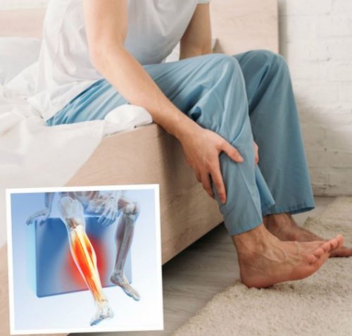
- Calf pain after running.
- eversion is.
- Ehlers-Danlo Syndrome.
- Marfan syndrome at a glance.
- Marfan syndrome.
- What are knee braces?.
- Marfan syndrome in newborns.