Since uterine prolapse is the final stage of organ movement into the vagina, symptoms develop gradually over a long period of time.
![]()
- Anatomical terms for movement – Anatomical terms for movement
- classification
- Abnormal movement
- classification
- Abnormal movement
- General movement
- flexion and extension
- adduction and adduction
- Cartilage thermolysis is a procedure in which the T-cartilage is exposed to high temperatures. This reshapes the cartilage so that it can bend according to the surgeon's wishes.
- Planes and axes in biomechanics
- Designations of body movements in biomechanics
- Damage to the medial collateral ligaments of the ankle
- MRI
- Video about our clinic for orthopedics and traumatology
- What will the doctor see during the gynecological examination?
- Possible complications
- 7.1 Infection
- 7.2 Constipation
- 7.3 Vascular disorders
- 7.4 Disorders of the urinary tract
- Arthrodesis surgery to correct an overpronated foot
- Intimate vaginoplasty after pregnancy and childbirth
- causes
- Treatment of lower eyelid ectropion
- Prognosis and prevention of lower eyelid exophthalmos
Anatomical terms for movement – Anatomical terms for movement
The movement, the process of locomotion, is described using specific anatomical terms. Movement includes the movement of organs, joints, limbs and certain parts of the body. The terminology used describes this movement by its direction in relation to the anatomical position of the joints. Anatomists use a consistent set of terms to describe most movements, although other, more specific terms such as arm, leg, and eye movements are required to describe the uniqueness of movements.
In general, movement is classified according to the anatomical plane in which it occurs. diffraction i stretch are examples of Angular movement in which the two axes of the joint move towards or away from each other. rotation Movements can also occur in other joints, e.g. B. in the shoulder joint, and are called internal or external. Other terms like Height i deepeningdescribe the movement above or below the horizontal plane. Many anatomical terms are derived from Latin terms with the same meaning.
classification
Movements are classified according to the anatomical planes in which they occur [1], although a movement is usually a combination of different movements that occur simultaneously in several planes. [2] Movements can be classified according to the type of joints involved:
- Slide Movements take place between flat surfaces, such as: B. in the intervertebral discs or between the wrist and the metacarpal bones of the hand. [1]
- Angular movement Movements occur through the synovial joints and increase or decrease the angles between the bones. [1]
- rotational movements Movements cause the structure to rotate along the longitudinal axis, e.g. B. Turning the head to look in any direction. [3]
In addition, the movement can also be divided into:
- linear movements (or Interpreter movement) that move along a line between two points. A straight line movement Movement is movement along a straight line between two points while a curvilinear Movement is movement along a curved path. [2]
- Angular movement movement (or Rotary movement Angular motion (or rotational motion) occurs when one object rotates around another object, increasing or decreasing the angle. The different parts of the object do not move at the same distance. Examples of this include knee movements in which the lower leg changes its angle relative to the thigh, or ankle movements. [2]
The study of movement is called kinesiology. [4] For a categorical list of human body movements and the muscles involved, see List of human body movements.
Abnormal movement
prefix hyper- is sometimes added to describe movements outside the normal range, e.g. B. at Hypermobility, Hyperflexion or Hyperextension. Range of motion describes the total range of motion that a joint can perform. [5] For example, if a body part, such as a joint, is hyperextended or 'bent backwards' due to hyperextension, it can be described as overstretched.. Hyperextension increases stress on the joint's ligaments and is not always caused by voluntary movement. It can be the result of accidents, falls or other causes of injury. It can also be used in surgery, for example to temporarily dislocate joints during surgical procedures. [It can also be used as a method to force a person to do something, e.g. B. to enable a police officer to make an arrest.
classification
Movements are classified according to the anatomical planes in which they occur, although a movement is usually a combination of different movements that occur in multiple planes at the same time. The movements can be classified according to the type of joints involved:
- Slide Movements take place between flat surfaces, such as: B. in the intervertebral discs or between the wrist and the metacarpal bones of the hand.
- Angular movements Angular movements occur in the synovial joints and cause the angle between the bones to increase or decrease.
- Rotary movements During rotational movements, the structure is rotated about its longitudinal axis, e.g. B. when you turn your head to look in either direction.
In addition, the movements can also be divided into the following areas:
- Linear movements (or Translational movements Movements that move in a line between two points. Linear curvilinear is a movement along a curvilinear path.
- Angular movement (or Rotary movement Rotational motion occurs when one object moves around another object, increasing or decreasing the angle. Different parts of an object do not move the same distance. Examples of this include the movement of the knee, in which the lower leg changes its angle relative to the femur, or the movement of the ankle.
The study of movement is called kinesiology. For a categorical listing of human body movements and the muscles involved, see the list of human body movements.
Abnormal movement
prefix hyper- is sometimes added to describe movements outside the norm, for example at Hypermobility , Hyperflexia or Hyperextension . Range of motion describes the entire range of motion that a joint can perform. For example, if a body part, such as a joint, is hyperextended or 'bent backwards' due to hyperextension, this can be described as overstretched. . Hyperextension increases tension on the joint's ligaments and is not always the result of voluntary movement. It can be the result of an accident, a fall or other causes of injury. It can also be used in surgery, for example to temporarily dislocate joints during surgical procedures. It can also be used to relieve pain, to force a person to do certain actions, such as: B. to enable a police officer to take a person into custody.
General movement
These are general terms that can be used to describe most body movements. Most terms have a unique counterpart, which is why they are considered in pairs.
flexion and extension

Flexion and extension describe movements that affect the angle between two body parts. The terms are derived from Latin words with the same meaning.
diffraction describes a bending movement that reduced reduces the angle between a segment and its proximal segment. Bending the hand at the elbow or clenching the hand into a fist are examples of flexion. When a person sits, their knees are bent. If a joint can move back and forth, such as B. in the following case. Neck and torso: Flexion is a forward movement. When the chin rests on the chest, the head and torso flex as the person leans forward. An arm or hip flexion is a forward movement of the arm or leg.
stretch is the opposite of flexion and describes an extension movement enlarged increases the angle between body parts. For example, when you stand up, your knees are stretched. If a joint can move forward and backward, such as: B. the neck and torso, an extension is a backward movement. Extension of the hip or shoulder causes the arm or leg to move backwards. A backward movement of the distal segment also causes extension in the other joints of the upper limbs, the elbow and the wrist. An exception is the knee, ankle and wrist, where the distal end must move forward to be considered upright. Hyperextension – is any stretch that exceeds 180 degrees and becomes reflexive.
For the toes, flexion means they curve downward and extension means they stretch or lift upward.
adduction and adduction

Cartilage thermolysis is a procedure in which the T-cartilage is exposed to high temperatures. This reshapes the cartilage so that it can bend according to the surgeon's wishes.
- The average duration of the procedure is about five minutes per eye.
- There are no seams, nothing to expose
- No cuts.
- The shape of the cartilage is restored rather than the 'excess' cartilage being removed.
The only downside is that some of my patients experience a prolonged period of swelling and inflammation after surgery. This is normal since the operation is a type of burn. The average rehabilitation time of tissue after surgery is about 14 days.
*The rehabilitation period is the time it takes for the eye or periocular tissue to heal and return to normal appearance.
Although the procedure is quick and not very traumatic, it is performed under general anesthesia. All steps must be performed precisely and quickly, which is why the patient must be sedated. The other good news is that the anesthesia duration is usually less than 15 minutes. This means that your pet can go home just 15-40 minutes after the operation 🙂
If your pet needs treatment for a bulging third eyelid cartilage, contact an ophthalmology veterinarian in your city or nearby with a referral. This way you can get the best treatment for your friend and the best results! If you are from St. Petersburg or the Leningrad Region – call the numbers on this page, we are always ready to help your animal!
Preoperative photo. T-cartilage bent and broken Photo before surgery Photo shortly after surgery. The white areas are the burn site
Post-operative photo
Planes and axes in biomechanics
All five important elements function in relation to the axes and planes of the body. Axes are the straight lines that run perpendicular to each other through the body. These lines are the main areas of rotation/rotation of the body.
There are three axes in our body: transverse, longitudinal and central. The transverse axis – is the horizontal line at waist level. The longitudinal axis is the vertical line that runs from the center of the head to the feet. The central axis runs diagonally from the hips to the shoulders.
There are also three body planes: sagittal, frontal, and transverse. The sagittal plane divides the body into a right and a left side. Movements such as flexion and extension take place in this plane.
The frontal plane is the back of the body and the front of the body. Movements such as adduction/reduction take place in this plane. The transverse plane divides the body into an upper and a lower part, in which rotational movements take place.
The combination of the two levels creates an oblique movement at the same time.
Designations of body movements in biomechanics
The basic movements that we perform when training on sports machines and equipment take place within the axes and planes mentioned above.
The names of these movements are often included in the device descriptions. For a better understanding of the terms, we should decipher the most common ones:
- dorsiflexion – reducing the angle of the ankle;
- Plantarflexion – increasing the angle of the ankle joint;
- Inversion – rotation of the ankle joint with the sole of the foot toward the other foot;
- Eversion - rotation of the ankle with the sole of the foot towards the opposite foot;
- Liberal Pivot – rotation of the body away from the medial axis;
- Medial Pivot – rotation of the body in the direction of the medial axis;
- Pronation – rotation of the forearm with the palms facing down;
- Supination - rotation of the forearm with the palms facing up;
- Retraction – raising the arm forward at the shoulder joint;
- Retraction - Raising the arm backwards at the shoulder joint.
Knowing your biomechanics not only allows you to be more aware of your body, but also allows you to choose the right gear and exercise equipment. Equipment made with correct biomechanics in mind makes an athlete's movements safe and effective during training. Get to know your body and improve the quality of your exercises!
Damage to the medial collateral ligaments of the ankle
Damage to the medial collateral ligaments of the ankle occurs when the foot and ankle are subjected to loads that exceed their physiological limits of resilience and range of motion and result in overstretching or tearing of the ligament.
An ankle medial collateral ligament injury is a very serious and extensive injury to the ligament on the inside of the ankle.
Ligaments are special anatomical structures that connect bones together and are involved in the movement of the joint. A joint is the connection of at least one bone to another bone. Ligaments provide stability to the joints. The inner collateral ligament of the ankle is also called the deltoid ligament.
The deltoid ligament is the most important ligament for stabilizing the ankle joint. It consists of a superficial and a deep part. The superficial part of the deltoid ligament prevents straightening of the hindfoot, while the deep part prevents external rotation and lateral movement of the talus (one of the bones that make up the ankle joint).
For more information on the ligaments of the foot and ankle click here.
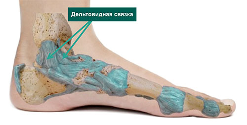
The medial collateral ligament of the foot and ankle and the deltoid ligament
An injury to the medial collateral ligament of the ankle occurs when the foot and ankle are stressed beyond their physiological limits of strength and range of motion, resulting in overstretching or tearing of the ligament.
The mechanism of injury is usually extension of the ankle (rolling or outward rotation) with or without torsion elements along the vertical axis.
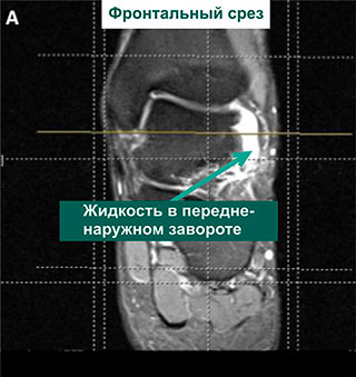
MRI
MRI provides high-quality static images of soft tissue. For normal ankle sprains, an MRI is usually not necessary. In severe injuries, it may be indicated to rule out other pathologies of the hindfoot. MRI is also helpful in assessing the dynamics of the healing process.
In severe ankle ligament injuries, MRI is used to diagnose:
- Cartilage damage – osteochondral damage
- Reactive bone lesions
- Ligament Damage – Damage to the inner ligament of the ankle
- Violation of the intercostal syndesmosis
- Pathology of the thigh tendon (tendonitis, tendon rupture)
- Any other pathology
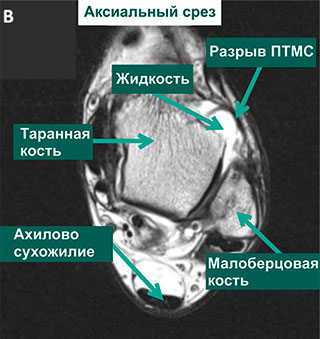
MRI of ankle showing evidence of PTMS tear
Video about our clinic for orthopedics and traumatology
What will the doctor see during the gynecological examination?
During the stool examination, the gynecologist will easily detect organ prolapse. In incomplete prolapse, the uterus is inside the vagina and the cervix may protrude from the genital tract.
The condition and appearance of the cervix also changes: it becomes swollen and pale, and microcracks, erosions, areas of epithelial malformation and bleeding may appear. The vaginal walls also look damaged and swollen.
Complete organ prolapse is diagnosed visually: the cervix is visible through the genital cleft. Such a condition is dangerous and can lead to complications.
The doctor then recommends the mandatory standard examinations:
- 1 flora and GN smear.
- 2 PAP test.
- 3 colposcopy.
- 4 Ultrasound examination of the pelvic organs and the urinary tract. Doppler ultrasound is used to assess blood flow.
- 5 If necessary, the following measures can be recommended: hysterosalpingography (examination of the patency of the fallopian tubes), MRI of the pelvis, consultation of a urologist, proctologist, examination of the rectum.
Danger!!! In case of complete uterine prolapse, only surgical treatment is indicated!
Possible complications
The risk of complications is directly related to early diagnosis, age, the presence of chronic diseases and the degree of progression. Complications are much more common in older women due to late diagnosis, self-medication and poor hygiene.
7.1 Infection
Damage and contact of the mucous membrane with the environment and underwear leads to maceration, which creates good conditions for the active development of pathogens.
There is pain, burning and cloudy discharge from the genital tract. The infection can spread upward with cystitis, appendicitis and pyelonephritis.
7.2 Constipation
Constipation due to strong pressure on the rectum, making the situation worse.
If the constipation occurred before the uterine prolapse, it is considered a causative factor. If the opposite is the case, it is considered a complication.
7.3 Vascular disorders
The pressure on the veins ultimately leads to a disruption of normal blood circulation in the pelvic organs, which manifests itself in the form of venous congestion, varicose veins of the limbs, hemorrhoids and tissue disorders.
7.4 Disorders of the urinary tract
These include urinary incontinence, urinary retention, loss of urine during the day and false urge to urinate.
Arthrodesis surgery to correct an overpronated foot
Often performed in growing children or in adults in combination with transposition of the longus flexor of the toe and forefoot reconstruction.
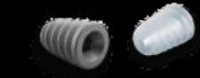
Titanium absorbable or tapered screws are used, which are screwed into the foot in a supinated position near the subtalar sinus.
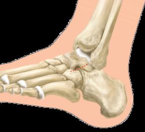
A minimal skin incision is made in the projection of the soft tissue in front of the anterior calcaneal process on the outer surface with a 1 cm long tip.
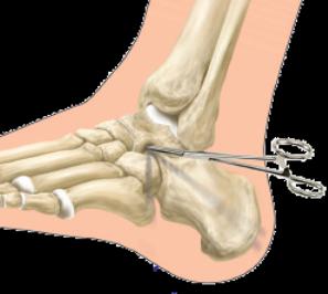
The soft tissue is carefully stretched towards the tarsal canal using the mosquito clamp.
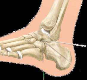
A guide pin is inserted through the soft tissue in the incisional projection into the tarsal canal, tilted 15 degrees posteriorly from the frontal plane.
A guide gauge in the form of a thread is inserted into the tarsal canal
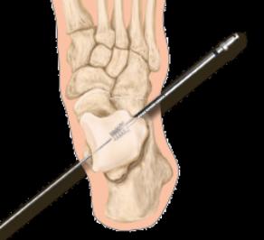
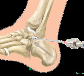
By changing the templates, the desired level of heel bone straightening is selected
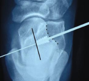
The intraoperative radiograph allows assessment of the level of screw insertion; the screw must not be placed medial to the midline of the calcaneus
A screw of the appropriate size is then inserted through the guide wire into the canal.
Intimate vaginoplasty after pregnancy and childbirth
The birth of a child is one of the most important and most anticipated events in a woman's life. Unfortunately, mothers sometimes experience certain physiological changes that affect their quality of life and cause a range of discomfort and dysfunction. One such problem is the changes in the pelvic floor after childbirth or, in simpler terms, the prolapse of the vaginal walls.
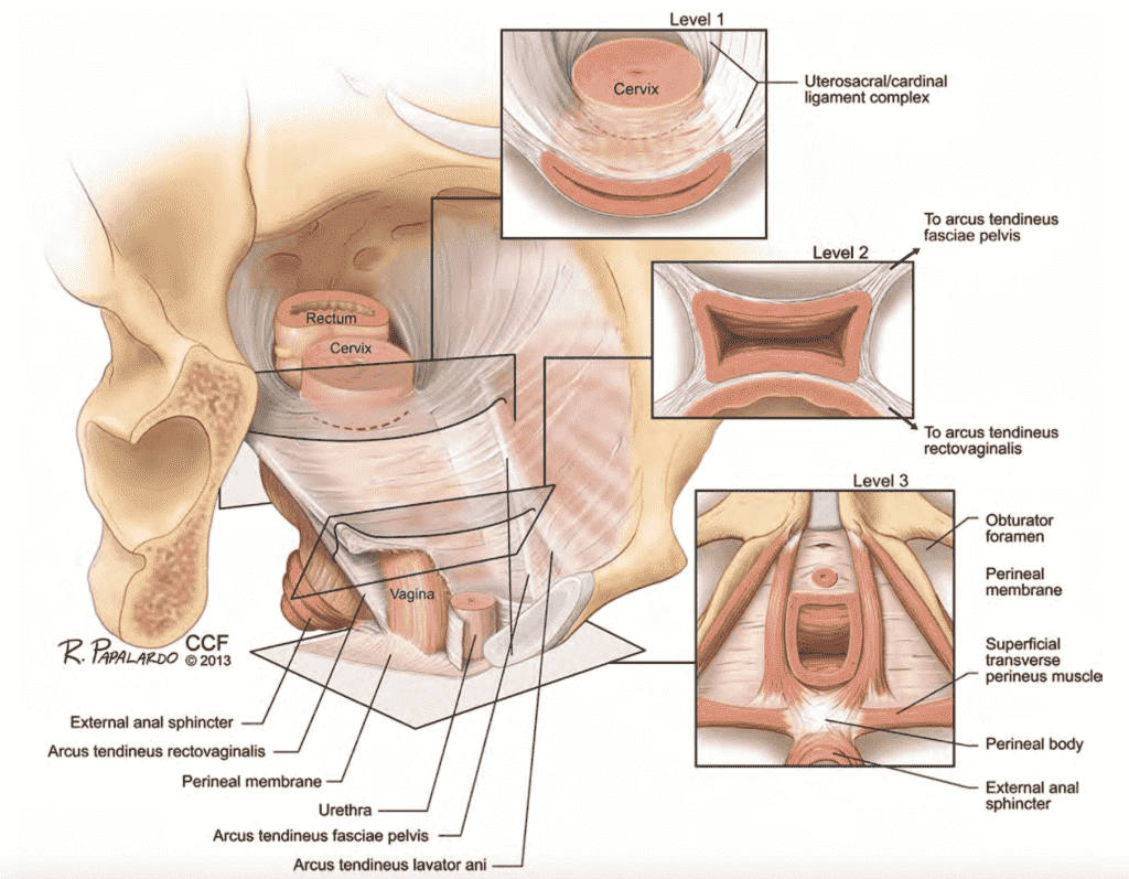
The pelvic floor is a complex muscle-fascia-fiber structure, which, on the one hand, must effectively resist intra-abdominal pressure and close the lumen of the urethra, rectum and partially the vagina, and on the other hand, determines the correct anatomical position of all pelvic organs, without which they cannot function properly can function. In this case, muscles and fascia work in a harmonious duo (they are even anatomically connected), and it is not possible to clearly say which structure makes a greater contribution (see Figure 1).
causes
During pregnancy and childbirth, both components of the pelvic floor are damaged to varying degrees. It is customary to distinguish between two groups of factors leading to pathology.
- The first group is predisposing factors. These are primarily genetic predispositions, that is, the characteristics of the genes that code for the composition and properties of the woman's connective and muscle tissue. Some women recover easily and completely from childbirth, while others take a long time and do not recover completely.
- The second group are the triggers. These are, first of all, the course of the birth, the way the process is managed by the staff and the woman in labor: how long the head stands, how timely the episiotomy is performed, whether the perineum recovers completely and so on.
Treatment of lower eyelid ectropion
The specific treatment for ectropion is blepharoplasty. During the surgical correction of the permanent eyelid, the ligaments are strengthened. If necessary, reconstruction with a skin flap is performed. In patients with paralytic ectropion, surgical intervention is not indicated until the underlying pathology has been treated.
Drug treatment is indicated if symptoms are mild or if there is a history of contraindications to surgery. To eliminate conjunctival dryness, moisturizing drops or gels are used. To prevent inflammation, topical nonsteroidal anti-inflammatory drugs (indomethacin) are prescribed.
Prognosis and prevention of lower eyelid exophthalmos
Surgical treatment of exophthalmos of the lower eyelids provides a favorable prognosis for life and ability to work. The prognosis for paralytic ectropion and all forms of the disease complicated by Lagotalmos is relatively favorable. The progression of complications of this pathology leads to reduced visual acuity up to complete blindness, leaving the patient disabled.
No specific preventive measures have been developed in ophthalmology. Patients are recommended to visit their ophthalmologist annually to ensure early detection of lower eyelid protrusion and timely and appropriate treatment. After blepharoplasty, the patient should be observed and examined by the attending physician twice a year.
Read more:- eversion of the foot.
- dorsiflexion.
- Medial and lateral side.
- Photo of the ankle.
- The flexor muscles of the foot.
- dislocation of the ankle.
- Additional joint components.
- The flexion of the foot is.