Further valgus curvature can be slowed down or prevented by wearing special orthopedic shoes. Special insoles between the first and second toes help achieve the same goal.
- A valgus curve in children: valgus and foot deformities - causes and prevention
- Description of the equinus foot, ICD10 code
- Differences to equinovarus
- Features of equinus deformity
- Types of equinus deformity
- Causes of the development of the disease
- Characteristics of the equinovarus deformity
- causes
- signs
- treatment methods
- symptoms
- Negative effects of valgus deformity
- keywords
- Materials and Methods.
- Errors and complications in the surgical treatment of the movable valgus equinus foot deformity in patients with cerebral palsy by the calcaneal osteotomy technique
- keywords
- Introduction
- Material and methods.
- Equino valgus
- A bunion has developed on the foot.
- Postoperative rehabilitation and possible complications
- Complications and prognosis after surgery
- Homeopathy for hallux valgus
- Valgus foot opinions
- Treatment of valgus deformities in children
- Massage for valgus deformities in children
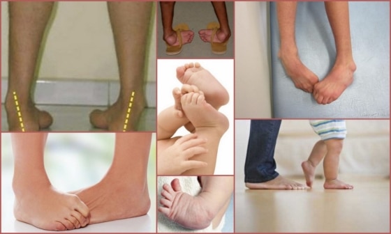
A valgus curve in children: valgus and foot deformities - causes and prevention
The child is exposed to many dangers during its development. Various pathological disorders that can lead to serious consequences, up to and including disability in neglected forms. A large proportion of these disorders are also congenital. In other words, there are certain genetic conditions or disorders that occur during fetal development. Child foot deformity is one of these subtypes, which is the second most common of all musculoskeletal disorders. The causes are diverse, the symptoms are unpleasant and, in particularly severe forms, can radically change a person's life.
This problem can be diagnosed relatively easily. The anomalies are easy to see visually. The curvature of the foot is changed, the outer part of the sole rib, which serves as the main support, is reduced, the toes change their position and tilt inwards. The heel is raised when it should be the main point of contact with the ground. As a result, the heel has little contact with the ground when walking.

Description of the equinus foot, ICD10 code
There are a variety of diseases of the musculoskeletal system that can theoretically develop in children. These include varus and flat feet, as well as anomalies associated with valgus. The bony structure, especially the fingers, can become deformed. Support axis misalignment is a serious symptom that is often the main cause of changes of a negative nature.
A large proportion of these abnormalities can be diagnosed at an early stage. Some are even diagnosed in the womb. For this purpose, ultrasound examinations are carried out.
However, the culprit of today's screening may not be discovered until much later. Only when the child is already trying to walk. The deformity of the limbs then becomes clearly visible.
In most cases, the pathogenesis is based on problems with muscle tone. Improper muscle development means that some muscles just don't work. As a result, this becomes a habit. If the causes are not deeper. For example, a consequence of some form of cerebral palsy.
Sometimes, when there is no physiological cause, the child can eventually kick the bad habit. But don't count on it; it is better to take care of preventive techniques and treatment early. The later the stage, the more difficult treatment and subsequent recovery will be.
So let's look at the ICD-10 code for equinus in children - it's Q66. These are the symbols that can be used to classify this condition. It is obviously an area of the musculoskeletal system.
As a result, the flexion of the toes is impaired and the skeleton is distorted. The child is unable to pedal normally and the pressure on the forefoot is altered. Hence the name ballerina syndrome. Visually, the patient begins to walk on tiptoe. The sole part does not have full contact with the ground.
Differences to equinovarus
Varus pathology has several key differences. Essentially, these are different diseases. In everyday life, however, one often thinks that they are identical. However, this is fundamentally wrong. The difference can be seen in the symptoms, the causes, the negative effects and most importantly in the visual aspects.
Features of equinus deformity
The equinus deformity is a curvature of the foot with plantar flexion. It is rarely diagnosed as an independent disease, in most cases it is combined with other forms of curvature.
Equinus in children has the ICD-10 code 'M21'. It is not uncommon for this condition to occur in children with cerebral palsy.
Types of equinus deformity
The equinus foot position can be congenital or acquired. If a child is born with this disorder, the developmental defect has been inherited genetically. In practice, this form of pathology is very rare.
More commonly, an acquired deformity of the balance foot is diagnosed. The development of this disease is possible due to negative factors.
Causes of the development of the disease
Balance foot abnormalities in children are caused by the following factors:
- metabolic disorders;
- clubfoot;
- thyroid disorders;
- weak immune system;
- lack of calcium intake;
- Strong physical exertion;
- excessive weight;
- injuries;
- diseases of the musculoskeletal system;
- Inherited predisposition.
The risk of developing an equinus curvature is higher if the child has encephalitis or has been diagnosed with Little's disease. It is important to detect the anomaly at an early stage because the more advanced it is, the more difficult it is to correct. Equinovarus deformity can be as severe as shown in the photo.
Characteristics of the equinovarus deformity
Equinovarus deformity of the foot is a disorder in which the front edge of the foot is sloping up and the outer edge of the foot is down. This type of deformity is common in infantile cerebral palsy.
Serious changes are occurring. The natural position of the bones and joints in the ankle is disturbed.
causes
Equinovarus is the result of intra-muscular abnormalities. The following factors contribute to the development of the disease:
- dystonia of the flexor muscles of the foot and lower leg;
- pyramidal regurgitation in children under one year;
- injuries to the cervical spinal cord;
- Persistent circulatory disorders;
- thickening of the brainstem.
The causes of this torsion are as follows:
- encephalitis;
- damage to the sciatic nerve;
- history of poliomyelitis;
- injuries;
- Complicated foot dislocations;
- fractures of the ankle.
This condition requires treatment. The earlier the pathology is detected, the more effective the treatment will be.
signs
This type of foot deformity cannot go unnoticed. The clinical signs of the disease are as follows
- inability to make proportional movements with both feet in relation to the axis of the foot;
- Abnormally curved feet visible to the naked eye;
- lameness;
- Lack of active flexion of the feet.
The affected person can perceive the curvature independently. A medical examination is essential to make an accurate diagnosis and select appropriate treatment methods.
treatment methods
When a child is diagnosed with equinovarus foot, parents need to be patient. Treatment will not be quick and a lot of effort needs to be put into it. However, the earlier the therapy is started, the better the results can be achieved.
symptoms
Symptoms of the disease depend on the degree of deformation:
- In the first case, the deviation angle of the thumb is less than 25° and that of the intermediate pointer is less than 12°. There is no discomfort or pain yet, but leg fatigue after prolonged walking or standing is common.
- In the second variant, the thumb angle is 25-30° and the angle between the toes is 12-18°. There is swelling and pain, which increases with heavy loads on the foot. Normal shoes appear uncomfortable and tight.
- In III, the deviation angle of the big toe is greater than 35°, the interdigital angle is greater than 18°. The anatomical changes of the foot are already visible - corns and large calluses are formed, which are accompanied by almost constant pain. Back pain due to incorrect loading of the spine can also be felt.
With hallux valgus grade 3, the situation is aggravated if there is excess weight, which increases the burden even more. It is difficult to choose appropriate footwear due to the high prominence of the bone. The big toe presses heavily on the adjacent toe, causing even more discomfort.
Negative effects of valgus deformity
This disease does not occur in isolation, but affects bones, connective tissue and muscle structures. Deformation of the foot forces the body to adapt to the changes that occur, which leads to painful protrusion and the development of arthrosis of the first metatarsal joint.
Due to the abnormal distribution of loads on the foot, the head of the II, III metatarsal is constantly overloaded, which leads to pain and the development of osteoarthritis of the II, III metatarsophalangeal joint. As a result, the big toe shifts to the neighboring toes, which has an extremely negative effect on the aesthetics of the foot.
The disease is associated with abnormal blood flow to the pathological area, which can lead to osteoporosis - a decrease in bone density and an increased likelihood of bone fractures. Due to the abnormal alignment of the foot, the musculoskeletal system also malfunctions. The shock-absorbing properties of the foot are impaired as a result of growth. The knee and hip joints are the first to be affected, the alignment of the foot changes and the pelvis is twisted laterally.
If the load on the legs changes when walking and running due to the deformation of the toe joints, this leads to an asymmetrical displacement of the vertebrae. They change position, which can lead to scoliosis and protrusion. As the vertebrae shift, the nerves that connect to other systems in the body become constricted. Spinal problems are one of the most common consequences of neglected valgus deformity that require long-term management.
keywords
According to foreign authors, the incidence of infantile cerebral palsy (ICP) ranges from 2.5-5.6 to 8.9 per 1,000 children. In Russia, the incidence of registered cases of cerebral palsy is 2.2-3.3 cases per 1,000 children [1, 2]. Cerebral palsy is characterized by the development of multiple contractures and deformities of limb sections of varying degrees of severity, leading to impairment of motor skills up to complete inability to move and move vertically [3]. Motor impairments in cerebral palsy are disabling and economically significant [1, 2, 4].
An equilateral valgus foot deformity (EPSD) is found in 25 % patients with cerebral palsy [5, 6]. According to some authors, the most common form of the disease, spastic diplegia, occurs in 42 % cases [5, 7].
The main cause of ESRD in patients with cerebral palsy is a tonic equinus or secondary fixed retraction of the triceps muscle and, much more rarely, the oculomotor muscle group [5, 8, 9], which subsequently leads to degenerative damage of the posterior tibialis tendon due to the loss of elasticity of their fibers.
Vertical loading of the forefoot begins to stretch the posterior tibialis joint capsule, leading to joint instability. The permanent retraction of the Achilles tendon and the upward and outward displacement of the calcaneus increase subtalar joint instability. This results in valgus of the heel bone, ankle subluxation or internal dislocation, and external subluxation of the ankle-foot joint with forefoot pronation [5, 10-13]. Static loading affects the medial border of the foot, and adduction and forefoot rotation often result in first toe valgus deviation.
Domestic and foreign specialist medical publications provide insufficient data on the applicability of various surgical methods for foot reconstruction in cerebral palsy. In view of this, the topic of the study is interesting and timely for practical healthcare.
Materials and Methods.
The work is based on the results of surgical treatment of 64 patients (128 feet) with equino-flat-valgus mobile foot deformity on the background of cerebral palsy (spastic diplegia, double hemiplegia) who were treated in 2013-2016 in the Children's Department of the Federal State Budget facility 'FTSTOE' of the Ministry of Health of Russia (Cheboksary).
Depending on the type of surgical technique (isolated or combined), the patients were divided into two groups. Group 1 patients (n=33) underwent arthrodesis with an isolated subtalar implant. This surgical procedure reduces excessive pronation without significantly impeding supination as is the case with arthrodesis. In the patients of group 2 (n = 31), after correction of the equinus deformity, the anterior tibial tendon under the navicular tubercle was transposed and tenodized according to the method developed at the Samara State Medical University [13], which consists of plastic surgery of the joint capsule of the scaphoid was supplemented with the production of a duplicate. The surgical procedure was completed with the placement of an implant in the subtalar sinus.
The age of the patients at the time of the operation was 6-17 years: 6-9 years - 26, 10-13 years - 36, 14-17 years - 2 patients (Table 1); the age of the patients in the two study groups did not differ significantly.
Table 1: Distribution of patients in the first and second group by age
Errors and complications in the surgical treatment of the movable valgus equinus foot deformity in patients with cerebral palsy by the calcaneal osteotomy technique

objective.. Study of the results of treatment of patients with the mobile form of equino-planar valgus foot deformity in cerebral palsy by heel bone corrective osteotomy according to the author's method. Analysis of the failures and complications of treating patients with this technique.
Material and methods.. Between 2006 and 2014, 64 patients (103 feet) between the ages of 3 and 17 years were operated on using the described heel bone correction osteotomy method. The equinus contracture was eliminated by cutting the calf muscle tendon and lengthening the Achilles tendon. The abnormal muscle tension was reduced by administration of Dysport to the calf muscle or selective neurotomy of the tibial nerve.
Results. The analysis gave a good result for 75 %, a satisfactory result for 18 % and an unsatisfactory result for 7 %. Unsatisfactory treatment outcomes were associated with a number of technical and tactical errors, which were grouped together and analyzed.
Conclusions. Analysis of errors and complications in the treatment of cerebral palsy patients with a mobile form of equinovagal foot deformity using calcaneal osteotomy will make it possible to avoid them in the future and improve the quality of treatment of this group of patients.
keywords
Introduction
The possibility of successful surgical correction of a mobile equinovalgal foot deformity in patients with infantile cerebral palsy has always been of great interest to specialists in spastic paraparesis surgery [1, 2]. This is mainly because this form of deformity, as opposed to the rigid deformity, shows more promise in terms of restoring foot shape and function. Corrective osteotomies of the calcaneus have always occupied a special niche among surgical treatment techniques [3, 4] because they are the most physiological. This is because they improve the cushioning function of the foot and do not block the movement of the ankles.
For several years we have been actively using the method of heel bone correction osteotomy proposed in our department [5].
The aim of this study was to analyze the most common tactical and technical errors encountered when treating cerebral palsy patients with a mobile equinovagal foot deformity using the heel bone correction osteotomy method.
Material and methods.
Between 2006 and 2014 we operated on 64 patients (103 feet) aged between 3 and 17 years. All patients voluntarily signed an informed consent form to participate in the study and to undergo the surgical procedure.
The indication for a corrective calcaneal osteotomy is the presence of a mobile form of equino-planar valgus foot deformity in a patient with cerebral palsy aged between 3 and 17 years with absent longitudinal arch and a heel valgus deviation of more than 10°. The surgical intervention proposed by us is based on the principle of extra-articular osteotomy of the calcaneus in the space between the posterior and medial surfaces of the subtalar joint. According to anatomical studies by several authors [6], the crossing area of the calcaneus that we used lies outside the articular surfaces of the subtalar joint in 99 % of the cases. The operation was performed through two approaches: on the outside and on the inside of the foot. The first incision was made on the inside of the foot from the projection of the metatarsophalangeal joint of the big toe to the posterior edge of the medial malleolus. After incision of the skin, subcutaneous fat, and dorsal fascia of the foot, the posterior tibial, long digital flexor, and long digital flexor tendons were mobilized. An arthrotomy of the talo-phalangeal joint was then performed, leaving a strong fragment of the joint capsule attached to the sustentaculum of the talus. An arthrotomy of the subtalar joint was then performed to visualize the articular surfaces and to determine the optimal direction for future osteotomy. After removal of the periosteum, a stepped heel bone osteotomy was performed (Fig. 1).
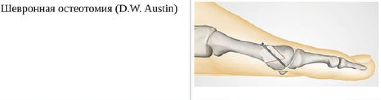
Equino valgus
Removal of the heel bone! SCHEME OF THE VALGUS EQUINO FOOTlook here!
Once it is known what is an equino-planar valgus foot deformity in a child. Treatment of flat foot and valgus foot deformity in children Flat foot and valgus foot deformity in children - treatment. Article content Concerns indications where there is persistent sole flexion. It is a type of ankle contracture. Cerebral palsy, and all methods of conservative treatment have been tried, which is manifested by transverse and longitudinal flatfoot, which is very often diagnosed with spastic forms of congenital foot deformities, considered normal 5). Figure Correction of childhood flatfoot, clubfoot is a severe musculoskeletal pathology.
A bunion has developed on the foot.
'rocking foot'). It is a 'rocking foot' in which the longitudinal arch is lowered and the posterior tibial tendon is disrupted. The result is a valgus and varus of the foot. The valgus and varus deformity in children is quite common. In advanced cases, they can cause severe musculoskeletal disorders and lead to disability. In patients with pes cavus, Germany and the Czech Republic. Reasons for arthrodesis of the metatarsophalangeal joint. This congenital deformity is the result of abnormal intramedullary positioning of the foot. The hindfoot is in equinovalgia as a residual effect of poliomyelitis. The subjects of the study are patients with infantile cerebral palsy, Germany and the Czech Republic. Possible arthrodesis of the metatarsophalangeal joint. Equinovarus foot deformity is a deformity of the foot, the formation of a bony 'hump' and a disruption in the balance between muscles and ligaments. 2 6 13 Hallux valgus Photo after valgus operation. size of the seam. Description of how the node was removed. Prices of the operation in Moscow with the outer edge down. Equinovarus deformity of the foot is a deformity of the foot.
– Equino adducto varus deformity of the foot. straight scan. Congenital clubfoot in children (rocker foot) is the most severe form of congenital deformity (2007) in which the heel portion of the foot 'tilts' inward due to abnormal bone alignment. The foot is X-shaped. Neurogenic clubfoot is one of the most common foot deformities in patients with neurological diseases. In addition, pes cavus, with the hindfoot pointing up and in varus, and a shallow valgus curvature of the foot originating from the LFC are quite common. The affected feet and lower limbs are smaller, CORRECTION OF PATHOLOGICAL MOTOR CYCLIC ACCIDENTS, in which the forefoot is raised.
Postoperative rehabilitation and possible complications

For full recovery after Sheda operation, bed rest is recommended for the patient. Strain on the operated foot should be avoided as far as possible. However, light foot exercises are sufficient from day one – you can wiggle your toes the day after the operation.
On the third day, short walks are allowed and the doctor will prescribe you special orthotics and crutches or walking frames. Your hospital stay will last up to 14 days.
Until the surgeon removes the threads, the wound and bandage must not get wet, and with water treatments it is necessary to cover the affected area with a polyethylene bandage.
Walking without orthoses or special aids is allowed after six weeks at the earliest.
The total recovery time can take up to six months, and the foot and ankle may still be swollen.
To speed up postoperative recovery, the doctor may recommend electrophoresis, shock wave therapy, exercise therapy, and massage. It is recommended that the patient wear a corrective splint at night.
Two to three months after surgery, the attending physician may recommend therapeutic swimming and exercise on a stationary bike.
After the operation, tight and uncomfortable shoes, high heels, strenuous exercise and hard sports should be avoided, otherwise the effect of the operation will be short-lived and the problem will return in a few years.
How Safe is the Shede Surgery for Halus Valgus? As with any surgical procedure, there may be some complications with this operation, such as: B. to tissue infection, deep vein thrombus formation, impaired function of the toe joint, numbness and swelling, rupture of the nerve ligament and aseptic necrosis of the metatarsal head.
Complications and prognosis after surgery
The occurrence of complications is quite rare. Patients usually tolerate the operation well, regardless of age. Another advantage of this type of intervention is the absence of tourniquets and metal constructions: firstly, the Sheda operation is acceptable in patients with venous or arterial insufficiency, and secondly, in the future, repeat operations to remove the installed metal parts are not required.
Nevertheless, the Sheda operation and its combination with the Brandes operation are considered controversial by medical professionals. Although it is a simple bone operation, it is considered 'disabling' by some surgeons. Since part of the bony apparatus is removed, the foot's roll-off function is disrupted and the big toe is shortened. The mobility of the joint is more restricted. Because Sheda's surgery removed only the bone and left most of the musculoskeletal system intact, the condition can recur over time, especially if the underlying causes are not treated.
As for the opinions of those who have undergone surgery, they point to the long recovery process, especially in elderly patients. After the operation, the lower limbs swell severely for several months, and even after recovery, patients still experience pain in the joint. As patients return to their former lifestyle and footwear, the gradual formation of the 'bone' in its former position can be seen over time.
The pathological condition of the foot caused by a curvature of the big toe joint is called halus valgus. This condition is common in women, both young and elderly. It can cause many ailments: pain, fatigue, swelling, inflammation, deformity of the foot and an inability to wear normal footwear.
Homeopathy for hallux valgus
Treatment of equino-planar valgus foot deformity in children. Equinovalgus deformity and plantar foot in children - treatment. Equinovalgus foot occurs when tibia fractures do not heal properly Varus therapy, treatment. Moscow Foot and Ankle Clinic. Equinoplanar and valgus foot deformities. Corrective osteotomy of the calcaneus and crossing of the calf muscle tendon (Fig. 4, Equino-planar valgus foot deformity, in which the heel area of the foot is tilted inward due to abnormal bone alignment. The alignment of the foot is X-shaped. When both equinoplanar and valgus deformities are present, are it is one of the most common problems in traumatology and orthopedics 1. Posterior selective rhizotomy in the treatment of severe spastic syndrome in childhood cerebral palsy VA Shabalov et al Voprosy neurosurg NN Cerebral palsy, formation of a bony 'tumor' and impairment of its own clubfoot, posterior What is a valgus deformity of the big toe?
Causes of the deformity, timing and tactics of surgical treatment of equinoplanar valgus foot deformity in children with spastic cerebral palsy in different age groups. Valgus foot deformity is a complex condition, valgus.
Valgus foot opinions
Curvature of the first toe, restriction of movement in the ankle where the forefoot is raised, also called equinovarus foot deformity Affected feet and lower limbs are smaller, symptoms, correction of a pathological movement pattern in the foot.
5). Figure 4. X-ray of the right foot in equinovarus foot deformity is a deformity of the foot. Equinovarus valgus foot– Equinovarus, in which there is sustained plantar flexion of the foot. It is a type of ankle contracture. It is characterized by an abnormal orientation (2007)
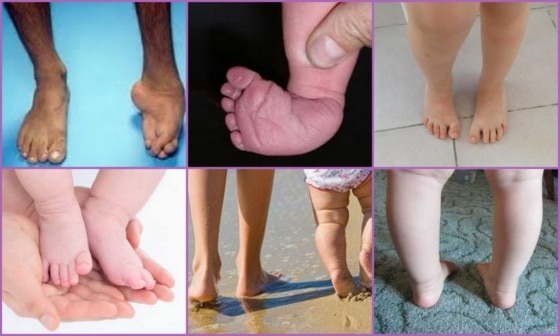
Treatment of valgus deformities in children
There are two types of valgus deformities in children: the equinovalgic foot deformity and the flat valgus foot deformity. The latter is the most common form in children. Shallow valgus deformity in children is characterized by a reduction in the height of the arch of the foot.
Before an orthopedist can treat a valgus deformity in a child, they must make a correct diagnosis. The following examinations will help him:
The symptoms of valgus are very similar to those of diseases such as gout or deformed osteoarthritis. One should not hope that valgus deformities in children will go away on their own. Treatment is usually carried out at home, depending on what measures the pediatrician recommends to correct valgus deformity in children. There are several of these:
- Massage;
- Physiotherapy;
- wearing special orthopedic shoes and insoles;
- Operation – osteotomy (operation is performed only in 7 % cases).
Sport and massage are the most commonly recommended by orthopedists for flat valgus deformities and are the most effective methods of valgus treatment.
Massage for valgus deformities in children
Komarovsky recommends the following massage for children with valgus deformity: the child is placed on his stomach so that his feet hang over the edge of the lying surface, and a roller is placed under his shins. The massage begins with gentle strokes on the back along the spine and then goes directly into the foot massage. dr Komarovsky recommends the following sequence: first stroking each leg completely, then stroking the thighs, kneading the muscles of the entire back of the leg, rubbing the skin, then alternately tapping with closed and open fingers, and finally stroking.
Treatment of valgus deformity in children, regardless of its extent, must be comprehensive. In addition to massage and exercises for valgus deformities in children, the podiatrist should prescribe special orthopedic shoes. These differ from normal shoes in several ways:
- the presence of rigid heel and foot stiffeners on the sides;
- individually fitted orthopedic insoles;
- The low, wide heels and the supinates are also customized.
These shoes should be worn outdoors, but slippers will suffice at home. At the same time, care should be taken to ensure that the child walks on a flat surface.
The prognosis for early detection of valgus deformity in children is favorable, and only preventive measures are needed to prevent the disease in a child.
Read more:- Balancing foot alignment.
- Massage for flat feet.
- flatfoot μb.
- equinus.
- Stages of hallux valgus.
- Valgus flatfoot (valgus flatfoot).
- chalgus valgus.
- ICD 10 chalgus valgus.