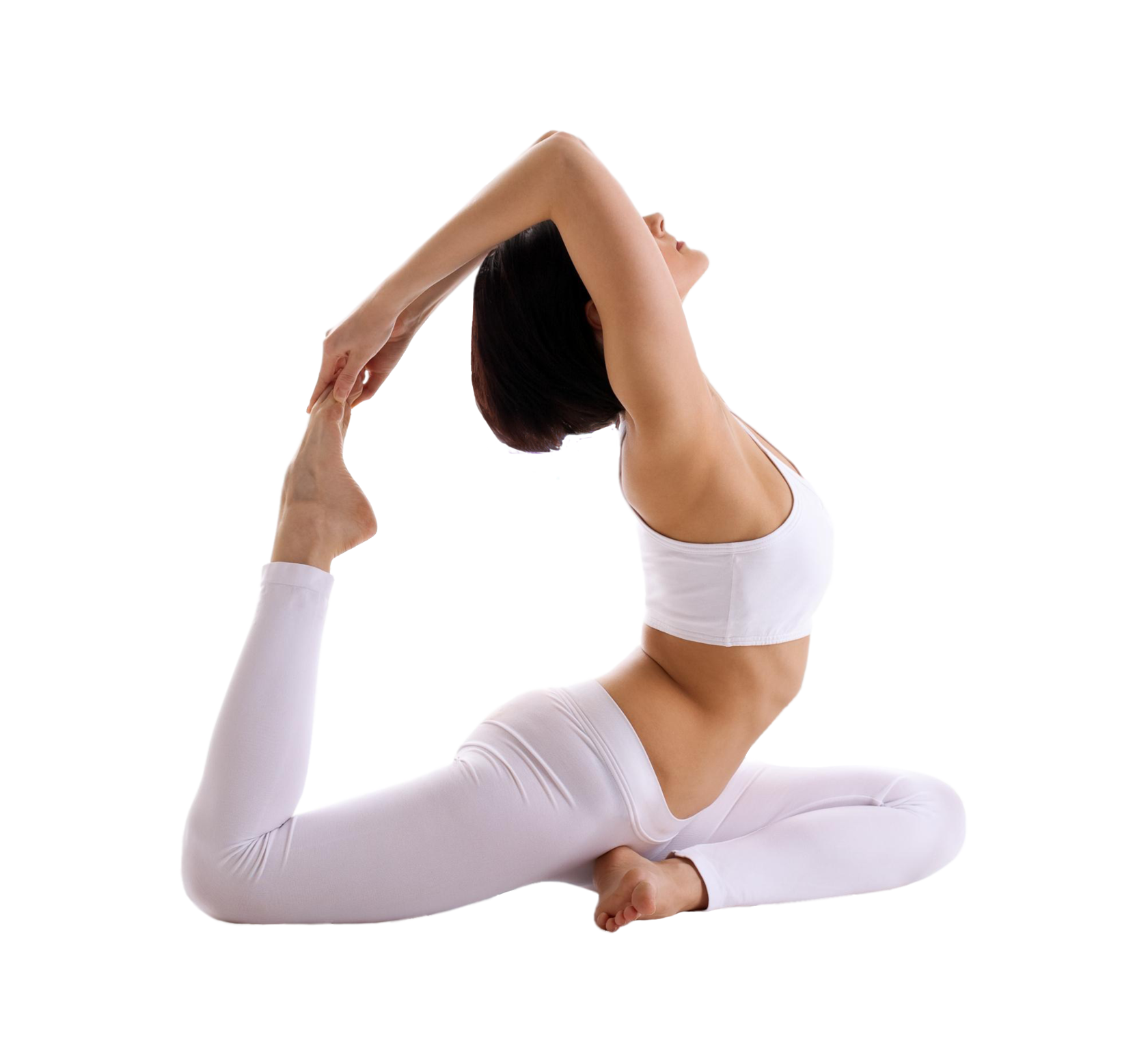
Basic course for personal trainers – For trainers who want to supplement their knowledge with basic information. An incredible amount of useful material that will take you to the next level.

- dorsiflexion
- mechanism of formation
- Intertrochanteric Ligaments (Syndesmosis)
- Achilles tendon (AC)
- Our courses, conducted by both novice and experienced trainers, offer income enhancement and career development.
- Low frequency magnetic therapy
- Ultraphonophoresis with hydrocortisone
- Achilles tendon.
- Thompson compression test (Achilles tendon compression test)
- Hoffa sign.
- Achilles tendon tap test
- ankle
- Lateral and medial stability test of the ankle
- Extended drawer test
- Why dorsiflexion is important for running
- Causes of poor dorsiflexion
- Why is dorsiflexion important for running?
- Causes of poor dorsiflexion
- What is dorsiflexion?
- Winter trail running gear to beat the cold
- Why is that so important.
- pathology
- Other causes of plantar heel pain.
- Planes and axes in biomechanics
- Designations of body movements in biomechanics
dorsiflexion
In recent decades, the number of vehicles has increased dramatically, and as a result of accidents, combined and multiple injuries to the injury structure, including hip injuries, have increased.
The biomechanics and blood flow of the hip joint are designed in such a way that bruises do not disappear. A distinctive feature of injuries to this joint is the possibility that various complications can develop over a long period of time.
Traumatic hip dislocations account for 5-8 % of all dislocations. Hip dislocations mostly occur in young and middle-aged men.
The long-term result of the treatment depends to a large extent on the family doctor and a correctly performed first aid. Road traffic accidents play a major role in the development of traumatic dislocations. To tackle the treatment of this pathology, the establishment of hip trauma units in primary and secondary hospitals, as part of a nationwide project, plays an important role.
The hip joint is formed by the head of the femur and the acetabulum of the pelvic bone. It's kind of a ball joint - it's cup-shaped.
The strong tendons and ligaments of the hip joint lead to traumatic dislocations when the injury type is high. Traumatic dislocations of the hip joint result from the indirect application of a significant force. In this case, the femur and knee joint form a kind of lever with the fulcrum, which is located intra-articularly.
The flexion of the hip joint is a prerequisite for the dislocation, since during extension the joint is blocked by the tension of the strong ligaments and further movements, especially rotation, are only possible with the help of the pelvis.
mechanism of formation
The victims of this injury are primarily drivers and passengers of cars and motorcycles who do not use seat belts. The typical mechanism of injury is when the knee of the bent leg hits the vehicle's dashboard hard upon impact with an obstacle. As a result, when the knee hits the dashboard, the following injuries occur: contusion of the distal femur, fracture of the kneecap, fracture of the ankle with the knee extended.
The second, less common cause is a fall from a height in elderly patients.
Of the more than 100 patients observed by the author, several suffered a dislocation from a fall from a height and one was hit by a car. In addition, one patient suffered this injury as a result of falling from a horse at a racecourse, which was previously the main mechanism for this injury in the historical literature. All others were drivers and passengers sitting in the front seat of passenger cars.
The type of dislocation depends on the degree of hip flexion, the presence of adduction or abduction, and external or internal rotation at the time of injury.
For example, a posterior dislocation typically occurs with hip flexion of at least 45°, adduction, and internal rotation. With any type of dislocation, the femoral head and the entire lower extremity assume a specific position. Hip flexion is found in all types of dislocations, but the degree of flexion is different in each type. The posterior dislocations are also characterized by adduction and internal rotation. Adduction and internal rotation are less pronounced in hip dislocations than in sciatic dislocations. With posterior dislocations, greater rounding of the gluteal region is noted.
Anterior dislocations also require flexion, but in combination with abduction and external rotation.
Isolated dislocations occur in only 10-20 % of all dislocations. But even among these, CT scanning of isolated femoral dislocations has shown that in 13 % of the cases they are associated with an anterior fracture of the femoral head combined with a fracture of the posterior rim of the acetabulum.
Intertrochanteric Ligaments (Syndesmosis)
The ligaments of the intertrochanteric syndesmosis are the intertibial ligament, anterior intertibial ligament, posterior intertibial ligament, and transverse ligament. The anterior longitudinal ligament of the syndesmosis is three times weaker than the posterior ligamentwhile the rear band can withstand a pulling force of up to 30.0±2.3 kg. However, the most stable structure of the syndesmosis is the interosseous membrane, the strength of which is twice the combined strength of the anterior and posterior ligaments.
With a violation of the intertibial syndesmosis, the clinical picture is characterized by a pronounced and persistent swelling of the lower leg and foot; severe injuries require surgical fixation of the tibia because of tibial diastasis and plateau instability.

Achilles tendon (AC)
The Achilles tendon (AT) is the strongest and most powerful tendon in the human body, capable of withstanding significant static and dynamic loads. It is the common tendon of the superficial calf muscles and the deep cambium.
The AC fibers have a helical course like a rope, which gives them great strength and at the same time the ability to stretch slightly under physical stress, straightening this helix and thus cushioning the load.
Although the strength of AC is considerable, it has its limits: it is around 50 N/mm2.
AC strain (elongation) under stress around 3-5% is to be regarded as physiological, up to 8% as harmful. Elongation of the AC more than 8% will inevitably lead to micro and macro cracks.
The number of vessels supplying the AS decreases proximal to the calcaneus and reaches a minimum 4-5 cm from the calcaneus tubercle, where nourishment occurs only by diffusion from the synovial fluid. The AS is therefore a poorly supplied tissue, which makes it very susceptible to micro-injuries and the development of degenerative diseases.
The arch of the foot, together with the plantar fascia and the AC, form a single functional system that absorbs the impact of running and jumping. With a significant flattening or enlargement of the longitudinal arch of the foot (cavus foot), spatial discrepancies in the metatarsal structure, hyperpronation or hypersupination of the foot, the shock-absorbing properties of the foot are reduced and the load on the ACL increases accordingly, which leads to wear and the development of chronic diseases.
The pathogenesis of spontaneous ACL is multifactorial. It is caused by a number of exogenous and endogenous factors or a combination of these factors.
The most important endogenous factor are dystrophic-degenerative changes – in the tendon itself (tendinopathy), their covering (paratendinopathy), as well as in the tendon capsule (Achilles tendon bursitis, deep and superficial bursitis).
Our courses will conduct
for both novice and experienced trainers, offering income enhancement and career development.
The user purchasing services on the evotren.ru website, hereinafter referred to as the 'Customer', on the one hand, and Evotren LLC, hereinafter referred to as the 'Contractor', represented by the General Director FG Kapishev, acting on the basis of the Charter, on the other hand automatically entered into this Agreement (hereinafter referred to as the 'Agreement') when purchasing the Services of the Contractor below:
1. TERMS AND DEFINITIONS USED IN THE CONTRACT
1.1. customer – Natural person, private entrepreneur or legal entity, regardless of its organizational and legal form, who places an order with the Contractor in accordance with the terms of this Agreement by purchasing the Contractor's services.
1.2. contractor – The legal entity that provides services to the customer under the contract.
1.3. Services – Services providing access to the study of the distance learning materials listed in the descriptions of the information courses.
1.4. site – The contractor's information resource, located on the Internet at: edu.evotren.com
1.5. Personal customer account – Programming interface on the website for studying the information material and other necessary information, accessible to the customer after authorization with a login and password.
1.6. Order – An automatically generated document detailing the services requested by the customer. The order is made by filling out the necessary forms on the website of the realiser -www.evotren.ru.
1.7. Acceptance of Terms and Conditions – The acceptance by the customer of the terms of this contract by paying for the services either in cash or non-cash or by electronic means. Acceptance of the terms of the contract is deemed to have taken place when the customer has paid for the services in cash or without cash or by electronic means of payment.
Low frequency magnetic therapy
The same types of currents with lower frequency and greater modulation depth are used for myostimulation. The effect is.Analgesic and iostimulating.
Variable, pulsating and low-frequency magnetic field. The inductors (electromagnets or solenoids) are attached with no gap or pressure. The magnetic field has (depending on the type) an induction of 5 to 50 mtl. and a frequency of 10 to 100 Hz. Duration of treatment 15 - 30 min. EffectAnti-inflammatory, trophic and regenerating.
Ultraphonophoresis with hydrocortisone
Treatments are carried out using the unstable method. Appointment on the 3rd - 5th day after the injury, at an ultrasound intensity of 0.2-0.6 W/cm 2, 7-10 min. Effect.: Anti-inflammatory.
Immobilization of the injury site is performed with
- round plaster cast,
- other types of plaster casts,
- Immobilization, which limits movement of joints adjacent to the injury site.
Type and duration (2 to 8 weeksThe type and duration (2 to 8 weeks) of immobilization depends on the type and location of the injury.
Achilles tendon.
Thompson compression test (Achilles tendon compression test)
method. The patient lies on his stomach. The feet hang over the edge of the table. Therapist grasps the muscles of the upper third of the affected tibia with one hand and squeezes them firmly.
Evaluation. Normal contraction of the lower leg muscles results in rapid passive plantar flexion of the foot. The absence of such plantar flexion is indicative of an Achilles tendon rupture. In patients with a partial rupture of the Achilles tendon, this test is not always meaningful and depends on the extent of the rupture. Patients with a torn Achilles tendon cannot stand on their toes, especially the injured limb, and the Achilles tendon reflex is absent.
Remarks. This examination can also be performed in the prone position, with the leg flexed to 90° at the knee joint. In this position, the doctor grasps the muscles of the upper third of the tibia with both hands and squeezes firmly. Loss of soleus flexion is a sign of an Achilles tendon rupture (Simmond's test).

Hoffa sign.
Allows the diagnosis of a longstanding rupture of the Achilles tendon.
method. The patient lies on his stomach, his feet dangling over the edge of the table. The physician passively performs dorsiflexion of both feet.
Evaluation. With an old Achilles tendon rupture, the tension in the Achilles tendon is reduced and the affected foot can achieve greater dorsiflexion than the healthy foot. The patient is then asked to stand on tiptoe alternately on both feet. At the limb where the Achilles tendon is torn, the patient cannot stand as suggested.

Achilles tendon tap test
Diagnosed a ruptured Achilles tendon.
ankle
Lateral and medial stability test of the ankle
Assessment of damage to the collateral ligaments of the ankle.
method. The patient lies on his back. The physician holds the lower limb with one hand behind the knuckles. With the other hand, he grasps the lateral side of the metatarsal and performs supination, attempting to widen the ankle joint space on the lateral side. To assess the medial ligaments, grasp the metatarsal on the medial side and pronate, attempting to widen the medial joint space.
Evaluation. Damage to any of these ligaments leads to instability and increased opening of the medial or lateral joint space. Increased supination can be caused by damage to the anterior talofibular ligament and calcaneus. Increased pronation can result from damage to the deltoid ligament. Rotational injury in supination is the most common mechanism of ankle injury and almost always involves the anterior talofibular ligament. Children typically have greater ankle mobility, which should not be confused with a ligament injury.
Both feet must be compared during the examination. X-rays under stress are essential to document ligament injuries, particularly in the ankle.

Extended drawer test
Serves to assess the stability of the ankle.
method. The patient lies on his back. The physician holds the patient's tibia from behind with one hand and grasps the metatarsal bone with the other hand. The doctor moves the foot back at the ankle against the force of the hand holding the shin. In the second step, the doctor grasps the front of the shin and the back of the heel. The foot is then moved forward against the force of the hand holding the shin.
Why dorsiflexion is important for running
Dorsiflexion can help runners be more efficient in the following ways:
- Reduction of injuries: One wrong step in a repetitive movement like running can expose the runner to all sorts of injuries as the body begins to compensate. Since everything moves up the kinetic chain, runners should always try to improve dorsiflexion to avoid short- and long-term leg, hip, back, and neck injuries.
- Reduce the likelihood of falls: Poor dorsiflexion can increase the risk of falling because the foot doesn't land where it should, which is why dorsiflexion is also known as 'dangling foot'. This is especially true over short distances, as push-off is critical to speed and power.
- Shortened time to complete the run: Effective dorsiflexion allows runners to increase their speed by reducing the time their feet are on the ground. The more runners bend their ankles, the easier they get up and the more time they spend in the air. This can be the case for longer runs, e.g. B. marathons, shorten the target time by seconds or even minutes.
- The performance is increased: By simply lifting your foot 10-30 degrees, you can land on your midfoot. This is beneficial because you end up in the center of your mass. This gives you the extra mass you need to push off harder than if you landed closer to your toes.
What part of the foot should you land on when running?
Causes of poor dorsiflexion
They can worsen your dorsiflexion and make walking difficult in any of the following ways:
Damage to nerves: One of the most common causes of poor dorsiflexion is nerve compression in the leg. A pinched nerve in your spine can also change your gait.
muscle weakness: Lack of strength in the thighs, glutes, hips, and shins can lead to movement compensation, especially when one side is weaker than the other. Runners with a dominant right or left side tend to touch the ground with more force and lift the weight with more force on their preferred side.
Lower Body Injuries: Foot and leg injuries ranging from an ankle sprain or plantar fasciitis to the hip and back can change the way you move. Your body doesn't adapt when any of these connective tissues are damaged.
Genetically conditioned: Your genes may predispose you to problems with dorsiflexion, e.g. B. by the length of the legs and structural inconsistencies. The help of a chiropractor can help you.
Mobility problems: If you have a strained calf muscle or tendon, or if you have a build-up of lactic acid from intense cardio or strength training, your ability to run may be impaired.
Restriction of the ankle: Scar tissue in the joint can lead to movement problems. The joint acts as a natural hinge for your foot. If this joint isn't working properly, you'll be limited in your ability to lift your legs.
Diseases: Any spinal cord disease, muscular dystrophy, or multiple sclerosis can cause your foot to drag on the floor as you move.
Operations: Hip or knee replacement surgery can result in an abnormal gait. Working with a physical therapist can help ensure that this problem is temporary rather than permanent.
Why is dorsiflexion important for running?
Dorsiflexion can help runners be more efficient in the following ways:
- Reduction of injuries: Improper foot strike in a repetitive movement like running can leave the runner vulnerable to all sorts of injuries as the body begins to compensate. Since everything moves up the kinetic chain, runners should always try to improve dorsiflexion to avoid short- and long-term leg, hip, back, and neck injuries.
- Reduce the risk of falls: Poor dorsiflexion can increase the risk of falls in runners because the foot doesn't land where it should. This is especially true when sprinting, as foot strike is critical to speed and power.
- Reduce the time it takes you to run: With effective dorsiflexion, runners can increase their speed because they reduce the time their feet are on the ground. The more the ankles are bent, the lighter the feet become and the more time they spend in the air. This can shave seconds or even minutes off the finish time in longer races like a marathon.
- Increase in performance: By simply elevating your foot 10 to 30 degrees, you can land more on your midfoot. This is beneficial because you end up in the middle of your crowd. This gives you the extra mass you need to push off harder than if you landed more toe-off.
Causes of poor dorsiflexion
Any of the following can affect dorsiflexion and make walking difficult:
nerve damage: One of the most common causes of poor dorsiflexion is compression of a nerve in the leg. A pinched nerve in your spine can also change your gait.
muscle weakness: Lack of abduction strength in the hip, glutes, thighs, and lower legs can lead to movement compensations, especially when one side is weaker than the other. Runners with a dominant right or left side tend to touch the ground with more force and lift the weight with more force on their preferred side.
Lower Body Injuries: Foot and leg injuries, like ankle sprains and plantar fasciitis, as well as hip and back injuries, can change the way you move. Your body makes abnormal adjustments when any of these connective tissues are damaged.
Genetically conditioned: Your genetics may predispose you to dorsiflexion problems such as: B. due to leg length and structure differences. Chiropractic treatment can help here.
Mobility problems: If your calf or hamstring muscles are tight, or if you have built up lactic acid from intense cardio or weight lifting, your ability to run may be affected.
Restrictions in the ankle: Scar tissue in the joint can cause problems with movement. The joint acts as a natural hinge for the foot, and if this joint isn't working properly, you'll be limited in your ability to lift your feet.
Diseases: Any spinal cord disease, muscular dystrophy, or multiple sclerosis can cause the foot to drag on the floor when moving.
Operations: Hip or knee replacement surgery can cause abnormal gait. Working with a physical therapist can help ensure that this problem is temporary rather than permanent.
What is dorsiflexion?
Dorsiflexion is created by the muscles at the front of the shin (i.e. the front of the shin that lifts the foot). This relatively simple movement plays an important role in running technique and can cause complex problems in athletes. When running, the foot is the source of ground contact. Everything that happens in the foot and ankle transfers up the kinetic chain and can affect the knees, hips, lower back and more.
Dorsiflexion doesn't seem to be as important when jogging at low speeds, but the faster and closer to sprinting you are, the more important dorsiflexion becomes as you expend more force.
Winter trail running gear to beat the cold
Why is that so important.
Proper dorsiflexion results in proper foot strike and helps prevent injury. Dorsiflexing while running puts the foot in an ideal position that cushions the impact of landing and tightens muscles so you can move forward with the next step. This reduces the time spent on the ground with each step, allowing you to run faster and more efficiently. In individuals with poor dorsiflexion, the foot may be 'loose' or 'slack' due to ankle laxity, causing the toes to strike the ground, resulting in poor force distribution and contributing to injuries such as shin splints and runner's knees . Poor dorsiflexion also reduces the ability to harness and deploy posterior chain strength.
Unlike the larger and stronger calf muscles, dorsiflexion is less natural and non-existent in most recreational runners. Therefore, if you are trying to become a skilled runner or sprinter, focus on dorsiflexion. Poor dorsiflexion can be caused by several factors. One of them is the flexibility of the posterior chain of muscles in the lower limbs. Another cause of decreased dorsiflexion is limitation of the ankle itself, usually caused by a tight joint capsule or by scar tissue and adhesions in the joint. It may even be a secondary reaction to a previous ankle injury, e.g. a sprained ankle. If there is no soft tissue or mobility problem, it can also be because the anterior muscles are weak, tire quickly and are not used in the first place due to a lack of awareness or correct technique.
pathology
Continuous and persistent injury to the plantar fascia results in inflammation, swelling, and pain, particularly at its insertion point on the medial side of the tubercle.
– Normal pronation is accompanied by a decrease in the height of the medial longitudinal arch and a relative lengthening of the foot.
– Excessive pronation compensates for a varus hindfoot, flexible pes cavus and lower limb abnormalities. With excessive pronation, the relative extension of the foot during loading exerts constant pressure on the plantar fascia throughout the support phase, increasing the elongation of the component.
– Inadequate pronation occurs with an uncompensated varus hindfoot and a fixed pes cavus. Inadequate pronation in the mid-pronation phase will result in the plantar fascia being under constant tension throughout the support phase due to the extension mechanism.
– Being overweight increases the tendency for increased tension at the origins of the fascia, which amplifies the pathomechanical factors and increases the compressive forces on the fat pad of the heel.
– The decreasing thickness and elasticity of the pad with age, as well as vascular and rheumatic diseases lead to pain and spurs or biomechanical abnormalities.
It is believed that repeated excessive strain from walking or standing causes changes in the fascia in the form of acute or chronic inflammation. If the inflammation affects the entire fascia, it is called plantar fasciitis, if it only affects the heel, it is called heel pain. Chronic inflammation includes collagen necrosis, angiofibroblastic dysplasia (overgrowth of local fibrous tissue and blood vessels), chondroid metaplasia (conversion to cartilage), and possibly calcification, particularly at the site of origin. In some cases, the first branch of the lateral plantar nerve, called the Baxter nerve, may be affected by fibrous tissue.
When inflammation affects the periosteum of the medial aspect of the fibula node, enthesopathy occurs (the insertion of a ligament or fascia into the bony surface is called enthesopathy).
Tension on the periosteum of the heel from the short toe flexor and the proximal part of the plantar aponeurosis stimulates new bone formation in this area and a spur forms. A heel spur is initially very painful and later becomes asymptomatic as traction from the plantar aponeurosis decreases. 16 % of people with spurs have no pain, 50 % of people with heel pain have no spurs.
Other causes of plantar heel pain.
Subacromial bunion. Also known as 'police heel' and described as a cause of plantar heel pain. It is a congenital or acquired bursa in the superficial tissues of the plantar heel, the symptoms of inflammation are similar.
compression neuropathy. Compression of the medial heel nerve or the first lateral branch of the longitudinal nerve can cause persistent heel pain. The pain is medial, stabbing, electric, and may radiate to the lower limbs. Excessive pronation of the foot causes repeated microtrauma, chronic nerve fibrosis and paraesthesia. In acute cases, surgical decompression and excision is required.
S1 root radiculopathy. Compression of the proximal S1 root can result in rebound pain at the extension of the median plantar nerve.
Planes and axes in biomechanics
All five major components function in relation to the body's axes and planes. Axes are straight lines that run perpendicularly through the body. These lines are the main areas of twisting/rotational movement of the body.
There are three axes in our body: transverse, longitudinal and central. The transverse axis – is the horizontal line at waist level. The longitudinal axis is the vertical line that runs from the center of the head to the feet. The central axis runs diagonally from the hips to the shoulders.
There are also three body planes: sagittal, frontal, and transverse. sagittal plane divides the body into a right and a left side. Movements such as flexion and extension take place in this plane.
The frontal plane is the back of the body and the front of the body. Movements such as abduction/reduction take place in this plane. The transverse plane divides the body into an upper and a lower part, in which rotational movements take place.
The combination of the two levels creates an oblique movement at the same time.
Designations of body movements in biomechanics
The basic movements that we perform when training on sports machines and equipment take place within the axes and planes mentioned above.
Often the names of these movements are attached to the device descriptions. To properly understand the terms, let's decode the most common ones:
- dorsiflexion – reducing the angle of the ankle;
- plantar flexion – increasing the angle of the ankle;
- Inversion - rotation of the ankle with the sole of the foot toward the opposite foot;
- Eversion - rotation of the ankle with the sole of the foot towards the opposite foot;
- Liberal pivot - rotation of the trunk towards the medial axis;
- Medial Pivot - Rotation of the trunk towards the median axis;
- Pronation – rotation of the forearm with the palms facing down;
- Supination - rotation of the forearm with the palms facing up;
- retraction – raising the arm forward at the shoulder joint;
- Retraction - Raising the arm backwards at the shoulder joint.
Knowing your biomechanics not only allows you to be more aware of your body, but also allows you to choose the right gear and exercise equipment. Equipment made with correct biomechanics in mind makes an athlete's movements safe and effective during training. Get to know your body and improve the quality of your exercises!
Read more:- How to determine the type of pronation.
- Sneakers with hyperpronation.
- What is pronation and supination?.
- Pronation and supination in anatomy.
- pronation and supination.
- These are the pronator muscles.
- pronation.
- hip pronation.