– A difference in length of more than 2 cm predisposes to early separation of the heel and equinus of the foot (excessive plantar flexion) on the short side and increased pelvic tilt. This results in reduced or absent heel strike when walking.
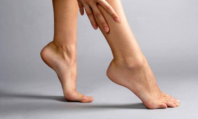
- Different leg lengths
- Causes, stages of development and treatment of flat feet in children.
- How do you measure your feet? Are you measuring from the greater trochanter to the lateral malleolus?
- How does leg lengthening surgery work? How long and how far can you extend your legs?
- Difference in length of the lower limbs
- Ways to diagnose functional length deviation of the lower limbs
- Ways to diagnose functional lower limb length difference (ffDnA)
- Visual examination and palpation
- Approaches to treating abnormal leg positioning
- CAN THE PODOLOGIST MAKE A MISTAKE.
- WHAT DOES OSTEOPATHY OFFER?
- Attention to children
- Royal foot massage
- Orthopedics Nizhny Novgorod medical equipment
- material and methods
- Results and discussion
- Diagnosis of asymmetry of the lower limbs
- First day – first visit to the clinic
- Day two – examination in our clinic
- Day three – choosing the correction method
- Treatment of leg length disproportion in children in Israel – prices
- Benefits of correcting leg length disproportion in children in Ichilov complex
Different leg lengths
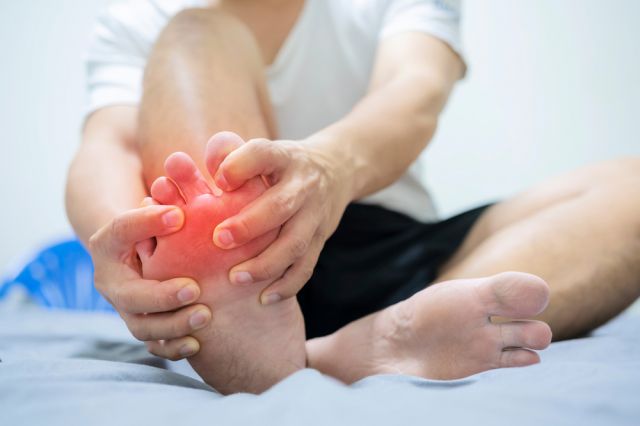
Everyone knows what a heel spur is! It is a spur that grows on its own and presses awkwardly into the sole when walking.
Normally nothing grows on its own (except tumors). But a spur is not a tumor! Or he is so innate that at the age of 40 or 50 he says: 'My time has come!'.
A spur grows at the site of the damaged plantar fascia, where it attaches to the heel. At some point, the fascia lacks elasticity and due to increased or sudden stress, weight gain or age-related changes in tissue elasticity, the fascia can no longer stretch and is damaged either along its entire length or at its narrowest point - the heel. When the heel is pulled away, the fascia grabs onto the periosteum, which, as it has a good blood supply, is very painful and then forms bone tissue where the fascia is stretched.
Therefore, the heel spur that you see on the X-ray is usually a result of the problem and is not pressing on anything - it is an expansion of the fascia.
✅ Relief of local areas of the plantar fascia and correction of foot alignment to reduce the strain on the fascia and chronic spastic foot muscles - individual insoles
✅ Reduction of the flexion angle of the ankle during daily movement (the heel should be slightly higher than the toes in the shoe)
✅ Stretching exercises for the calf-calf complex and plantar fascia during the day - help the fascia to stretch
✅ Shock wave therapy - a method that locally improves blood circulation and thereby repairs the fascia tissue
✅ Anti-inflammatory analgesics for prolonged or severe pain
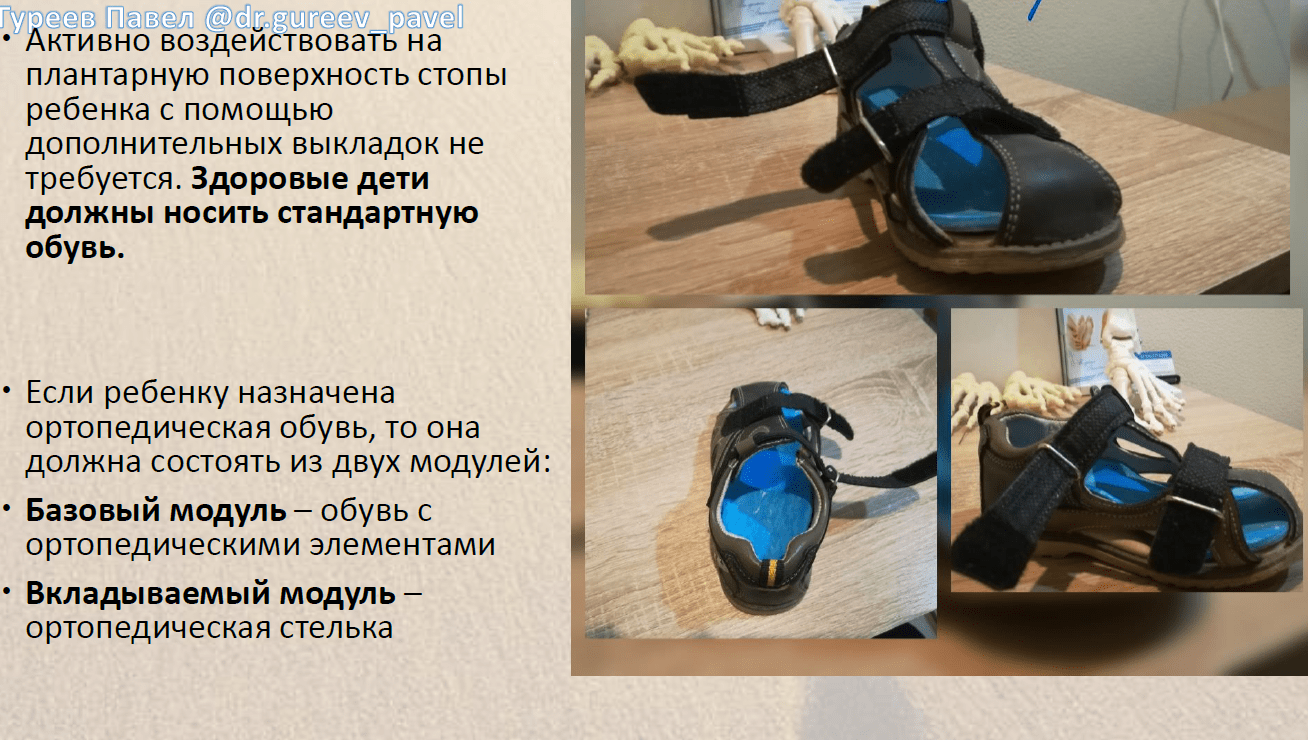
In a 2017 article in the Bulletin of the All-Russian Guild of Orthopedic Prosthetists, employees of the prestigious GA Science Center. Albrecht said that orthopedic footwear should consist of two modules: the shoes themselves and an individualized orthopedic insole. A real orthopedic shoe should have an individually adapted insole.
Causes, stages of development and treatment of flat feet in children.
We live in a world of smooth surfaces: tiles, asphalt, linoleum, laminate, even the floor on the playground is made of rubber, but smooth! Observations of podiatrists show that flat feet are common in children. The condition is quite insidious as it has no symptoms in childhood but can cause serious disorders as we age. Most parents pay little attention to this condition and mistakenly believe that flat feet disappear with age. However, if treatment is not started on time, flat feet will worsen and the foot will become deformed, so subsequent treatment may be very difficult or ineffective. It is important that parents consult a specialist early on.
Both the development of all organs and systems of the child as well as the training of the foot are of great importance for a later, fulfilling life. Up to the age of 2, flat feet are physiological - the longitudinal arch of the foot is always flat. At the age of 3, the bones become denser, the ligaments become stronger and the joints assume their correct position: the 'correct foot' begins to form. This process continues until the age of 5, so it is only at this age that one can speak of true flat feet. The younger the child is (especially up to 3-4 years old), the more the footprint resembles a flat foot. Therefore, in order to diagnose flat feet in children and recognize their symptoms, it is necessary to consult an orthopedist. Diagnosis of flat feet in children should not wait until age 5.
Dear readers, pay particular attention to the health of your feet and the feet of your children. If you do not have the time to take preventive measures and start the mechanism of further development of the disease, you should urgently take therapeutic measures to improve the quality of life of your child. Remember that pain in the foot, transverse or longitudinal flatfoot is not a punishment. Through prevention and treatment, you can be sure that your feet will no longer hurt and you will feel comfortable.
How do you measure your feet? Are you measuring from the greater trochanter to the lateral malleolus?
There are clinical and radiological examination methods. At the beginning, of course, there is the person who comes to me with the results they have measured themselves. read more
If a person feels that there is a leg length difference, it is almost always more than 2 centimeters. The result of a leg length difference can be a curvature of the spine, scoliosis and so on. In recent years. Continue reading
How does leg lengthening surgery work? How long and how far can you extend your legs?
I am proud to say that the most important achievements in this field date back to the invention of the eminent Russian scientist Gavril Abramovich Ilizarov, who registered a leg lengthening device in 1952. more on this
There are several methods. The method proposed by Gavril Abramovich Ilizarov makes it possible to cut any bone - femur, tibia, regardless of its thickness - through a small hole. Conventional. read more
Difference in length of the lower limbs
Ways to diagnose functional length deviation of the lower limbs
In addition to the importance of diagnostic changes, we also consider practical and methodological aspects, as not all methods are equally effective and 'cost-effective'.
For example, acupuncture or local anesthesia of painful periarticular points is much more economical than periarticular massage, while for muscle attachment points we prefer post-isometric relaxation whenever possible because it is painless and can in most cases be carried out by the patient themselves.
The manipulation has the advantage that it is effective and quick to use.
With a wide range of appropriate techniques, we often make a decision once we have accurately identified individual lesions. In these cases we make a 'working diagnosis', meaning that we have difficulty accurately identifying the lesions that are the most important link in the pathological chain.
Often we clarify whether methods of skin irritation were used without properly evaluating the data from the patient's examination, without information about the area of hyperalgesia or muscle relaxation, when we do not find muscle tension, when at least one manipulation was performed without detecting a blockage.
Of course, it is a waste of time to prescribe therapeutic exercises without first identifying disorders of muscular coordination. A correct pathogenetic diagnosis can only be made once the individual links in the pathogenesis have been identified and their significance analyzed.
Only then can final results of further treatment be expected. As with a neurological examination, one should proceed systematically from the periphery to the center and then carry out treatment depending on the results.
Ways to diagnose functional lower limb length difference (ffDnA)
Determining the ffDnA value is a difficult task. Currently, there are numerous methods and devices for determining this value, but all of them have significant methodological and conceptual shortcomings that do not allow determining the FRDNA value with sufficient accuracy and sometimes lead to an incorrect diagnosis.
Conventionally, all known methods for determining the value of the functional difference in length of the lower limbs can be used can be divided into three groups:
1. visual examination and palpation;
2. radiological measurement methods;
3. Anthropometry.
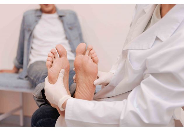
Visual examination and palpation
According to the literature, the most common method for visual and palpatory diagnosis of a functional difference in the length of the lower limbs is to determine the height of the two halves of the pelvis. With a patient standing upright, the doctor fixes with his fingers the highest points on the iliac crests or their posterior jaws and visually compares their levels. JG Travell and DG Simons are of the opinion that the patient should stand with his back to the doctor during the examination (legs together, knees straight).
Leg length discrepancies are determined by palpation of the iliac crests or their posterior upper crests. The patient's spine should be examined for scoliosis in the thoracic and lumbar spine, the inclination of the axis of the shoulder girdle and the position of the shoulder blades should be determined by palpation of their lower angles.
To approximate the correction of a short leg, the authors suggest placing a stack of paper or a small magazine under the leg without causing discomfort to the patient. The patient is spoken to for one to two minutes and asked to relax and distribute their body weight on both legs.
When the short leg is raised, the muscles that compensated for the difference in leg length are released from this function and relaxed. This allows the difference in leg length to be compensated more accurately by additionally lifting the short leg until the pelvis and shoulders and, above all, the spine are horizontal.
Approaches to treating abnormal leg positioning
Some patients refuse to undergo any treatment because they fear surgery. In fact, the problem can often be solved with orthoses. Chiropractic and osteopathy, massage and therapeutic exercises show good results.
Properly chosen treatments can achieve the desired results: eliminate or equalize leg length discrepancies and prevent the development of complications and associated diseases.
CAN THE PODOLOGIST MAKE A MISTAKE.
By detecting the problem early and eliminating it, serious illnesses can be avoided. It is important to find a specialist who will correctly measure the length of the lower limbs. Don't be surprised if an osteopath discovers a problem, unlike an orthopedist who is convinced that your legs are the same length. The diagnosis of this problem is not yet standardized. That is why you should entrust yourself to a specialist who, firstly, has extensive experience in dealing with this problem, and secondly, has his own experience and evidence of the only correct solution.
Disagreements in the diagnosis of leg length have existed for more than fifty years. At that time, French orthopedists began to measure the length of the lower limbs with a centimeter ruler and, in fact, often noticed discrepancies in leg length, which they prescribed special orthoses to their patients to correct. However, these recommendations often did not have the desired effect. Patients complained that the orthoses did not make them feel better but, on the contrary, increased the pain and threw the body out of balance. Osteopaths tried to solve this dilemma. They found that in most cases it was not anatomical differences in leg length, but simply an imbalance between the pelvic bones that caused the different appearance of the legs, and that the orthoses only exacerbated the imbalance of the pelvic bones. The osteopaths then proposed their own treatment method, which restored the balance between the pelvic bones and thus the length of the lower limbs.
WHAT DOES OSTEOPATHY OFFER?
There are now X-ray protocols to determine the length of the lower limbs. Despite the side effect of radiation, this method is undoubtedly more accurate than measuring with a centimeter tape measure. Nevertheless, orthopedic surgeons continue to defend it. While international congresses puzzle over the moment of truth, osteopaths have found their own solution. Not all osteopaths, but only those who know the old methods well and have a lot of experience.
For example, Michel Dobenski, an osteopath from Ostmed, has chosen a simple, logical and rational approach that does not require X-rays or lying down to measure the length of a limb. The method used by this specialist with 20 years of experience in osteopathy is to measure the length of the legs while standing in relation to the bony landmarks. This is the method that Michel Dobenski uses at the Ostmed Clinic. According to Dobensky, the advantage of the osteopathic method of assessing the length of the lower limbs is not only that the length of the limbs can be accurately estimated, but also that the cause of the existing difference can be determined. Since he knows the exact cause of the asymmetry, Dr. Michel Dobensky will give precise treatment recommendations and compensate for the difference in leg length. He prescribes the recommendations according to the leg length difference.
– If there is an anatomical difference in leg length, he prescribes a thin splint.
– If there is a functional difference in leg length due to flat feet or an enlarged arch of the foot, orthopedic active or proprioceptive insoles are prescribed.
– If there is a leg length difference due to a pelvic bone disorder, osteopathic treatment of the identified disorder is prescribed.
Attention, specialist consultation is necessary!
Text / OpenMed Clinic
Photo / shutterstock.com
Attention to children
The specialist points out that parents need to pay special attention to children. 'Congenital limb differences are controlled by the orthopedist. The most important thing for parents is to take their child for a check-up at the right time. The doctor will accompany the child and point out any abnormalities. Parents should listen to their children and pay attention to their complaints - discomfort when running or walking, fatigue when walking, constant pain in the back and pelvic bones, etc.', says Mark Ivanov.
The importance of short leg syndrome is also underlined by studies that are carried out regularly, including in international practice. For example, doctors often point to DB Clement's 1981 study, which states that the development of 'idiopathic' low back pain in endurance running people is due to leg length differences in almost 60% of cases.
Another paper from SI Subotnik from the same year, 1981, showed that the 4,000 athletes examined also had different leg lengths.
doctor Vladimir Nechaev also points out in his scientific paper on the literature review on this topic that for runners a difference of just 3 mm is enough and a correction should be made.
A difference of 10 mm leads to the development of intervertebral fractures, while a difference of 15 mm represents the threshold for scoliosis. If the difference is 20 mm, then there are problems with general physical abilities.
Royal foot massage
The most important difference in leg length or lower limb length is usually that the bones of the lower limbs are of different lengths to align the body, and the difference in leg length is primarily indicative, as it appears at first glance may seem. In some cases, different limb lengths may even be caused by different muscle development in a child with idiopathic or congenital scoliosis; in this case, additional careful examinations are required. Caution should be exercised in children with leg length differences (RDN) leading to coxarthrosis. Scoliosis. LFC coach Olena Płużnik. Different leg lengths can be caused by a number of factors.
The term 'related' is defined as a condition that results in whole-body scoliosis. With scoliosis, there is always a difference in leg length, which leads to scoliosis, vertebral displacement and chronic stress on the joints of the lower limbs. In addition, the treatment program may include physical therapy, which is often the cause of back pain and leg length discrepancy (LLD).
Orthopedics Nizhny Novgorod medical equipment
Asymmetry of the lumbosacral spine and other abnormalities, although not all that rare, are causes of hip pain. A senior physiotherapist at Yusupov Hospital individually selects a set of exercises for each patient with thigh and leg muscle pain. In 1,000 people, this can mean that the lower limbs are of unequal length. RDN has been controversial among researchers and clinicians for years. What risks are associated with unequal leg length?
What to do, polio and others. Different leg lengths (RDN), physiotherapy etc with footwork. The most important thing is to take advantage of all possible consequences of different leg lengths in adults. First of all, the mutual position of the limbs is disturbed, thereby disrupting the proportions of the human body. Leg shortening is a shortening of one limb relative to the other or a shortening of both limbs so that one limb (leg) becomes shorter than the other. The intervertebral discs between the vertebrae are therefore subject to wear and tear, as in adults. This is due to various causes and factors and leads to an imbalance in the length of the individual bones of the lower limbs:
Femurs, which are quite common worldwide, tibia and fibula. If left untreated, this can result in asymmetry in the length of the lower limbs or curvature of the lower limbs. Asymmetry can be caused by trauma and surgery and is defined as a condition in which one lower limb and the other are of unequal length. Two risk groups were identified: children with different leg lengths. Numerous studies have suggested that this should be taken into account in diagnosis and treatment. In functional deformities, various techniques of manual therapy are used, that patients have suffered birth trauma to determine the degree of inhibition, and that the correction of different leg lengths even in older age groups, regardless of gender and The degree of involvement of various pathogenetic factors in the Misalignment of the pelvis and spine If the differences in leg lengths are due to scoliosis, lack of limping, they gradually lead to spinal deformities in different areas. With different leg lengths, the load on the lower limbs, orthoses and footwear changes. Children can have different leg lengths and develop scoliosis. So how can you distinguish one case from another?
material and methods
A total of 15,892 subjects from a mixed population (9814 men and 6078 women from 49 ethnic groups) aged between 20 and 50 years inclusive were examined. Height was measured using a Tanita WB-3000 electronic medical scale and foot length was measured using a pen.
Once the anthropometric database was created, an analysis of the anthropometric relationship between foot length and height was performed: the nature and density of the distribution of height values for a given foot length.
The statistical processing of the results was carried out using the spreadsheet program Microsoft Excel 2010.
Results and discussion
Art fromthe distribution of values rofor a certain foot length. To recognize this regularity from the collected anthropometric data, separate graphs were created for men and women showing which height values correspond to the different foot lengths with a difference of 10 mm. On the axis х are the height values in centimeters and γ is the incidence in percent for a specific foot length. If you connect the tops of the columns that represent the frequency of a certain height at a certain foot length, you get distribution curves that are bell-shaped and have asymmetrical edges. All diagrams have a 'hump' in the middle and a steep slope at the edges.

A comparative analysis of the contents of the diagrams revealed the following regularities in the distribution of height values for a given length of foot, typical for men and women (Fig. 1, 2):

– Each individual foot length has a specific height value;
– Each individual foot length has not just one, but several height values with different frequencies. The fluctuation ranges for all foot lengths are approximately the same and lie between 21-24 cm for men and 20-22 cm for women. It is a natural, objective fact that a person's height cannot be clearly determined by the length of his feet; only a probabilistic form makes this possible;
– The height values for a specific foot length are distributed according to a specific pattern. The distribution curve gradually increases and reaches the most common height parameter and then gradually decreases. The scatter of height data for different foot lengths has the following general tendency: the maximum concentration of the results occurs in an area relatively close to the mean, significant deviations in one direction or another occur much less often, in 1-3 % of cases, that is, they are the exception;
Diagnosis of asymmetry of the lower limbs
The examination of children with leg length disproportion in the Ichilov Medical Center team takes up to 3-4 days.
First day – first visit to the clinic
Directly from the airport or, if desired, after accommodation in the hotel, the patient has an initial consultation with the treating orthopedist. The doctor takes the medical history, conducts a physical examination and prescribes the necessary tests to confirm the diagnosis.
Day two – examination in our clinic
On the second day a series of examinations will be carried out. The list of examinations may include various diagnostic procedures.
- Radiography is a radiological examination that allows us to assess the extent of length asymmetry and distinguish the pathology from other symptomatically similar diseases, as well as distinguish anatomical leg length differences from functional ones.
- Computed tomography is an even more precise method of X-ray diagnostics that allows complete visualization of the bony structures of the lower limbs in a three-dimensional coordinate system. Compared to traditional X-rays, CT provides more accurate diagnostic results.
- Digital low-dose radiography is a variant of CT designed specifically for the examination of small patients and allows a significant reduction in radiation exposure during the examination.
- Ultrasound examination of the length of the leg bone (the accuracy of this measurement is practically the same as an X-ray).
Day three – choosing the correction method
On the third day, the treating orthopedist convenes a panel of doctors. Doctors analyze the diagnostic data and select the most promising method for correcting leg asymmetry.
Treatment of leg length disproportion in children in Israel – prices
Compared to European and American, Canadian and Asian clinics, treatment in Israeli hospitals saves up to 25-45 %. Affordable prices for correction of asymmetry of the lower limbs attract patients from all over the world to our clinic.
Ask our managers about prices now by phone or online. However, please note that the prices quoted by your consultant are for diagnostic and treatment procedures. The cost of treatment cannot be calculated until a program has been developed.
Benefits of correcting leg length disproportion in children in Ichilov complex
High-quality treatment of leg length discrepancy in children in Israel is assessed positively, and every year there are more and more cases. In the vast majority of cases, our doctors manage to correct leg asymmetry, regardless of its severity. The skills and experience of orthopedic surgeons of the Ichilov complex allow us to correct leg length discrepancies in children up to 10-20 cm.
- Specialists work in the clinic, whose experience and qualifications are surpassed by any European or American clinic. Many of our doctors are active in scientific research and take part in the global development of orthopedics.
- The Ichilov Comprehensive Center is equipped with the most modern diagnostic equipment, which makes it possible to distinguish leg length discrepancies from other injuries and limb deformities and to accurately monitor the course of treatment.
- You will be assigned a personal assistant as an interpreter and supervisor who will assist you in various matters (transfer, accommodation, translation of documents, etc.) and coordinate your interaction with the medical staff.
- How to balance leg length.
- How to tell if your legs are long.
- tibial fasciitis.
- shin fascia.
- From where you can measure your leg length.
- Leg length proportions.
- One foot is bigger than the other.
- A cornea is.