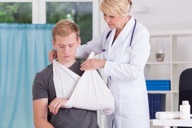
This can be done in different ways. The most common form of immobilization is a plaster cast, but in some cases other methods are also possible: soft Desaut bandage for shoulder injuries, skeletal traction (see) for hip injuries. The duration of immobilization after a shoulder injury is approximately 3 weeks, and for a hip injury it is up to 4 weeks. Longer immobilization (1-1.5 months) is required for the collarbone, shinbone and foot.
: X-ray image.")
- Dislocated joints: symptoms and reduction
- Premium care: What modern diapers can do
- Symptoms of a dislocated joint
- Common symptoms
- Types of joint dislocations
- causes
- Symptoms of a shoulder dislocation
- Patenatomy
- classification
- Diagnosis of habitual dislocation
- Treatment of habitual dislocation
- causes of injury
- Which doctor should I see?
- Our specialists
- Treatment of dislocations, joint subluxations and their reduction with MANUAL-DELUX
- Types of dislocations
- Dislocation symptoms.
- Congenital dislocations
- Pathological dislocations
Dislocated joints: symptoms and reduction
Joint dislocations are a common dislocation in trauma surgery that is characterized by a complete displacement of the articular surfaces of the bones relative to each other. This dislocation is most commonly caused by trauma. In some cases, it can be caused by weakness of the joint capsule, musculoskeletal system and changes in the articular surfaces. In this case one speaks of a habitual dislocation. In this article, we'll look at the main symptoms that occur with a joint sprain and the methods to fix them.

Premium care: What modern diapers can do
Symptoms of a dislocated joint
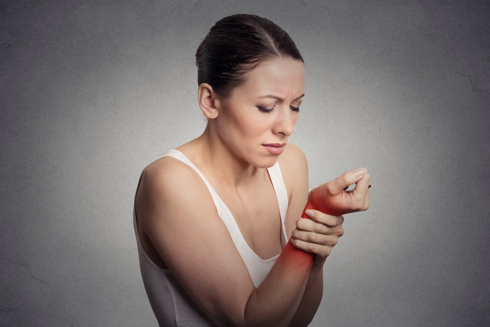
The main clinical symptom of acute traumatic dislocation is an acute pain syndrome with a characteristic clicking or popping sound.
The joint itself is swollen and deformed. Bleeding may occur from the skin in the injured area. In some cases, the skin beneath the lesion is pale and cold. Sometimes the compression or damage to the nerve trunks caused by the dislocation leads to sensory disturbances.
Severe limitation of motor activity is an obligatory symptom.
A habitual dislocation is a repeated episode of displacement of the articular surfaces of bones in a certain area relative to each other. Gradually, dislocations become more common due to the progression of pathological changes in the joint.
Clinically, this pathology is also accompanied by pain, although it is much less than with a traumatic dislocation. In some cases there is no pain at all.
Common symptoms
Symptoms of pathology vary depending on the type of dislocation. Experts have compiled a list of common symptoms that occur with all types of dislocations:
- Redness of the skin over the injured joint.
- Significant pain that increases with the slightest movement.
- The examination shows a changed shape and size of the injured joint.
- Significant swelling of the tissue in the area of injury.
- Loss of sensation due to impaired innervation of the limb.
- Restriction of movement.
- High fever.
A common specific symptom of dislocations is the appearance of spring-like resistance when attempting passive movement.
Types of joint dislocations
In traumatology, several types of joint dislocations are distinguished:
- By location – a dislocation characterizes the lower extremity or upper vertebra.
- By origin – acute dislocations (up to 3 times in one joint, fixed by X-ray), habitual dislocations (after 3 acute injuries in one joint), pathological dislocations (in diseases, neoplastic lesions of the joints), congenital dislocations (consequence of congenital injuries).
- Depending on the degree of damage, a distinction is made between complete and incomplete dislocations. A complete dislocation results in complete separation of the joint components. In incomplete dislocations there is partial contact of the articular surfaces.
- The integrity of the skin in the area of injury is disrupted - either open or closed.
- Depending on how long ago the injury occurred – fresh (up to 3 days), not fresh (up to 4 weeks), old (more than 4 weeks).
Treatment and rehabilitation depends on the location of the injury.
causes
A traumatic shoulder dislocation usually occurs as a result of indirect trauma, such as: B. Falling onto a bent or raised arm. The capsule of the shoulder joint tears and the head of the humerus shifts in the direction of the tear. In some cases, an anterior shoulder dislocation is caused by a direct blow to the back, while a posterior dislocation is caused by a direct blow to the shoulder joint from the front.
Depending on the etiology, traumatology and orthopedics distinguish between primary (traumatic) shoulder dislocations, voluntary, congenital, habitual and pathological dislocations.
- The habitual shoulder dislocation results from inadequate regeneration of the rotator cuff following a traumatic dislocation.
- Pathological dislocations can be caused by tumors, osteoarthritis, tuberculosis, osteochondropathy, osteodystrophy, etc.
A shoulder dislocation can be combined with a fracture of the humeral head, the anatomical or surgical humeral neck, a tear of the small or large humerus, a fracture of the socket, acromion or scapular process, as well as injury to nearby tendons, vessels and nerves. If the dislocation is combined with another injury, it is called a complicated shoulder dislocation. Depending on the direction of dislocation of the humeral head, a distinction is made between anterior, posterior and lower shoulder dislocations. Anterior dislocation of the shoulder is the most common (3/4 of all dislocations). The second most common dislocation is the inferior shoulder dislocation (approximately 20 %).
Symptoms of a shoulder dislocation

Traumatic dislocations of the humerus are associated with acute pain at the site of injury and deformation of the shoulder joint (it becomes angular, sunken or concave). Movement of the joint is impossible. During passive movements there is a characteristic spring-like resistance.
In an anterior shoulder dislocation, the head is displaced forward and downward. The shoulder is in a forced position (sideways or flexed, extended and stretched). During palpation, the humeral head is not in its normal position and can be felt in the anterior region of the armpit (in anterolateral dislocations) or below the shoulder process of the scapula. Anterior and anterolateral shoulder dislocations are sometimes associated with a tear of the humeral tuberosity and a fracture of the shoulder process of the scapula.
If the lower shoulder joint is dislocated, the head is displaced into the armpit. Vessels and nerves run through the armpit. When the head compresses the neurovascular bundle, numbness of the skin and muscle paralysis occur in the area innervated by the compressed nerve. Posterior shoulder dislocation is characterized by a displacement of the head toward the scapula.
Patenatomy
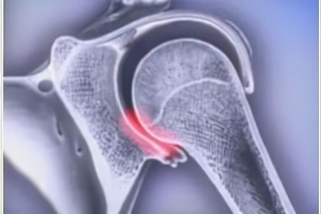
A joint is a movable connection of two or more bones covered by a synovial membrane, separated by a joint space, and connected by a capsule and ligaments. There are different types of joints (ellipsoidal, block-shaped, spherical, saddle-shaped), but regardless of their shape, they are all composed of congruent (fitting together, complementary) surfaces.
Due to this structure, the joint surfaces slide against each other during movement and the joint acts like a hinge. The muscles that attach to the bones above and below the joint are responsible for movement. Tight muscles pull the bone in a specific direction, while the joint capsule and ligaments prevent the ends of the joint from moving too far apart. During a sprain, the ends of the bones that make up the joint are displaced. The surfaces are no longer 'aligned' and movement is no longer possible.
There are three main mechanisms by which a dislocation can occur. Traumatic - as a result of increased muscle tension, a direct impact or a violent blow in indirect trauma, excessive displacement of the articular ends of the bones occurs. The impact is too strong, the joint capsule cannot withstand it and tears, and ligaments can also tear. Pathological - as a result of various pathological processes, the strength of the capsule and ligaments decreases and they lose the ability to hold the articular ends of the bones in the correct position even under slight pressure, so dislocation can occur during normal, unforced movements . Congenital – due to abnormal development of the joint structures (bones, ligaments, capsule), the joint surfaces initially do not fit together or are not held in the correct position.
classification
Orthopedic traumatologists distinguish between total dislocations, in which the joint ends are completely separated from each other, and subluxations, in which there is partial contact between the joint surfaces.
- Congenital sprains are caused by malformations of the joint components. The most common are congenital hip dislocations; congenital knee and patellar dislocations are less common.
- Acquired sprains are caused by injury or illness. Traumatic dislocations are the most common. The upper limbs are affected 7-8 times more often than the lower limbs.
Traumatic dislocations are further subdivided:
- Depending on the age of the injury: fresh (up to 3 days after injury), not fresh (up to 2 weeks after injury), frozen (more than 2-3 weeks after injury).
- With or without injury to the skin and underlying soft tissues: open and closed.
- Based on the presence or absence of complications, a distinction is made between uncomplicated and complicated fractures with nerve or vascular injuries and periarticular or intraarticular fractures.
A distinction is also made between uncontrollable dislocations, which include dislocations with soft tissue interpositions that prevent closed reduction, and all chronic dislocations.
There are also two different groups of pathological dislocations:
- Paralytic dislocation – the cause of development is the paralysis of a muscle group, as a result of which the pulling force of the antagonist predominates.
- Habitual dislocation – is a recurrent dislocation that occurs due to weakness of the joint capsule, muscles and ligaments and/or due to changes in the configuration of the articular surfaces. It is most commonly caused by premature displacement of the joint following repair of an acute traumatic dislocation. Habitual dislocation occurs less often in diseases affecting bones and ligaments (osteoarthritis, osteomyelitis, poliomyelitis and some systemic diseases, including hereditary diseases).
Diagnosis of habitual dislocation
If you suspect a habitual dislocation, you should immediately consult a traumatologist or surgeon. The doctor will examine you and take a medical history. Most patients complain about the consequences of trauma and limited mobility. During the examination, the specialist conducts a test: he checks the mobility of the joint and tries to reproduce the dislocation, which is successful in most cases.
The main and mandatory diagnostic method is an X-ray of the joint in two projections. MRI and CT images are reliable diagnostic methods with a high level of detail. They show the condition of cartilage, soft tissue, joints and bones.
Additional examinations are usually recommended if surgical treatment in hospital is required:
- General urine and blood tests;
- Rhesus factor and blood group test;
- coagulogram;
- testing for HIV, syphilis and hepatitis;
- blood chemistry;
- ECG;
- Ultrasound of the damaged joint - this allows problems within the joint to be identified without dangerous radiation.
The final diagnostic and treatment step is arthroscopy. This is a minimally invasive surgical procedure that involves making several small punctures to gain access to the joint. A miniature video camera is inserted into the joint cavity. This allows the doctor to look at all the tissues of the joint in detail and determine the cause of the habitual sprain.
X-rays of the damaged joint before and after reduction are sufficient to certify incapacity for work or to undergo a medical examination by the military or other authorities.
Treatment of habitual dislocation
The first step is to reset the joint and realign the joint components. Pharmacological treatment is not very effective for habitual dislocation. Medication is prescribed if the pathology arose as a result of premature loading of the injured limb during the treatment of primary dislocation.
One of the most popular drugs with proven effectiveness is Artracam. It is prescribed after a joint dislocation to relieve pain, heal and regenerate joint and cartilage tissue. It contains glucosamine, which is essential for restoring and preventing cartilage and joint destruction. Artrakam effectively relieves pain, combats age-related degenerative changes, accelerates healing of micro-injuries and normalizes blood circulation in damaged tissues. Artrakam is packaged in convenient sachets containing a powder for the preparation of a therapeutic suspension. It should be taken in one serving daily for 40 days. Two packs of Artrakam are sufficient for one treatment.
The product can be taken not only for joint diseases and injuries, but also for their prevention. It is particularly recommended for risk groups: older people, competitive athletes, people with a sedentary lifestyle and overweight people.
In other cases, surgical intervention is required. The surgeon's job is to repair damaged ligaments and cartilage and restore normal mobility and range of motion to the joints. Surgical intervention is always planned, since the condition of a patient with a habitual dislocation is not particularly dangerous.
The most common surgical procedure is minimally invasive arthroscopic surgery. These procedures are minimally invasive, allow for a shorter rehabilitation period and allow joint mobility to be completely restored within a few months. When restoring the integrity and function of damaged tissue, fixation is achieved with medical staples, screws, anchor sutures, and total sutures. Materials are often used that can be reabsorbed after the tissue has healed without leaving scars. If arthroscopy is not possible, open surgery can be performed.
causes of injury
A dislocation can occur under any circumstances. Very often it is preceded by an injury in which the joint capsule and ligament are torn. In many cases, a sprain can also be caused by one of the conditions mentioned above:
The above diseases can affect people of all ages and genders. As these diseases progress, the joint surfaces become weakened, deformed and decomposed, resulting in dislocation. This injury is often caused by a sudden muscle contraction and a fall onto an outstretched or semi-extended limb. This most often happens during sporting activities. The most traumatic activities are considered to be:
Skiers and ice skaters are also at risk. In addition, the symptoms of a sprain are very well known to those who are professionally involved in dance, especially modern movement. The pathology can be not only acquired, but also congenital. In this case, the dislocation occurs during the intrauterine development of the child. It is caused by abnormal changes in the development of the musculoskeletal system.
Which doctor should I see?
If an injury occurs, a specialist should be consulted immediately. The most common way to diagnose and treat a dislocation is:
Our specialists
The prices indicated on this page do not constitute a public offer. For more information about the cost of services and doctor's visits, please call 8 (495) 255-37-37.
Treatment of dislocations, joint subluxations and their reduction with MANUAL-DELUX
Joint dislocations and subluxations are diseases that are often not life-threatening, but significantly limit the quality of life.
- First and foremost, it is important to immobilize (immobilize) the joint. This can be done with a sling, bandage, cloth or splint to hold the injured joint in place.
- Cold can be applied to the joint in the form of ice, a cold water bottle, or something from the freezer (a piece of meat).
- You should then immediately consult a chiropractor or trauma surgeon for diagnosis and treatment (Reduction of a sprain or subluxation).
Reduction of sprains and subluxations
Joint sprains and subluxations should be treated as quickly as possible because swelling may worsen over time. alignment becomes more difficult After this time, the swelling gets worse and the joint becomes difficult to straighten. This procedure should only be performed by an experienced doctor after checking whether there are any contraindications to the joint, such as: B. Broken bones or damage to the blood vessels.
Correction of a dislocation (subluxation) of a joint in Kiev in 'MANUAL-DELUX' is carried out by an experienced chiropractor (osteopath/chiropractor) using painless and non-traumatic techniques.
It is important to know that self-reduction can lead to aggravation and possible damage to the joint structures!
Joint dislocations and subluxations are usually reduced by chiropractors without surgical intervention. In severe cases, it is worth seeing a surgeon who will carry out a special surgical procedure to treat the joint dislocation (subluxation).
Different techniques are used to correct dislocated or subluxated joints, depending on which joint is affected, how old the patient is, and what other factors are at play. The mechanism used is opposite to the mechanism of injury: osteopathic reduction according to ET Still and other osteopathic techniques, chiropractic reduction, Kocher reduction, Janelidze reduction, Hippocrates reduction and other techniques.
Types of dislocations
Dislocations can be classified according to the degree of displacement, the origin and the size of the dislocated joint.
In terms of the degree of displacement, a dislocation can be complete - when the joint ends are completely separated from each other - or partial - when the articular surfaces are partially in contact. A dislocated joint is one that is further away from the trunk. Exceptions include the vertebrae (a dislocated vertebra is the one above it), the clavicles, and the shoulder (a dislocation can be anterior or posterior).
Depending on their cause, they can be congenital or acquired. Congenital dislocations are caused by abnormalities in the child's fetal development. Hip dislocation (dysplasia) is a common hip dislocation, while knee dislocations are less common. Acquired sprains are caused by trauma or disease (arthritis, osteoarthritis, osteoarthritis, poliomyelitis, etc.).
They can also be open or closed. Closed dislocations pass without tearing the skin and tissue over the joint, while open dislocations can result in a wound. Damage to muscles, blood vessels, bones, tendons or nerves makes a sprain complicated. A habitual sprain occurs when the sprain can occur again with even a slight impact due to poor treatment. Pathological dislocation, characteristic of the hip and shoulder joints, occurs when a pathological process destroys the articular surfaces.
When the muscles surrounding the joint are paralyzed, it is called a paralytic sprain.
In addition, modern medicine distinguishes more than ten types of sprains that can affect the following joints in the following parts of the body:
Dislocation symptoms.

The symptoms of a sprain depend on the type of sprain. For example, a congenital hip dislocation is associated with gait disturbances because abduction is limited, the buttock folds are asymmetrical, and one leg becomes shorter than the other, resulting in a limp. If the hip is dislocated on both sides, the gait becomes a 'duck'.
A congenital dislocation of the knee joint is associated with severe pain, gait disturbances, inflammation and stiffness. Traumatic sprains are manifested by swelling, stiffness and pain in the joint.
However, if we talk about general symptoms, the following, among others, are observed.
- Redness appears in the area of the affected joint;
- There is severe pain that increases with every movement of the limb;
- The injured person can visually recognize the deformation of the joint because the dislocation not only changes the size of the joint but also its shape;
- There is severe swelling at the site of the dislocation;
- Some people experience loss of sensation in the limbs (in the case of nerve damage);
- impairment of motor function;
- Fever;
- Fever followed by chills, etc.
Congenital dislocations
Congenital dislocations Are the result of intrauterine malformations that cause deformation of the articular ends of the bones. Congenital dislocation of the femur is the most common (Figure 10), affecting 0.2-0.5 % of all infants - 5-7 times more common in girls. Unilateral congenital V is 1.5 times more common than bilateral.
The success of the treatment of congenital V. largely depends on its early detection.
Radiological evidence of a congenital V. In young children, linear and angular indices of the relationship between the femur and the acetabulum, such as the Schenton line, the vertical Ombredann line and the angle between the horizontal and the plane of the so-called roof of the acetabulum (see hip joint ), are taken into account.
The femoral head is underdeveloped and the hip socket is flattened (x-ray).

Congenital V of the femur in adults can also be diagnosed based on the anatomical features of the proximal femur (underdevelopment of the femoral head combined with valgus deformity of the femoral neck and front) and the flattening and enlargement of the acetabulum (Fig. 11).
Detection of congenital hip dislocation in newborns allows early treatment and good results with functional treatment methods (Vilensky, Volkov, CITO et al.)
Late discovery of a congenital dislocation significantly complicates treatment, requires surgical techniques, and is associated with poorer functional outcome.

Figure 12: Pathological dislocation of the left thigh in tuberculosis. You can see the damaged femur, the condyle head and the outline of the hip socket (the femoral head is displaced). X-ray image.
Pathological dislocations
Pathological dislocations Pathological dislocations often arise as a result of pathological processes in the joint cavity or at the ends of the joint (Fig. 12), which cause deformation of the ends of the joint (osteochondral tuberculosis, arthrosis and others) and have symptoms similar to trauma B.

Fig. 13: Pathological subluxations of the metacarpophalangeal joints I - IV and the interphalangeal joint I in polyarthritis (X-ray).
Often pathological veins and subluxations are caused by the destruction of the articular surfaces in mono- and polyarticular arthritis (Fig. 13) and in osteoarthritis.
Abnormal contortions can also occur in flaccid limbs (poliomyelitis) and in some forms of neurogenic arthropathies (see). Treatment of a pathological dislocation depends on its genesis and consists in restoring joint function through a complex of therapeutic measures (e.g. arthroplasty with subsequent reconstructive therapy for coxarthrosis).
I. Joint – see article by joint name: I. Shoulder – see shoulder joint; I. Forearm – see elbow joint; I. Tibia – see knee joint, etc.; I. Fingers and toes – see hand, foot; I. Clavicle – see clavicle; I. Patella – see kneecap; I. Jaw – see temporomandibular joint. Temporomandibular joint – see temporomandibular joint.
Bibliography: Babich BK Traumatic dislocations and fractures, Kiev, 1968, bibliography; Barta O. Congenital hip dislocation and its early conservative treatment, translation from Hungarian, Budapest, 1972; Bogdanov FR and Levenets VN Treatment of traumatic dislocations, Proceedings of the 3rd All-Union Conference on Surgery and Orthopedics, p. 63, Voronezh, 1969; Volkov MV, Ter-Egiazarov GM and Yukina GP Congenital dislocation of the hip, Moscow, 1972, bibliography; Kaplan AV Closed injuries of bones and joints, Moscow, 1967, bibliography; Kurbatov AI Pathological burn dislocations, Orthop. and Traumatology, ¹ 3, p. 50, 1968; Maykova-Stroganova VS and Rochlin DG Bones and joints in X-ray imaging, L., 1957, bibliography; Mitelman Yu. N. X-ray examination of large joints in patients in orthopedic and traumatological clinics, Kiev, 1962, bibliography; Reinberg SA X-ray diagnosis of bone and joint diseases, Vol. 1-2, M, 1964; Watson-Jones R. Bone fractures and joint injuries, translated from English, M., 1972; Farshatov MN and Glebov Yu. I. On the treatment of open dislocations of limbs, Vestn. chir., vol. 88, no. 4, p. 70, 1962; Bruckner H. Fractures and Dislocations, B., 1974; Pollen AG Fractures and dislocations in children, Edinburgh-L., 1973, bibliogr.
Read more:- How much does shoulder dislocation surgery cost?.
- This is what a dislocated leg looks like.
- pelvic subluxation.
- Pronation of the shoulders - what is it?.
- Anatomy of the Lisfranc joint.
- Dislocation of a bone in a joint.
- How do you treat a sprained ankle?.
- dislocation of the foot.