The ArthroMedCentre joint and spine clinic has been treating sports injuries for many years, using the innovative MBST technique to repair damaged tissue.

- Sprained ankle ligaments
- Physical examination
- Characteristics of the injury
- clinical signs
- Diagnosis of an ankle sprain
- How a strain is diagnosed
- methods of investigation
- diagnosis
- First aid
- First aid for sprained ankles
- Symptoms of an ankle sprain
- Causes of ankle ligament injuries
- Types of ankle injuries
- What is a fracture?
- Treatment of dislocations and fractures
- Where does it hurt?
- Diagnosis of a torn ankle ligament
Sprained ankle ligaments
This is a very common injury in all age groups.
The ankle ligaments are strained or torn when these relatively strong anatomical elements are subjected to a load that exceeds their tensile strength. This is a common injury in all age groups. Depending on the severity of the ligament injury, sprains or strains can be mild or severe.
Most ankle ligament injuries are relatively harmless and can be treated at home by resting the injured limb and applying ice packs. However, if there is significant swelling of the ankle joint and a reduction in the support function of the limb or you are afraid of standing on your foot, you should definitely consult your doctor.
If left untreated and not properly rehabilitated, more or less severe ankle ligament injuries can lead to hypermobility of the joint, making the ankle vulnerable to further injury. Repeated ligament injuries can lead to more serious problems, including chronic pain syndrome, degenerative damage, and chronic instability.

An ankle ligament injury is an injury to one or more of the ligaments that stabilize the ankle joint.
Ligaments are strong connective tissue anatomical structures that connect one bone to another. The ligaments of the ankle joint hold the bones that make up the joint in an anatomically correct position, thereby stabilizing the joint.
Most ankle ligament injuries are characterized by damage to the external collateral ligaments. These can be mild sprains, individual fiber tears in the thickness of the ligament, or a complete tear of the entire ligament.
Physical examination
Diagnosis of an ankle ligament injury is based on a thorough clinical examination of the foot and ankle. This process can be relatively painful.
- Scan. The doctor carefully feels the ankle joint to determine which ligament is damaged.
- Determination of the extent of movement. The doctor assesses the mobility of the ankle joint in various directions, which is of course restricted if there is severe swelling.
If there is no fracture, the doctor can assess the severity of the rupture based on the swelling, pain and bleeding.

During the clinical examination, the doctor will carefully examine the soft tissues around the lateral malleolus and identify the area of greatest pain.
Characteristics of the injury
Every step a person takes puts strain on the ankle joint. Careless foot movement or a fall can cause serious injury. A tear in the ankle ligaments causes discomfort and significant pain when attempting to move the ankle. With rest and conservative treatment, the joint's previous mobility can be restored.
The ankle is a complex block-shaped joint that connects the shinbone to the foot. Its high mobility is due to its ability to not only flex and extend the foot, but also abduct it. Mobility is provided by ligaments located on all sides of the joint.
A ligament tear occurs during sudden movements when the connective tissue is stretched beyond its normal extent. Partial or complete damage to the fibers leads to loss of the ability to move independently. Statistics show that one in five people in the world has suffered a severe sprain.
Any type of sprain can be considered a torn ligament, and even the most minor injuries cause microtears in the connective tissue. A complete tear is associated with severe pain, swelling and complete limitation of movement.
Ligaments located on the outside of the joint are most often injured when the leg is externally rotated. The delta band on the inside is stronger and less likely to tear.
clinical signs
Severe pain occurs at the time of injury as the ligament is stretched to its limits. The crack is accompanied by a loud clicking sound, similar to the sound of a plucked string. The affected person can no longer move independently and the ankle loses its function.
There will be slight swelling in the first few minutes after the injury. After a few hours, the ankle will be covered in swelling that will be more visible directly at the injury site. A hematoma forms a few centimeters below the joint. At first a small bruise appears, but within a few days the bruise covers the entire surface of the ankle. The swelling and bruising will disappear within three weeks.
First aid consists in taking painkillers (Ketanov, Baralgin, Next), cooling the joint and applying a tight bandage to prevent severe swelling and relieve pain. After the injury, diagnostics should be performed at a trauma center to rule out a complicated ankle fracture.
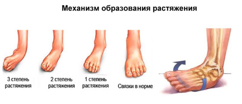
In traumatological practice, three degrees of ligament tears are distinguished:
- The first degree is classified as easy. The band maintains its anatomical integrity. If a force is applied that stretches the ligament against its natural, physiological direction, the connective tissue fibers are micro-damaged. At this point, there will be moderate pain and a small collection of fluid around the ankle. Hematomas do not usually occur with minor injuries. The joint's mobility remains, but is painful. It is still possible to walk independently, but with a limp and with the help of a stick or crutch.
- The second stage is characterized by rupture of collagen fibers. The pain becomes more severe, the leg is severely swollen and subcutaneous bleeding is visible. Severe pain occurs when attempting to take a step and movement is severely restricted. The joint becomes hypermobile and the damaged ligament no longer holds the foot in a physiological position.
- The third stage occurs when the connective tissue is completely torn. The injury is accompanied by a loud crunching sound with stabbing pain. The affected person falls and can no longer step on the injured leg. Swelling and bruising occur immediately, indicating a serious injury. The joint becomes unstable and the foot is not immobilized.
Diagnosis of an ankle sprain
The doctor will inquire about the circumstances of the injury and perform a physical examination.
Doctors also move the ankle in different directions to determine the severity of the sprain. However, if the victim is in severe pain and has severe swelling or muscle spasms, the examination is usually postponed until x-rays can be taken to detect fractures.
To assess the ankle, the doctor will gently palpate the ankle to determine where the pain is most severe. If touching the skin over the ligament causes severe pain, you probably have a torn ligament. If touching the skin over the bone causes pain, you may have a broken bone. Swelling and cramps make it difficult to assess the condition of the ankle. In such cases, the doctor may immobilize the joint with a splint and examine it again after a few days.
The diagnosis of a sprain is based primarily on the results of the examination. However, sometimes the doctor will order an X-ray to rule out a fracture if the
How a strain is diagnosed
A complete ligament tear does not cause any specific symptoms. A dislocation or fracture can be incorrectly suspected. The following symptoms are observed:
- Pain at rest and when moving;
- swelling of the ankle joint;
- smoothing the contours of the ankle joint;
- instability of the joint;
- fracture when ruptured;
- numbness of the limbs;
- Tingle;
- Swelling;
- Hematoma.

There are no signs of intoxication. The swelling occurs immediately after the ligament tears. In the first few hours she has no clear boundaries. The swelling occurs directly at the site of the ligament tear. Over time it increases and spreads to the ankle joint. The severity of the swelling is directly related to the severity of the injury. The swelling of the extremity lasts 2-3 weeks.
A complete rupture is always characterized by bruising. This is due to vascular injury in this area. Hemarthrosis may occur. Blood collects in the joint cavity. The bruise is large. Every experienced doctor has seen pictures of injuries like this. They are characterized by acute restriction of movement.

The ankle becomes unstable because the ligaments can no longer fulfill their primary function.
The contours become smoother. Congruence (the correct alignment of the bones in the joint area) is impaired. The affected person has great difficulty supporting himself on his leg. The ligaments are usually torn on one side. When pressure is applied to the lower leg, severe pain occurs. A limp may occur. In advanced cases, complications can occur. An infection often occurs.
methods of investigation
If left untreated, the consequences can be very serious. You may be unable to work for 1-2 months. An experienced doctor must first rule out a fracture or dislocation of the ankle. For this purpose, the following investigations are carried out:

If hemarthrosis is significant, a diagnostic puncture may be necessary. The extent of damage to the ligamentous apparatus can be determined using MRI. It is one of the most precise diagnostic methods. The advantages of an MRI scan are:
The procedure is not suitable for people with pacemakers, metal implants or aneurysms. There is no radiation exposure. Since not all hospitals have an MRI scanner, X-ray examination is the most common method of detecting a fracture and ruling out other injuries (fractures, dislocations).
diagnosis

An orthopedist/traumatologist diagnoses and treats ligament tears. First, the doctor visually examines the problem area, examines it by palpation, asks the patient to perform several movements and analyzes the mobility of the joint, determines the type and intensity of the pain and takes the medical history. The subsequent instrumental diagnosis allows an objective assessment of the anatomy of the joint:
Based on the diagnostic results, a differential diagnosis for bone fractures and ligament tears is made, as these diseases manifest themselves almost identically.
First aid
Proper first aid will help slow the progression of pathological changes and increase the effectiveness of treatment. If it is not possible to go to a trauma center, you should take action yourself.
First aid for torn ligaments includes:
- complete immobilization of the injured joint (no or minimal movement of the injured limb, use of a cane or crutches for support);
- applying cold to the injured area (ice pack or frozen food wrapped in a towel);
- Applying a compression bandage (moderately tight).
If the patient is unable to see a doctor immediately, painkillers can be taken.
First aid for sprained ankles
In the event of an ankle sprain, timely first aid has a great impact on the regeneration of the tissue of the injured ligaments and determines the time until the joint function is completely restored.
- First, the injured ankle should be cooled with ice wrapped in a towel for 10-15 minutes and the procedure repeated after 15 minutes. Such treatment not only relieves pain, but also prevents the swelling from spreading (under the influence of cold, blood vessels reflexively contract).
- The injured ankle should be left alone and no splint should be placed on the foot until a diagnosis is made. The foot must be fixed with a house or transport rail. Immobilization of the area should be maintained until examination by a trauma surgeon or traumatologist.
- Place a pad under the foot to elevate it.
- Give a pain reliever (oral or by injection) if possible.
Symptoms of an ankle sprain
Symptoms of an ankle sprain depend on the extent of the injury and the number of tendons involved in the mechanism of injury. A complete tear of the ankle ligaments only occurs as a result of a sprain. This condition is characterized by excessive mobility in all projections of the joint. The main symptoms of a sprained ankle are:
- Severe pain that occurs at the time of injury (fall, impact, etc.);
- swelling around the injured area (medial or lateral ankle);
- bruising;
- extensive hematomas;
- localized increase in temperature (in the area of the injury).
When trying to move, the sufferer continues to experience painful sensations.
Repeated sprains of the ankle ligaments result in complete rupture of the tendon. A lengthy and complicated rehabilitation is then required. That's why the experts at ArthroMedCenter recommend seeing a doctor at the first signs of a sprain. To avoid re-injury and the development of another pathology, comprehensive treatment, including rehabilitation, is useful.
Causes of ankle ligament injuries
Ankle ligament injuries are not random events. They are the result of the presence of a number of risk factors. Their pathogenic effects can affect the integrity of various tissues.
The ankle joint has a fairly complex structure. It consists of three bones. At the tip are the distal ends of the tibia and fibula. These are connected to each other by a cartilaginous anastomosis. If this anastomosis breaks, the entire joint deforms. From below, the joint is formed by the talus bone. This bone forms another joint with the heel bone - the subtalar joint.
The joint is surrounded by a dense joint capsule containing synovial fluid. There are ligaments and tendons nearby. The ankle joint is involved in walking, running and jumping. It takes on part of the damping load.
Damage to the ankle ligaments is very likely in people with the following risk factors:
- Excessive body weight – each additional kilogram increases mechanical pressure on the articular surfaces;
- sedentary lifestyle – the muscles around the ankle joint do not function fully and do not supply the ligament fibers;
- heavy physical work with constant standing, lifting and carrying loads;
- disturbed metabolic processes in the body;
- Impairment of blood supply to the lower limbs, usually obliterative arteritis, varicose veins of the lower limbs, atherosclerosis, vasculitis, diabetic angiopathy, etc;
- unsuitable footwear for daily wear and sports;
- Flat feet, clubfoot and other types of foot abnormalities;
- Deforming osteoarthritis in the knee and hip;
- Postural abnormalities and curvatures of the spine, leading to an imbalance in the distribution of shock-absorbing forces and an increased risk of rupture of the anastomosis between the tibia and fibula;
- Valgus or varus deformities of the tibia, big toe, etc.
Types of ankle injuries
There are many different types of ankle injuries. A closed ankle injury is, for example:
- Dislocation of the ligamentous apparatus (or microscopic tears in its fibers);
- Dislocation of the distal condyles of the tibia and fibula with tear of the cartilaginous anastomosis;
- Achilles tendon injury;
- Damage to the supporting ligament and development of fasciitis;
- Bone spur (growth) on the heel bone (heel spur);
- Fracture of the ankle bone (often caused by a fall on the heel from a height);
- Fractures and fractures of the distal ends of the tibia and femur;
- the penetration of blood into the joint cavity and the development of hemarthrosis.
Any, even partial, injury to the ankle ligaments requires the intervention of a traumatologist. If treatment and rehabilitation are not done quickly, ligament separation can occur. Such a patient can only be helped by a surgeon. Surgery is required to repair the damaged ligamentous apparatus. And after the procedure, a long rehabilitation period is required.
What is a fracture?
In simple terms, a fracture is a break in the continuity of a bone caused by mechanical damage. There are several types of fractures:
- Closed (the soft tissues are intact).
- Open (soft tissue damaged, bone fragments outward).
- Not displaced (no displaced bone fragments).
- Shifted (bone fragments are shifted).
- Pathological (caused by a bone disease – cancer, osteoarthritis, osteoporosis, etc.).
- Sensation of pain (can be of different types).
- The limb is in an unnatural position.
- Mobility is impaired - you can only bend it a little, in the part where there is no joint.
- Bone splinters are visible in the wound (if the fracture is open).
- There is severe swelling at the site of the injury, which developed very quickly.
- Traces of blood are visible at the site of the injury and there is a hematoma.
- Pain increases when the limb is rotated about its axis, a symptom of axial overload.
Treatment of dislocations and fractures
Both injuries require appropriate assessment and treatment plan. Therefore, trauma surgeons prescribe X-rays to determine the extent of the injury (less commonly CT, MRI or ultrasound). Only after taking a picture of the injured area, taking the medical history and asking the patient about the treatment will the surgeon decide on how to proceed.
In severe cases, a bone prosthesis may be indicated; in milder cases, a cast or bandage may be indicated. For mild sprains, the doctor may prescribe ointments to relieve pain. Doctors also often prescribe the following medicines for bone sprains and fractures:
On our website you can see all the medicines available in pharmacies in your city and their prices by entering the name of the medicine in the search box and doing a search.
Where does it hurt?
Torn ankle ligaments can lead to subluxation and dislocation, and this injury is often associated with fractures. The consequences of inadequate treatment of a torn ankle ligament can affect the patient for many years. The most common symptoms are joint instability or the development of osteoarthritis, in which the articular cartilage breaks down prematurely. All that is needed is a correct diagnosis.
[10], [11], [12], [13], [14]
Diagnosis of a torn ankle ligament
Diagnosis of any injury begins with interviewing the patient. The doctor will assess the mechanism of the injury. With ankle injuries, the patient hears a cracking or snapping sound, feels pain, and sees that there is bleeding into the joint, swelling, sometimes the size of a chicken egg. No change can be seen on x-rays if there is no ligament tear. If the ligament is torn, you can see bone fragments that have come loose with the ligament. Therefore, an MRI scan is the most informative method for investigating a suspected ligament tear. If an MRI examination is not possible, an ultrasound examination is carried out.
[15], [16], [17], [18], [19], [20]
Read more:- Damaged ligaments of the ankle photo.
- Injury to the ligaments of the ankle.
- Rupture of the ligaments of the ankle.
- Partial tear of the ligaments of the ankle.
- dislocation of the ankle.
- Injury to the ankle.
- Structure of the human ankle.
- Treatment of torn ligaments in the ankle.