The therapy not only has a positive effect on muscles, ligaments and joints, but also on the nervous system and promotes a positive attitude. Read more

- equinovarus foot deformity
- Causes of Equinovarus Foot Deformity.
- Advice and support from an orthopedic surgeon at the Perseus Orthopedic Center. Tel. 8 (495) 469-99-05
- Clubfoot in children
- causes and symptoms
- What is the difference between Equinovarus
- CAUSES OF FOOT BALANCE
- COMPLICATIONS OF THE EQUINUS FOOT
- Possible complications
- Errors and complications in the surgical treatment of mobile equino-planar valgus foot deformity in patients with infantile cerebral palsy using the technique of calcaneal correction osteotomy
- keywords
- Introduction
- Material and methods.
- prophylaxis
- Development of disorders
- Are there neurological problems in children?
- Clinical features.
- What shoes to wear at Equinus
- Possible complications
equinovarus foot deformity
Equinovarus foot deformity is a pathological foot position in which the forefoot is raised and the outer edge of the foot is lowered. Equinovarus deformity is a foot deformity commonly diagnosed in spastic forms of cerebral palsy. Cerebral palsy is a group of heterogeneous clinical syndromes resulting from non-progressive impairment of motor function and posture.
Equinovarus foot deformity is a severe form of musculoskeletal deformity.In most cases the deformity is bilateral. An equinovarus foot deformity can be:
Causes of Equinovarus Foot Deformity.
The equinovarus foot deformity is caused by a varus or equinus foot. It can also be caused by loss of eye muscle function. It is important to know that with equinovarus foot deformity, the paralysis can spread to the anterior tibial muscle group.
Main Causes of Equinovarus Foot Deformity:
- damage to the sciatic nerve;
- Damage to the oculomotor nerves (after stab and gunshot injuries);
- encephalitis;
- Poliomyelitis;
- Little's disease;
- Complicated and inadequately treated dislocations of the foot;
- Purulent and destructive processes on the foot;
- Dislocations and fractures of the ankle or other joints of the hindfoot.
Equinovarus deformity of the foot is manifested by the following clinical signs:
- Absence of active proportional movement about the anteroposterior horizontal axis of the foot;
- Presence of the characteristic components of the deformity (plantar flexion, varus, inward rotation of the distal end of the foot);
- Lack of active flexion of the foot.
Advice and support from an orthopedic surgeon at the Perseus Orthopedic Center. Tel. 8 (495) 469-99-05
- Orthopedic Surgery
- Consultation with a podiatrist
- online consultation
- Visit a specialist
- Manufacture of footwear
- Orthotics, traction insoles
- Orthoses and braces for the lower limbs
- Stabilization in cerebral palsy
- Insoles for clubfoot.
- With reinforced arch support
- For dynamic splints
- For children
- teenagers
- Adult
- Home
- Supportive footwear for prostheses and aids
- For diabetics
- Tactical shoes, military shoes, outdoor and hiking shoes
- Custom insoles
Clubfoot treatment with orthopedic footwear. Treatment of club feet. Perseus. 12

2 Complicated orthopedic footwear made from impression and contouring
Shoes with inserted tunic, stiff buttocks and lengthened ankle boots, lengthened heels: P-45, P-48, P-14


Equinovarus ankle leather corset
The splint is designed to fit hard-soled orthopedic shoes.

Clubfoot in children

Clubfoot is a deformity of the distal part of the lower limbs.
It includes equinus, varus, pes cavus, and anterior adduction. Clubfoot is more common in boys than girls.
Clubfoot in childhood is congenital. In newborns, this defect occurs with a frequency of one in a thousand. Acquired club feet occur as a result of nerve disease or trauma.
Massage and incremental affusions are required for effective treatment of clubfoot.
The best treatment results are achieved when treatment is started in infancy. Success depends on age and the severity of the deformity. Orthopedic shoes are prescribed for mild deformities when a cast is not indicated, or after removal of the cast to prevent recurrence of the deformity. During a child's active growth, foot deformity may recur. If treatment is started in a timely manner, the prognosis is favorable. Clubfoot is a developmental disorder that can be diagnosed soon after birth. In the early stages, clubfoot infants can be treated in a short time. If clubfoot is neglected or inadequately treated, the child may lose the ability to walk normally and become disabled. If you suspect this condition in your child, you should immediately contact an orthopedist who can make the correct diagnosis.In our center you will receive the necessary advice on clubfoot treatment and can confirm or allay your fears. It takes about two months to correct this anomaly in a child. The main factor that can delay clubfoot treatment for a long period of time is the severity of the deformity. In this case, surgical intervention can be avoided. Chiropractic adjustments, orthopedic shoes, and a gradual change in casts can help. Clubfoot is a developmental disorder that can become apparent as soon as a child is born. In the early stages, clubfoot in infants can be treated in a short time. If clubfoot is neglected or not treated in a timely manner, babies can lose their ability to move normally and become disabled. If you suspect this condition in your child, you should immediately contact an orthopedist who can make the correct diagnosis. At the Perseus Orthopedic Center you can get the advice you need to prevent and treat clubfoot. If treated promptly, the prognosis is favorable.
causes and symptoms
There are many possible causes. This is the reason why this condition is so common. A child's fragile bones deform easily, the muscles are not yet strong enough, and injuries do not heal well. And adapting to new stresses on the body is a very demanding process. The child rarely does this, if only because he doesn't understand the motivation.
If we consider the most common causes of its occurrence, the following variations can be distinguished in the development of an equinus deformity in children (ICD Q66):
- Injuries of various kinds. Almost any injury to the sciatic nerve can lead to changes in gait. From small to very noticeable and severe deformities. This also applies to almost all fractures of the lower limbs. This is particularly true for the ankle joint. Even a simple dislocation can have negative consequences. This also applies to a possible tear or complete rupture of a tendon in the ankle area. In such cases, surgery is also recommended for adults.
- Severe inflammatory processes in the ankle joint. This can be the result of both a simple injury and illness. For example, osteoarthritis is very harmful in this area.
- Infection. This is both a serious inflammation and a change in the structural elements of the tissue. If the focus of the lesion is near the ankle joint, ballerina syndrome is equally likely to develop. Encephalitis and poliomyelitis are considered the main causes.
- Abnormal recovery from injury. There are two factors here. The first is that the bones cannot articulate properly. And that's a good reason. The second option is somewhat rarer, but much worse. This is a false casting. If the worker was not the best professional.
- The most dangerous cause of the Q66 IBC or balance foot is the impact of cerebral palsy.
- Any type of endocrine disorder can also cause this abnormality. And they express themselves very seriously. Primarily any thyroid disease and especially the well-known diabetes.
- But there are also more prosaic reasons. A genetically caused weakening of muscle mass. Roughly speaking, bad genetics. Strengthening needs to be worked on. If the diet contains too little calcium, bone tissue becomes softer. And then it changes slightly. And, most unfortunately, such a serious problem can arise simply from uncomfortable footwear. That's why it's always best to buy special shoes for your child, like those offered by our OrthoPanda project. Otherwise, you could unintentionally provoke serious problems.
What is the difference between Equinovarus
Let's look at these aspects in table form.
parameter equinus Equinovarus Expression An anomaly manifested by a change in the arch of the sole. This condition is associated with the name 'horse foot'. This is a common variant. There is plantar flexion (flexion of the sole of the foot). The lateral side goes up to the shinbone and the outside side goes down. It is somewhat reminiscent of a club foot, but in a very severe form. Visual aspect This symptom is immediately noticeable. The affected person begins to literally walk on tiptoes. It relies on the fingers and joints. This in itself is very dangerous and leads to loss of function in these parts of the body. The affected person walks on the edge of the sole, with the foot assuming a very unnatural position. Causes and Causes The anomaly often begins during the child's development phase. It is an acquired disorder that can be avoided through preventive measures. In most cases it is a congenital form. Only treatment is suggested, prevention is not common. CAUSES OF FOOT BALANCE
The deformity of the balance foot in children is congenital or acquired. The main cause is damage to the long flexor tendons of the foot. In most cases, the sciatic nerve, which supplies the muscles of the lower leg and foot, is damaged. The tone of the muscle structures is reduced and the calf muscles shorten, with the toe pointing down and the heel pointing up.
Other triggers include:
Equinus deformity in ICD 10 refers to congenital anomalies and is coded number 66 for congenital foot deformities. The ICD-10 code for equine foot deformity therefore looks like Q66.
 COMPLICATIONS OF THE EQUINUS FOOT
The equinus deformity in children is often combined with transverse flatfoot, which is due to a misalignment of the foot. The child shifts the body weight to the first half of the foot and the arch of the foot gradually lowers while the heel remains elevated.
When equinus is combined with transverse flatfoot in children, treatment should primarily focus on the underlying cause, equinus deformity.
If left untreated, it leads to longitudinal-transverse flatfoot in children, in which the foot flattens in both length and width. However, the main symptoms occur in patients over 45 years old.
Possible complications
Equinus deformity of the foot is a serious condition of the lower limbs that requires early diagnosis and correction. In advanced cases of the disease, complications occur. Common consequences of pathology are:
- The child has difficulty communicating. He is ashamed of his disability and does not want to communicate with his peers. The child develops into a withdrawn, depressed and anxious being.
- The equinus foot leads to delayed physical development.
- Children often suffer injuries while walking. Parents must understand the difficulty of the situation and monitor the child's movements.
- Uneven foot pressure leads to the development of flat feet.
- The function of the knee joint is impaired.
- In advanced cases, muscle loss occurs in the lower limbs.
- The spine suffers from an abnormal gait. Scoliosis and lordosis develop.
- The rough skin on the outside of the foot is a cosmetic blemish that is often accompanied by constant pain.
- The gross deformity prevents normal movement. The person affected becomes disabled. He or she can only walk with the help of crutches.
Walking on tiptoes is the first warning sign of a possible illness. Even a mild degree of balance disorders leads to pathological changes in the spine and shins.
Over time, problems develop in the cardiovascular and respiratory systems. A monthly examination at the pediatrician can prevent the development of serious pathologies and diseases of internal organs! Abnormal deformities are common in children. Equinus is expressed by a raised and a lowered forefoot.
Errors and complications in the surgical treatment of mobile equino-planar valgus foot deformity in patients with infantile cerebral palsy using the technique of calcaneal correction osteotomy

The aim of the study.. Study of the results of treatment of patients with the mobile form of equinoplanar valgus foot deformity in infantile cerebral palsy using heel bone correction osteotomy according to the author's method. Analysis of errors and complications in the treatment of patients with this technique.
Material and methods.. Sixty-four patients (103 feet) aged 3 to 17 years were operated on between 2006 and 2014 using the described method of heel bone correction osteotomy. The equinus contracture was eliminated by cutting the calf muscle tendon and lengthening the Achilles tendon. Abnormal muscle tone was reduced by administration of Dysport to the calf muscle or selective neurotomy of the tibial nerve.
Results. The analysis showed a good result at 75 %, a satisfactory result at 18 % and an unsatisfactory result at 7 %. The unsatisfactory results were related to a series of technical and tactical errors that were grouped and analyzed.
Conclusions. Analysis of the errors and complications in the treatment of cerebral palsy patients with a mobile form of equinovalgal foot deformity using calcaneal corrective osteotomy will make it possible to avoid them in the future and improve the quality of treatment of this group of patients.
keywords
Introduction
The possibility of successful surgical correction of mobile equinovalgal foot deformity in patients with infantile cerebral palsy has always been of great interest to specialists in spastic paralysis surgery [1, 2]. This is mainly because this form of deformity, in contrast to rigid deformity, is more promising in terms of restoring foot shape and function. Corrective osteotomies of the heel bone have always occupied a special position among surgical treatment techniques [3, 4] because they are the most physiological. This is because they improve the cushioning function of the foot and do not block the joint movements of the foot.
We have been actively using the corrective osteotomy of the heel bone proposed in our department for several years [5].
The aim of this study was to analyze the most common tactical and technical errors encountered when treating cerebral palsy patients with a mobile equinovagal foot deformity using the heel bone correction osteotomy method.
Material and methods.
Between 2006 and 2014, we operated on 64 patients (103 feet) between the ages of 3 and 17 years. All patients voluntarily signed an informed consent to participate in the study and to undergo the surgical procedure.
The indication for corrective osteotomy of the calcaneus is the presence of a mobile form of equino-planar valgus foot deformity in a patient with cerebral palsy between the ages of 3 and 17 years, with an absent longitudinal arch and a heel valgus position of more than 10°. The surgical procedure we propose is based on the principle of extra-articular osteotomy of the calcaneus in the space between the posterior and medial surfaces of the subtalar joint. According to anatomical studies by several authors [6], the crossing area of the calcaneus we used lies outside the articular surfaces of the subtalar joint in 99 % cases. The operation was performed via two approaches: on the outside and on the inside of the foot. The first incision was made on the inside of the foot from the projection area of the metatarsophalangeal joint to the posterior edge of the medial ankle joint. After incising the skin, subcutaneous fat, and dorsal fascia of the foot, the tendons of the posterior tibialis muscle, long toe flexor, and long finger flexor were mobilized. An arthrotomy of the talo-phalangeal joint was then performed, leaving a strong fragment of the joint capsule attached to the sustentaculum of the talus. An arthrotomy of the subtalar joint followed to visualize the articular surfaces and determine the optimal direction for future osteotomy. After debridement of the periosteum, a step osteotomy of the calcaneus was performed (Figure 1).
prophylaxis
To avoid the occurrence of foot deformity, one should:
- Have a preventive examination carried out by a specialist.
- Wear properly fitted footwear made from natural materials.
- Eat a nutritious and balanced diet.
- lead an active lifestyle.
- In case of injury, seek medical attention.
- When walking, pay attention to the correct position of the child's feet.
- Keep body weight within normal limits.
An equinus deformity, even if mild, needs to be treated immediately. Malposition of the lower limbs affects the entire body: the leg joints and the spine, which in turn can lead to problems in the respiratory, cardiovascular and other systems. Correct and early treatment can prevent many problems in the future.
Development of disorders
The development of pyramidal insufficiency depends on the changes in muscle tone and the extent of damage to the nervous system (NS).
For example, central paralysis (incomplete loss of voluntary movements - central paresis) leads to disinhibition of neurons in the spinal cord. The pyramidal tract begins to send a large number of nerve impulses to the muscles, resulting in increased reflexes and muscle tension. There is also a tremor.
Flaccid paralysis (paresis) or peripheral paralysis occurs when a neuron and its extensions in the spinal cord are damaged. The muscle tension is reduced, even to the point of complete immobility of the muscles. Reflexes decrease or disappear completely and hypotrophy of the innervated area occurs.
Clinical paralysis of the lower limbs - the legs tremble and tense, the tone increases, the gait changes. The leg can hardly be extended, the foot is tilted to one side and there is a feeling of stepping ('houndstooth').
Clinical paralysis of the upper limbs - hands tremble and picking up objects requires great effort due to increased muscle tone. Hypotrophy or muscle wasting begins to develop.
Causes in adults:
- Inflammation in the brain (meningitis, encephalitis);
- Changes in brain hemodynamics (heart attack, stroke);
- head injuries causing disruption of nerve impulse transmission;
- Benign and neoplastic tumors.
Causes of illness in children:
Are there neurological problems in children?
The following neurological problems may well be caused by a diagnosis of pyramidal insufficiency:
Clinical features.
Symptoms of pyramidal insufficiency in children:
- trembling of arms, legs and chin;
- tilting the head back;
- Difficulty holding objects
- Squeezing the toes when standing
- movement on tiptoes;
- Poor toe control;
- Impairment of gait, coordination and eye movement;
- language impairment;
- low intelligence.
Symptoms of pyramidal insufficiency syndrome in adults:
- muscular hypertonicity of the limbs;
- High blood pressure;
- partial or complete immobilization of body parts;
- cramps, spasms;
- decreased reflex activity;
- overweight;
- sexual dysfunction.
In children under 2-3 months, pyramidal insufficiency is not a cause for concern as there is natural muscle hypertonicity. However, when the condition is diagnosed in older children, it is very worrying because it leads to movement disorder syndrome (MDS). In these children, not only do their righting reflexes develop and develop, but their motor skills are also delayed.
Dr. Komarovsky explains what pyramidal insufficiency is:
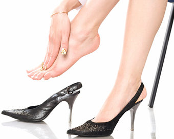
What shoes to wear at Equinus
According to the anatomical structure, the human foot consists of two arches: the transverse and the longitudinal arch. These are responsible for evenly distributing the load on the feet. Both arches, muscles, ligaments and tendons provide stability and cushioning.
A deformed foot suffers from high stress. When the foot is in an equine position, the distribution of body weight between the lower limbs is disrupted. Tight shoes that are not appropriate for the season worsen this condition.
The footwear must meet certain criteria:
- You must have a stiff back,
- they must provide good support for the back of the foot,
- Heels no higher than 2 cm are acceptable,
- and they have to be exactly the right size,
- Experts recommend buying shoes made from natural materials. Such shoes do not cause allergic reactions and do not affect the respiratory processes of the skin,
- Shoes that are too tight or too wide should not be worn
- Shoes should have a low sole. Flat soles promote flat feet,
- Platform soles, wedges, interfere with the normal flexion of the foot.
Possible complications
Equinus deformity of the foot is a serious condition of the lower limbs that requires early diagnosis and correction. In the advanced stages of the disease, complications arise. Common consequences of pathology are:
- The child has difficulty communicating. The child is not aware of his disorder and does not want to communicate with his peers. The child grows up withdrawn, depressed and anxious.
- The equinus foot leads to delayed physical development.
- Children often suffer injuries while walking. Parents must understand the difficulty of the situation and monitor their child's movements.
- Uneven foot pressure leads to the development of flat feet.
- The function of the knee joint is impaired.
- In advanced cases, muscle atrophy of the lower limbs occurs.
- The spine suffers from an abnormal gait. Scoliosis and lordosis develop.
- Rough skin on the outside of the foot – a cosmetic defect that is often accompanied by persistent pain.
- A large deformity prevents normal movement. The person becomes disabled. She only walks with the help of crutches.
Walking on tiptoes is the first warning sign of a possible illness. Even mild equinus leads to pathological changes in the spine and shins.
Over time, problems develop in the cardiovascular and respiratory systems. A monthly examination at the pediatrician can prevent the development of serious pathologies and disorders of internal organs! Pathological deformities – a common occurrence in children. Equinus is expressed by a raised and a lowered forefoot.
- equinus.
- Equino valgus.
- equinovarus foot deformity.
- Foot.
- Paraparesis - what is it?.
- Clubfoot in 7-year-old children.
- Why does a child develop clubfoot?.
- What to do if your child has clubfoot?.