A balanced and healthy diet, hardening of the body, wearing good shoes, moderate physical activity, outdoor walks and swimming should also not be forgotten.

- Congenital clubfoot in children - the most effective treatment
- What is clubfoot?
- ICD-10
- classification
- Main causes of clubfoot
- symptoms of the disease
- Who Treats Clubfoot?
- When should the treatment start?
- A visit to the orthopedist is the first thing to do
- Regular gymnastics – physical therapy for clubfoot
- Classification: Types of clubfoot in children
- Symptoms of clubfoot in children
- Causes of Clubfoot
- Diagnosis and Symptoms of Clubfoot
- Causes of Clubfoot
- When should treatment start?
- What to do if a child walks with an abnormal clubfoot?
- Immobilization with soft bandages and elastic orthoses
- Ponseti treatment
- physiotherapy
- Injecting medication into the calf muscle
- How to correct pathology at home?
- gymnastic exercises
- massage
Congenital clubfoot in children - the most effective treatment
Congenital clubfoot ranks second to hip pathology among all congenital diseases of the musculoskeletal system. Between 0.6 and 6.8 children per 1,000 are born with clubfoot, in the Russian Federation the figure is 1 to 3 per 1,000 newborns.
Turner Science and Research Center for Pediatric Traumatology and Orthopedics. The Turner Science and Research Center for Pediatric Traumatology and Orthopedics has been dealing with the treatment of this developmental defect since it was founded and has always played a pioneering role. Since 2000, NICT has been treating clubfoot using the Ponseti method, recognized by the international orthopedic community as the 'gold standard' for treating this pathology.
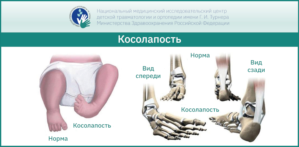
What is clubfoot?
Congenital clubfoot is a complex malformation in which the external shape of the foot is a manifestation of the skeletal, articular, soft tissue, nervous, and vascular systems of the lower limbs.
Clubfoot can be detected by ultrasound as early as 3 months of pregnancy, but there are cases where the parents are not 'surprised' until after the birth. In the maternity ward, the initial diagnosis can be made by a pediatrician. In any case, the pediatric orthopedist can only determine the degree of malformation, the need for treatment and the postpartum treatment plan. In larger cities, children are examined by a pediatric orthopaedist in the maternity ward in the first few days after birth.
ICD-10
clubfoot (Equinovarus foot deformity is one of the most common musculoskeletal abnormalities (33-38 %). It is usually bilateral. It is twice as common in boys as in girls.
The causes are not fully understood. Risk factors for clubfoot are thought to include fetal abnormalities, lack of amniotic fluid, smoking, alcohol, and drugs. The development of the foot bones, muscles and nerves of the lower leg is impaired due to fetal impairment. Possible secondary clubfoot due to pathology of other parts of the musculoskeletal system.
classification
Traumatology and orthopedics classify the following types of clubfoot:
- Idiopathic clubfoot. It is characterized by a drop in the waistline associated with an abnormal positioning of the neck, equinus (balance foot) in which the heel is pulled up and the foot is arched towards the sole, abnormal positioning of the forefoot in relation to the hindfoot, abnormal development of the Articular surfaces of the foot, shortening of the calf muscle, abnormal development of the tibial vessels in the front part of the tibia.
- Postural (positional) clubfoot. The heel and talus bones are unchanged. The articular surfaces are normally developed and subluxated.
- Congenital clubfoot in combination with congenital neuropathy and myopathy. The foot deformity is secondary and is caused by developmental anomalies in other parts of the musculoskeletal system (multiple curvatures of the limb bones, bilateral congenital hip dislocation, etc.).
- Syndromic clubfoot. Combination of the previous form of clubfoot with a non-medical pathology (amniotic stenosis, renal anomalies, etc.).
Main causes of clubfoot
Depending on the cause of clubfoot in children, a distinction is made between congenital and acquired defects. The prerequisites for the development of the first type of pathology include:
- Severe poisoning during the first trimester of pregnancy, when the nerve endings are forming;
- Viral infections with an increase in body temperature above 38 degrees;
- alcohol consumption, uncontrolled medication, smoking, drugs;
- vitamin and micronutrient deficiencies;
- Excessive stress during pregnancy;
- multiple pregnancy.
Acquired clubfoot can occur due to rickets or bone dysplasia. It can be caused by abnormal development of ligaments and muscles, micronutrient deficiencies, tumors or mental disorders. It can also be caused by excessive strain on the musculoskeletal system, excessive strain on the lower back muscles, wearing poor quality footwear, and severe injuries to the lower limbs.
Congenital spastic clubfoot in children develops against the background of pathological changes in the central and peripheral nervous system of the child. Causes of clubfoot include cerebral palsy, a congenital short Achilles tendon, ligament and muscle weakness.

symptoms of the disease
The different forms of clubfoot in children are immediately recognized by experienced professionals - an inward or outward arching of the foot, flexion of the sole, the legs in the child's foot area appear underdeveloped.
Congenital clubfoot is characterized by fibula atrophy, skin lesions, calluses, corns, and mucous membranes on the surface of the foot. Feet tire easily when walking, and there is a painful feeling when moving.
Symptoms of clubfoot in children in the acquired form:
- footprints on a flat surface parallel to the ground;
- Toes turned inward when moving;
- Difficult gait - child seems to be brushing feet on floor;
- The knees 'look' inwards.
The main symptoms of clubfoot are most noticeable when the lower limbs are relaxed and the child is asleep. Muscle tissue and ligaments cannot keep up with the growth of the skeletal system.
Who Treats Clubfoot?
Treatment of clubfoot should be carried out by an orthopedist who is familiar with the Ponseti method, and no one else. Unfortunately, even today there are specialists from distant countries who are not familiar with modern methods and use barbaric methods or completely useless ones Prescribe treatments such as massage, physical therapy, etc. Nowadays this is very rare as parents search the internet for information and if they suspect something is wrong they switch doctors. However, there are other cases.
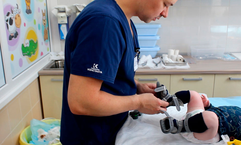
Clubfoot treatment for children is carried out at the KONSTANTA Clinic in Yaroslavl by pediatric orthopaedists and traumatologists - Dr. M.Vavilov and Dr. I. Gromov - performed at a high level. The treatment is financed at the patient's expense or free of charge from the charity fund.
After an individual consultation, identification of the problems and discussions with the parents, the doctor selects a treatment tactic, after which therapy can begin.
See 'What to do' for next steps for parents of children diagnosed with congenital clubfoot.
When should the treatment start?
The sooner the better. Ideally, treatment should begin at 3-4 weeks of age. However, situations vary, so treatment may start sooner or later depending on the child's and family's other issues.
The Ponseti method was proposed by the orthopedic surgeon Ignacio Ponseti in the USA in the 1940s. In Russian reality it has been used for 18 years. Before that, other techniques of home treatment according to VJ Vilinsky, TS Zatsepin and others were used. This included long casts of six to 12 months, extensive surgery, casts again, and heavy shoes at the end. After all these procedures, the child was issued with a disability card. The foot was more or less corrected but limited in mobility and the patient usually had painful walking and running difficulties and difficulty in choosing footwear.
The Ponseti method is a conservative, gentle treatment of clubfoot, which allows for a full recovery of the patient without consequences. Their main advantages are:
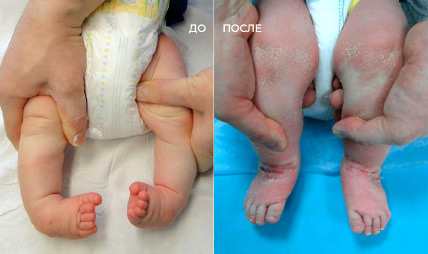
- Minimal surgical intervention and therefore low traumatization of this method.
- Relatively short length of stay of the child in plaster cast (2-2.5 months).
- High success rate of 98 %.
- The feet are completely healthy, the patients do not feel any pain after the procedure and can wear normal shoes.
A visit to the orthopedist is the first thing to do
To reduce fears and worries, an appointment with an orthopedist should be made.
The doctor will assess the condition of your child's feet and make the necessary therapeutic recommendations that can stop the progression of the pathology.
Common orthopedic advice for treating and preventing the development of clubfoot is usually
- a massage course;
- some sports suitable for children with this anatomical defect, such as B. dancing or gymnastics;
- a set of therapeutic physical exercises aimed at eliminating clubfoot;
- the use of massage mats at home;
- the use of orthopedic insoles and/or orthopedic shoes.
All these recommendations are aimed, on the one hand, at strengthening and stretching the muscles and, on the other hand, at activating or relaxing them, depending on the orthopedist's recommendations. It is important to remember that the course of massage - its duration and specificity - should only be prescribed by the doctor. As for the sport, you can choose any sport you like, but pay attention to the injury rate of each sport. Now it is important to 'tune' the child's body for proper development, so doctors recommend dancing, gymnastics (non-Olympic sports), swimming, etc.
Let's tell more about these methods of clubfoot treatment and correction that you can do yourself at home.
Regular gymnastics – physical therapy for clubfoot
The aim of clubfoot exercises is to strengthen and activate the relaxed muscles and, on the contrary, to relax the overly tight muscles.
When performing these exercises, it is important for the parents to observe and try out the child:
We will introduce a number of exercises in this article.
Be sure to discuss these exercises with your orthopedist!
1. the 'rubber band'. Starting position: sitting on the floor, arms behind you, legs straight. Place a wide elastic band around the front of your feet.
Extend your foot to the side. Keep your heels closed. Try to push your toes outward so that you straighten your foot. Hold this tension for ten seconds to one minute at a time.
2 'Block' . Starting position: Stand with the front foot on a 10-15 cm high bar, heel down, hold on to the crossbar of a wall bar or backrest.
Raise your heels to the floor while stretching the back of your shin and Achilles tendon three times. Return to the starting position. Repeat this eight times.
3 – 'fishbone' . Starting Position: On the mat, draw a straight bar with 'branches' sloping to the sides and with a frequency corresponding to the spacing of the small steps. Then place your feet on the branches so that your heels touch the middle, that is, the middle strip, and your front foot tilts to the side. At this point, look down to position your feet correctly.
4. the 'goose step'. The starting position is a pronation position with the feet extended to the sides. Hands free or at the waist.
5. 'Heel Walk'. Walk for twenty seconds.
6. 'Foot rotation'. Starting position: sitting on a chair, put your right foot on your left knee, put your hands on your right knee to fix your right foot. Make clockwise circles with your right foot until you tire easily. With your left foot, make counter-clockwise circles until you tire easily.
Classification: Types of clubfoot in children
With a clubfoot on one side, one leg is shorter than the other. There are the following degrees of severity:
- easy – full range of motion in the ankle is possible;
- moderate – the movement of the joint is somewhat limited, and hindrance and springiness are felt when trying to balance the position;
- severe – the bones and ankle are deformed and it is impossible to put the foot back in the right position with manual correction methods.
Severe cases involve dysfunction of the muscles and nerves of the foot, and other developmental abnormalities may also be present.
Depending on the changes in the bones and joints of the foot and the presence of other accompanying problems, the following types of clubfoot are distinguished:
- Idiopathic: associated with a reduction in foot waist;
- Positional (postural): There is subluxation of the joint between the talus and heel bone, but the bones themselves are properly developed;
- Syndromic: in combination with other developmental disorders, e.g. B. congenital kidney defects;
- Clubfoot secondary: caused by neuro- or myopathy.
Symptoms of clubfoot in children
As a rule, the pathology is already evident at birth: the plantar area is directed inwards and upwards, and movement in the ankle is limited. When the child is already walking, it only supports itself on the outer edge of the foot. The child steps on the foot on which it is resting. The gait is shaky and uneven.
If left untreated, the bony misalignment worsens and subluxations occur in the small joints of the foot. The outer edge of the foot skin becomes thicker. The muscles of the lower leg that are not involved in walking gradually weaken and atrophy. As a result, the functioning of the knee and pelvic joints is impaired. The later treatment is started, the more difficult and poor the correction of the impairment.
The main symptoms of clubfoot in children include:
- a plantar flexion (flexion of the sole of the foot) at the ankle;
- the sole is turned in and the outer edge hangs down;
- the forefoot is turned inward;
- the muscles of the lower limbs are underdeveloped and atrophy in older children;
- A shaky gait is noticeable;
- In unilateral cases, one leg is several inches shorter than the other.
The children tire quickly when walking and standing, they get bruises on their skin and aching pains in their legs.
Causes of Clubfoot
Congenital clubfoot has its own code in ICD 10 (the tenth version of the International Classification of Diseases prepared by physicians to facilitate diagnosis and systematize data) – Q66. Other congenital foot abnormalities are also included in this group. Specialists consider a hereditary predisposition as one of the causes of this anomaly. It is known to occur from time to time in different generations in members of the same family. Much more often, however, the development of such a foot deformity in an infant is the result of external factors. This includes:
- Fusion of the amniotic fluid (amnion) with the lower limbs;
- pressure of amniotic fluid on baby's foot;
- Compression of the baby's foot from the umbilical cord wrapped around it;
- Pressure on the foot from the muscles of the uterus when there is a lack of amniotic fluid and its protective function is reduced;
- pressure on the baby's foot from a tumor in the uterus;
- Presence of abnormalities in the newborn due to dysfunction of the spinal nerves;
- Infections, including toxoplasmosis, contracted by the mother during the first trimester of pregnancy.
Important!!!
The emotional state of the woman is also important - prolonged, chronic stress is dangerous. Long-term observations show that the number of children born with congenital clubfoot increases sharply in the war and post-war years, when psychological stress is unavoidable.
However, there is no clear link between these causes and clubfoot in newborns. A woman can survive illness and stress during pregnancy and give birth to a healthy child.

Diagnosis and Symptoms of Clubfoot
Thanks to modern diagnostic methods, medical professionals can determine the presence of clubfoot long before the baby is born. It is not difficult for an experienced specialist to detect clubfoot on an ultrasound scan of a pregnant woman. When a child is born with this pathology, clubfoot is diagnosed almost immediately. The following symptoms are characteristic of this condition:
Their severity varies. They depend on the severity of the disease, which is divided into three groups:
- Lightweight – ankle mobility is not restricted, foot alignment can be corrected with light hand pressure;
- Moderate – the ankle is clearly restricted, there is some elasticity combined with resistance when trying to correct the foot;
- Severe – there is a severe deformity of the foot and ankle that cannot be corrected by manual manipulation.
There is also a definition of clubfoot depending on the suspected etiology (cause of occurrence). There are three forms of congenital clubfoot:
- Typical. The exact cause of this type of pathology cannot yet be determined. It is characterized by dysplasia (abnormal development, underdevelopment) of the ankle and abnormal anatomical structure and localization of the muscles and ligaments. With this form, one-step correction is impossible even in the first days after birth – a long, gradual treatment of clubfoot in the newborn is necessary;
- Positional Clubfoot – caused by shortening of muscles and ligaments without damage to bones and joints. It is caused by abnormalities in fetal development. Treatment of this form is less complicated and allows complete healing without permanent damage;
- secondary – arises from various congenital anomalies of the neuromuscular system. The clinical picture as well as the treatment options and results are directly related to the primary disease;
Causes of Clubfoot
Why was the child born with clubfoot? This question worries many parents.
Clubfoot is not a malformation but a developmental defect. Typically, a healthy fetal leg deforms into a clubfoot in the second trimester of pregnancy, as increased collagen is deposited in tendons and ligaments during this time. And this process can be influenced by dozens of factors: stress, chronic diseases, alcohol, genetic predisposition.
For example, if one parent has clubfoot, the child's risk is 3-4 %, if both parents are affected, the risk is 30 %.
This malformation is rarely discovered during an ultrasound scan before the 16th week of pregnancy.
When should treatment start?
The treatment of congenital clubfoot should always be started conservatively: with step plaster! Therefore, conservative treatment should be started as early as possible - 7-10 days after birth. Immediately after birth, the soft tissues have greater elasticity, so correcting the foot by the age of 1 can be much easier than later in life.
The elasticity of the tissue decreases over time. This does not mean that clubfoot treatment is impossible, but it is possible that further steps are necessary.
- From the age of 9 months, conservative treatment loses its effectiveness.
- By the age of 3.5-4 years, the skeleton of the foot is deformed, which drastically increases the likelihood of recurrence;
- Surgical treatment is indicated once bone growth is complete.
- At any age, it is important to wear special footwear and exercise.
What to do if a child walks with an abnormal clubfoot?
As soon as the diagnosis is made, orthopedists immediately begin therapeutic measures aimed at correcting the pathology. Treatment depends on the child's age, the type and severity of the disease. The following methods will help improve the condition.
Immobilization with soft bandages and elastic orthoses
Toddlers first receive a comprehensive treatment with massage and exercise. Bandages are then applied. After surgical intervention, this technique gives positive results when the disease is mildly expressed.
If the pathology is detected only at a later age, a special plaster shoe is used. The shoe is worn intermittently for two months. After seven days, the shoe is removed and the doctor orders a massage. The shoe is then worn for another week.
Ponseti treatment
Parents often ask the podiatrist what to do if the child is already 1 year old and has a clubfoot. At this stage, Ponseti therapy is the most effective method:
- First, the toddler is put on a plaster cast for 2 months.
- Then the Achilles tendon is surgically shortened.
- A mobile orthosis is then put on to relieve the ligament joint.
physiotherapy
Physical therapy and other treatments are necessary to correct the ankle. The treatment normalizes blood circulation in the affected areas of the legs.
The orthopedic surgeon always prescribes: electrophoresis, magnetic therapy and paraffin compresses.
Injecting medication into the calf muscle
If clubfoot is diagnosed at birth or at the age of 2-3 years, Botox injections are done every six months. This relaxes the muscle and corrects the foot.
If the abnormality was diagnosed due to a nerve disorder, doctors prescribe medications containing proserine and strychnine to improve nerve impulse conduction.
How to correct pathology at home?
Mild cases can be treated at home under the constant supervision of a podiatrist. The doctor prescribes complex procedures.
gymnastic exercises
Manipulations of the newborn are carried out by a specialist in the presence of the mother at the beginning of treatment. They can then be repeated at home, observing certain points:
- Work each foot for at least 7 minutes for 10 reps.
- Be sure to correct the child's behavior so that they do not turn their feet inward during treatment.
- Warm up the muscles with massaging movements.
- Bend and straighten your legs at your knees.
We advised to correct clubfoot before age 2 because it becomes more difficult to treat after age 5-6.
The following exercises are necessary:
- Make circular movements with your feet.
- Walk on heels and crouch.
- Raise your feet at a 90 degree angle.
- Drawing with a stick or pencil placed between the thumb and second toe.
- Pick up a towel from the floor.
- Stretching the spine with a bar.
- In older age, skateboarding and horseback riding are very effective.
massage
This treatment should be carried out by a professional who is familiar with the anatomy of the skeleton, because during massage the muscles are relaxed or, on the contrary, tightened.
The therapist uses hand movements (if the child has clubfoot in a newborn or one-year-old) such as stroking, tapping, light pressure, and pinching.
To avoid later complications, treatment should be started as early as possible. Parental negligence and inaction can lead to serious health problems:
Read more:- Congenital clubfoot.
- 1 year old child with clubfoot.
- What is clubfoot?.
- The Ponseti method.
- Clubfoot in 7-year-old children.
- Why does a child develop clubfoot?.
- Ponseti breeches.
- Clubfoot Treatment.