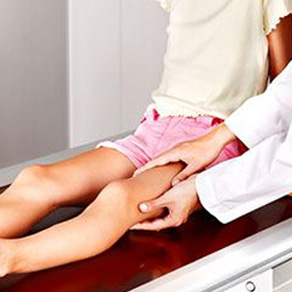
Between tendons, ligaments and other joint structures are special fluid sacs (bursa) that reduce friction during movement and thus prevent mechanical damage to the joint protrusions.
- Soft tissue tumors in children. Information for Parents.
- Classification of soft tissue tumors.
- Causes of synovitis
- classification
- Symptoms of juvenile bone tumors
- Are bone tumors in children always a warning sign?
- symptoms of the disease
- complications
- Wrist hypertrophy in a child
- Hemangioma overgrowth on the leg in a child
- diagnosis
- treatment of fibroids
- Conservative treatment
- surgical treatment
- When should you see a doctor?
- Who is at risk?
- lipoma
- fibroma
- Diagnosis of a hygroma
- surgical treatment
Soft tissue tumors in children. Information for Parents.
Soft tissue sarcomas in children are relatively rare. About 850 to 900 children and adolescents are diagnosed with rhabdomyosarcoma (RMS) or one of the other soft tissue sarcomas each year. Of these, 350 are cases of RMS. RMS is the most common soft tissue sarcoma in children 14 years of age and younger; other sarcoma types are more common in adolescents and young adults. Infants may have soft tissue sarcomas instead of rhabdomyosarcomas, but their tumors show a different histology, including infantile fibrosarcoma and malignant hemangiopericytoma, which do not occur in adolescents.
A heterogeneous group of malignant neoplasms (MN) that arise primarily in soft tissue and are predominantly mesenchymal in origin. The third most common group of childhood solid tumors (after CNS tumors and neuroblastoma). More common in boys (m:f 1.5:1).
The soft tissues of the body connect, support and surround other parts of the body and organs. Soft tissues include:
- Fat.
- bone and cartilage system.
- Fibrous tissue.
- muscles.
- Annoy.
- tendons
- Synovial tissue (tissue around the joints).
- blood vessels.
- lymph vessels.
Soft tissue sarcomas can occur anywhere in the body. In children, tumors most commonly form in the arms, legs, chest, or abdomen.
The types of soft tissue tumors that most commonly occur in children are.
- Rhabdomyosarcoma (RMS) : 57%
- Ewing's sarcoma family (extra-segmental Ewing's sarcoma/peripheral primary neuroectodermal tumors): 10%
- Synovial sarcoma: 8%
- Peripheral malignant schwannoma (malignant nerve sheath tumor): 4%
- Fibroma: 2%
- Undifferentiated sarcoma: 2%
Classification of soft tissue tumors.
The most common histological variants in children are RMS (61%), USS and PNEO (8%), synovial sarcoma (SS) (7%), neurofibrosarcoma (4%), fibrosarcoma (3%), and leiomyosarcoma (2%). The current subclassification of RMS distinguishes between embryonic and follicular types
The most common symptom of childhood soft tissue sarcoma is a painless lump or swelling in the soft tissues of the body. A sarcoma can appear as a painless lump under the skin, commonly on the arm, leg, chest, or abdomen. Initially, there may be no other signs or symptoms. When the sarcoma enlarges and presses on nearby organs, nerves, muscles, or blood vessels, it can cause signs or symptoms such as pain or weakness.
Other medical conditions can produce the same signs and symptoms. Of course, the symptoms described here in a child or adolescent do not always mean a soft tissue sarcoma or another malignant tumor. Contact your pediatrician if your child has any of the above problems.
The clinical presentation and the severity of the symptoms depend primarily on the location and extent of the tumor and are therefore very variable. Patients with head/neck RMS-like tumors (e.g., orbital tumor with initially painless exophthalmos) may suffer only mildly, while those with paraminomatous tumors with intracranial involvement may present with symptoms of pain, swelling, nasal and paranasal sinus obstruction, cranial nerve palsy (III, IV , VI, VII) and vomiting. Patients with RMS tumors in the genitourinary tract may complain of abdominal pain, hematuria, dysuria, constipation, and scrotal enlargement. Tumors on the extremities express themselves as painful or indolent swellings.
Causes of synovitis
The main trigger for this condition is trauma to the joint from falls, bruises, collisions, and other situations that damage one or more components of the joint. For example, bursitis in a child can be caused by any injury to the soft tissues, muscles and ligaments of the knee, including when blood enters the periarticular sac (cuts, abrasions, subcutaneous hemorrhage).
Other possible causes of bursitis in the hips, elbows and other joints in children may include:
- infectious and inflammatory processes in the tissues surrounding the joint (phlegmon, carbuncles, boils);
- osteomyelitis;
- Chronic diseases of an infectious nature, such as various urinary tract infections, tonsillitis, bacterial infections of the gastrointestinal tract;
- Excessive physical activity, especially of a monotonous nature;
- Injuries from sports, dance, or other intense physical activity;
- obesity, which increases the load on the joints;
- Chronic autoimmune and endocrine diseases associated with disorders of metabolism and absorption of micronutrients;
- Acute poisoning (poisoning by toxic substances, heavy metal salts, strong drugs);
- postural defects;
- Frequent hypothermia and overheating.
Children with allergic diseases, children with severe bacterial infections at a young age, children with frequent acute respiratory infections are at risk. Students who engage in extreme sports, cycling, martial arts, and dance are at increased risk of rebelling if they don't follow safe routines.
classification
Bursitis can be acute, subacute, chronic, or recurrent. Depending on the cause of its origin, a distinction is made between primary pathology, which is due to direct infection of the bursa or trauma to the joint capsule, and secondary pathology, which is due to chronic diseases and predisposing factors.
Depending on the type of effusion (synovial fluid), a distinction is made between hemorrhagic, purulent and serous forms. It can also be classified according to localization, e.g. B. Bursitis of the back of the knee, hip or shoulder bursitis, etc.
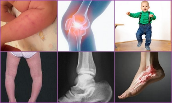
Symptoms of juvenile bone tumors
Symptoms of a bone tumor depend on its size, location, and stage. In the early stages, chondromas are an incidental finding when diagnosing other pathologies.
In addition, the symptoms of the disease can be absent for up to several years. However, there is also the reverse situation, in which chondromas appear almost immediately.
- For example, if the chondroma forms in the cartilage at the base of the skull, it quickly begins to press on the surrounding tissue, causing pain, dizziness, incoordination, and other very noticeable symptoms.
- When chondromas form on smaller long bones, pressure on that area can cause pain as the lesion presses on nerve endings.
- When long bones are affected, symptoms do not appear immediately. However, when they do occur, the child complains of joint pain. Since there are no signs of joint inflammation, the diagnosis is difficult.
- When chondromas are located on the bony structures of the rib cage, they often involve the ribs, causing a visible cosmetic defect. If a lump on the ribs is visible or palpable through the skin, a doctor should be consulted, otherwise pain will occur.
It is important to know that the thickening of the rib bones and their deformation in preschool children is also a symptom of advanced rickets.
One of the later symptoms of chondromalacia is a propensity for abnormal fractures. The bone replacement makes the spongiosa structures brittle and the growth of the child's bones leads to a partial fracture, even without trauma.
Symptoms of such a fracture include a grinding sound, pain, characteristic immobility, and swelling.
Are bone tumors in children always a warning sign?

Experts from the Indiana University School of Medicine conducted a retrospective analysis of a unique X-ray archive compiled between 1926 and 1942: children were X-rayed each year to examine age-related bone changes. Today this is forbidden: the radiation doses are too high for such examinations, but nobody knew that at the time.
In total, more than 25,000 X-rays were collected from 262 study participants, ranging from newborns to teenagers. And the researchers found that nearly 20 of the % children develop benign bone tumors during the growing season. Thus, 35 tumors were diagnosed in 33 participants, and since the X-rays were only taken on one side of the body, the result was multiplied by two to obtain a prevalence of 18.9 %, ie almost every fifth child develops such bone tumors.
Fibroadenomas, connective tissue growths that have not developed into bone tissue, were found most frequently. They appear before the age of five, i.e. at the peak of the formation of the skeletal structures. The good news is that nearly half of these masses disappear with age, and it's likely that the rest will also disappear after the study.
The scientists found enostoses and chondromas somewhat less frequently. The frequency of exochondromas, that is, tumors on the outer surface of the bone, was low, probably due to the use of X-rays as a method of investigation.
symptoms of the disease
Diagnosing the disease is usually not difficult. The neoplasm is clearly visible to the naked eye. To the touch, the cyst is dense but soft and elastic. However, if it is small, it can go undetected for many years.
Occasionally, the cyst begins to grow. When this happens, the child begins to feel the bump when bending the knee, which can be annoying. The increased pressure on the surrounding tissue causes swelling and pain.
As the cyst grows, the inflammation covers the entire knee joint. The following symptoms appear:
- Local temperature increase in the knee area;
- The joint becomes less mobile;
- The child's gait changes;
- The cysts can regress spontaneously.
The cyst may resolve on its own. For this reason, doctors often adopt a wait-and-see attitude and observe the knee joint. Complications can only be prevented by timely treatment.
complications
If left untreated, complications can occur. Large cysts can cause tissue compression, affecting nutrition in the lower limbs. This increases the risk of varicose veins and thrombophlebitis.
A complication of a hamstring cyst can be a rupture of the tendon. Although synovial fluid does not cause bacterial infection, its leakage into the ankle area leads to acute inflammation. This causes the following symptoms:
The effects of the rupture can last for several weeks. During this time, the uncomfortable symptoms prevent the child from leading a full life.
Wrist hypertrophy in a child
A hygroma of the wrist in a child does not cause pain at first, but it interferes with the proper functioning of the joints, and later it can cause severe pain if the child exerts a lot of force or regularly bends the wrist. This is due to the impairment of flexion and extension movements that prevent the child from leading a normal life. Therefore, a tumor resembling a cystic mass usually requires immediate surgical intervention.
Hyproma of the wrist in a child can be caused by monotonous movements or excessive use of the muscle groups of the hand. It often occurs in children who play the violin or piano, spend a lot of time on the computer, etc.
A hygroma in a child on the wrist looks like a tumor, reaching several centimeters in diameter. It is dangerous if the hygroma is near the radial artery - on the wrist under the hand. This complicates the operation because the radial artery must not be injured. If the surgery isn't done carefully, the child may sustain an injury to the artery, which then leads to reduced blood supply to the wrist.
Hemangioma overgrowth on the leg in a child
Leg hirgromas in children can occur around the knee, usually below the knee, or elsewhere. In medicine, it is not uncommon to find a nodule in the ankle area. It should be borne in mind that such knots are very painful, which affects children's activity and mobility. When developing a hirgrom on the foot, the child often complains of pain when moving, which should immediately alert parents. In this case, medical treatment is essential.
Hygromas in children on the leg usually arise as a result of severe overload, as well as systematic injuries to the tendons or joints of the leg. Swelling of the knee, for example, progresses quite quickly and can lead to many complications later. It is caused by the accumulation of excess fluid in the cavities of the joint capsule as a result of an injury to the knee joint or overstretching. The tumor of the hamstring in a child is caused by a muscle congestion and impedes the bending movements of the leg. As a result, the child has increasing difficulty walking, so this swelling requires immediate surgical intervention, that is, removal.
In the case of the hyalgroma on the child's foot, it is associated with the ankle. The tumor can also develop at the back of the metatarsal bone. Initially there is a small, prominent lump on the foot. It does not cause pain to the child, but if not treated in time, it can grow to a very large size. Such growth of the hirgroma naturally puts pressure on the nearby blood vessels and nerves in the foot, causing the child severe pain. The pain is greatly aggravated by various physical activities, wearing uncomfortable shoes and additional injuries to the foot. When the hirgrom is damaged, it can lead to severe inflammation of muscle tissue. For this reason, the hirgroma should be removed before its pathological course begins.
diagnosis
Diagnosis of hirgroma is usually based on medical history and characteristic clinical signs. X-rays may be recommended to rule out bone and joint pathology. In cases of doubt, an ultrasound examination, an MRI or a puncture of the Hirgrom can be performed. Ultrasound can not only identify the cyst, but also its structure (homogeneous or fluid-filled), whether there are blood vessels in the wall of the hirgroma, etc. If a lump is suspected, the patient may be referred for an MRI scan. This examination makes it possible to determine the exact structure of the tumor wall and tumor contents.
The differential diagnosis of hirgromas is made with other benign tumors and soft tissue tumors (lipomas, atheromas, epithelial trauma cysts, etc.) on the basis of localization, tumor consistency, and patient complaints. A hygroma in the hand area sometimes has to be differentiated from bone and cartilage tumors.

treatment of fibroids
Conservative treatment
Surgeons and orthopedic traumatologists treat the pathology. In the past, the fibroma was treated by crushing or kneading. Some surgeons practiced puncture, sometimes with simultaneous injection of enzymes or sclerosants into the hygroma cavity. Physiotherapy, mud, ointment dressings, etc. were also used. Some clinics still use these methods today, but the effectiveness of the treatment is not satisfactory.
surgical treatment
After conservative treatment the recurrence rate is 80-90 %, after surgical removal the risk of recurrence is only 8-20 %. Based on the statistics presented, the only effective method of treatment today is surgery. Indications for surgical treatment:
- Pain when moving or at rest.
- Limitation of the range of motion in the joint.
- Unsightly appearance.
- Rapid weight gain.
Surgery is recommended, especially if the hirgroma is growing rapidly, since removing a large mass is difficult. Hirgromas are often found near nerves, vessels, and ligaments. As the tumor grows, these masses begin to move and are more difficult to remove. Sometimes the operation is performed on an outpatient basis. However, during the operation, the tendon sheath or joint may be opened, so it is better to hospitalize the patients.
The operation is usually performed under local anesthesia. The limb is tied off by placing a rubber band above the incision. By bleeding and injecting an anesthetic into the soft tissue around the hirgmanen, the border between the tumor and healthy tissue can be sharply delineated. For complex or large lesions, anesthesia or regional anesthesia may be used. During the operation, it is very important to extract and excise the hematoma so that not even small areas of the changed tissue remain at the incision site. Otherwise, the hematoma may recur.
When should you see a doctor?
- If pain in the knee prevents the child from performing everyday activities;
- the knee is swollen and red;
- if the knee pain is caused by high fever, blockage or instability of the knee joint.
During activities that require running, jumping, and bending—such as soccer, basketball, volleyball, and ballet—the muscles of the thigh (quadriceps) pull on the tendon that connects the kneecap to the growth plate at the top of the tibia. The tension can be so great that the tendon indents the tibia, causing pain and swelling in the child.
In some athletes, the body tries to deal with the new bone growth, resulting in a nodule.
Who is at risk?
The main risk factors for developing Osgood-Schlatter disease are:
- Age. Osgood-Schlatter disease occurs during the growing period of puberty. Age ranges are gender specific, with girls entering puberty earlier. Osgood-Schlatter disease occurs in boys at 12-14 years and in girls at 10-13 years.
- Gender. Osgood-Schlatter disease is more common in boys, but recently that distinction has blurred as girls have started playing sports too.
- Sports. This condition is most common in activities that involve running, jumping, and quick changes of direction.
- Agility. Tightness in the quadriceps muscles can increase tension on the patellar tendon at the growth plate at the top of the tibia.
lipoma
Is a benign growth of adipose tissue. It is most commonly found on the extremities, but lipomas can appear in other locations as well. The growth is soft and consists of fatty tissue.
Fat deposits do not grow on the skin, only in the fat layer. They can be up to a few centimeters in size and are painless to the touch, but on rare occasions when they are growing rapidly, pain can be caused by pinched nerves.
Doctors recommend removing an adipoma, although it is very rarely malignant and is only a cosmetic defect. The reason is that its rapid growth can cause pressure and infection of the surrounding tissues.
fibroma
A benign growth composed of fibrous connective tissue. It looks like a lump under the skin. There are two types of fibroids: soft and hard. They can grow for years and are usually spherical in shape.
A fibroma is caused by a blockage in the sebum outlet, which prevents secretions from escaping. The tumor is benign, composed of fat and epithelial cells, and is thick to the touch. The size of the lump varies from one centimeter to the size of a hen's egg or more. The scab usually appears on the scalp, back, and face. The growth does not cause any discomfort, except for a cosmetic defect, and has a round shape with clear edges.
Diagnosis of a hygroma
A hygroma can be diagnosed by palpation by a podiatrist or surgeon. Examination reveals a small, round, smooth nodule 1 to 5 cm in diameter, soft and elastic to the touch. If necessary, the doctor will recommend additional and instrumental examinations, e.g. B. An X-ray of the joints.

- Conservative treatment includes puncture of the hirgrom, which is performed for therapeutic or diagnostic purposes. With a puncture needle, the nodule is punctured and its contents drained. Sclerosing agents are then injected into the cleaned defect and a pressure bandage is applied.
- The limb is immobilized with an orthopedic splint or cast for 7-8 days to prevent movement of the tendon (or damaged joint), which reduces synovial fluid production. A major disadvantage of this treatment method is the high recurrence rate.
- If necessary, physiotherapy (UV light) or paraffin compresses are prescribed.
surgical treatment
- Excision of the hirgroma – This method is considered to be an effective way to treat the hirgroma. It consists essentially in the complete excision of the thickened capsule, which does not take place up to the changed tissue, but with a correspondingly secure suture and subsequent suturing with the subcutaneous fatty tissue of the residual limb. The advantage of this method is that there are significantly fewer recurrences compared to other methods.
- In laser removal of the foot hirgroma, the tumor is heated with a laser until it is completely destroyed; the adjacent tissue is not attacked. This shortens the healing time and the biggest advantage is that no scars are left behind.
Indications for removal are rapid growth, significant tumor size, cosmetic defect, development of complications (abscess, inflammation, swelling, redness or severe pain). When surgically removing a nodule, all procedures are performed under local anesthesia, which eliminates any pain sensations.
Read more:- Bump on the side of the child's foot.
- disarticulation is.
- Root nodules in children.
- A child begins to have clubfoot between the ages of 1 and 5.
- Cubic Bone.
- What is the name of the ankle bone?.
- Front lower leg area.
- The child walks on its toes.