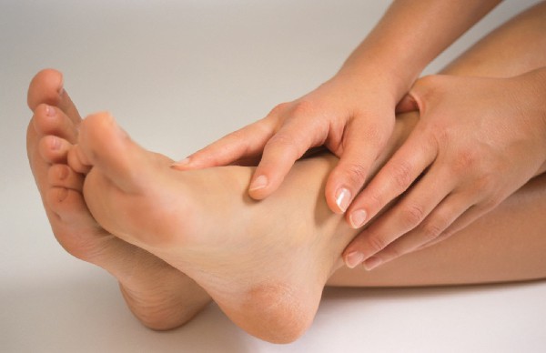
With plantar fasciitis, heel pain occurs more often after sleeping. After a while they get easier, but in the middle of the day, with long walks, the heel pain increases again.
- Haglund's deformity
- Symptoms of Haglund Syndrome
- Why does a heel bone fracture occur?
- Main symptoms and diagnosis
- Treatment of heel bone fractures
- Important points
- How do you treat sore heels?
- Physical therapy for heels
- Prevent heel pain
- Causes and symptoms of a bone fracture
- Principles of Diagnosis
- Indications for X-ray examination of the heel
- Preparing for surgery
- News
- Video about the laboratory
- Naise' gel
- Use of Naise during pregnancy and lactation
- opinions
- last comments
- Personal experience
Haglund's deformity
Haglund's deformity is a condition of the heel bone that presents as a dense bony lump or inflamed soft-tissue blister around the upper back part of the heel bone and the lower part of the Achilles tendon. The correct diagnosis requires a lot of medical experience: during the examination, the Haglund deformity must be distinguished from joint diseases of a different nature (rheumatoid arthritis, bursitis), which have similar symptoms. Occasionally, Haglund's deformity develops in people without any symptoms or defects in foot structure and function. The longer the disease lasts, the more difficult it is to treat.
Each doctor at ON CLINIC International Medical Center is highly experienced so that you can receive effective medical care and improve your health. You are always welcome in our clinics!
Symptoms of Haglund Syndrome
The most obvious symptom is a bump on the upper back of the heel. In some patients it is firm and painless when there is no inflammation. In other patients, inflammation causes a soft bump and pain in the heel and Achilles tendon area. Pain also occurs due to swelling and blistering of the skin in the heel area.
In Haglund syndrome, self-treatment often aggravates the situation and leads to tissue destruction in the heel area and even to a rupture of the Achilles tendon. Contact the experienced specialists at ON CLINIC. We can also help in difficult and neglected situations!
Free consultation with the head of the Center for Orthopedics and Traumatology, orthopedist-traumatologist, doctor of the highest category, candidate of medical sciences Samoilov VV for joint surgery.
joint endoprosthesis. Treatment of arthrosis in the ON CLINIC
Why does a heel bone fracture occur?
The heel bone is one of the largest structures of the foot that supports the weight that keeps the body upright. Above the heel bone is the ankle bone, which connects the foot to the lower extremity. In a violent heel fall or direct impact, gravity acts through the shin and onto the ankle bone. The heel block moves forward and hits the heel bone with full force, causing it to shatter into several pieces. These situations are most common in traffic accidents, sports injuries, and falls from heights. The following factors increase the likelihood of a fracture:
- older age;
- influence of alcohol or drugs;
- menopause in women;
- diet low in calcium and phosphorus;
- prolonged immobility (coma or paralysis);
- Practice of dangerous sports (mountaineering, motorcycling, racing).
Main symptoms and diagnosis
A heel bone fracture can be unilateral or bilateral when a person falls on both feet at the same time. Signs of injury appear within the first few minutes after injury, and if the patient is conscious, they will actively complain. However, if the victim is in pain shock, the symptoms are much more difficult to recognize. A heel bone fracture is suspected when the following symptoms are present:
Acute malaise immediately after injury. The pain increases when trying to step on the injured foot, remove shoes or clothing, or put pressure on the arch of the foot.
The patient is unable to use the injured leg for support, resulting in impaired gait. There may also be reflex flexion of the fingers, which is a protective response to the injury. Attempting to straighten them and return the limb to the correct position increases the discomfort, and with displaced fractures the patient may need long-term rehabilitation.
There is massive hematoma and swelling on the heel, causing the contours of the foot to appear flattened. An open injury creates an open sore on the surface of the heel that shows splintered bone and bleeding blood vessels.
Immediately after the fracture, an X-ray of the heel bone is always taken in several projections to confirm the diagnosis. In complicated cases, an MRI or CT scan is recommended to determine the treatment strategy.
Treatment of heel bone fractures
The question of whether a treatment of intra-articular fractures of the heel bone Surgical or non-surgical fractures are controversial.
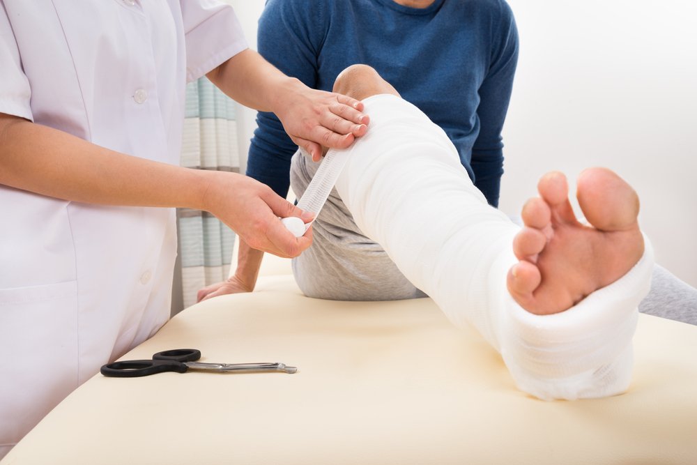
Extra-articular fracture of the heel bone is treated symptomatically with protection, rest (avoiding heavy lifting), a pressure bandage (which also provides protection), and ice, and an elevated position of the limbs (treatment regimen [ PRICE A fracture is a violation of the integrity of the bone. Most fractures result from a only significant force applied to a normal bone. In addition to fractures, there are also injuries to the musculoskeletal system. Read more information ]). Once the swelling has gone down, a plaster cast is put on.
Important points
If heel bone fractures are not recognized and treated in time, they can lead to long-term disability.
Because these fractures are usually caused by high axial loading of the feet, other injuries (eg, compression fractures of the thoracic and lumbar spine) are common; other complications include compartment syndrome (in up to 10 % of cases).
It is controversial whether intra-articular fractures of the calcaneus should be treated surgically or non-surgically.
When diagnosing a heel bone fracture, it should always be checked for a lumbar spine fracture.
Extra-articular fractures of the heel bone are treated symptomatically with PRICE and a cast.
Copyright © 2023 Merck & Co, Inc, Rahway, NJ, USA and its subsidiaries. All rights reserved.
How do you treat sore heels?
Heel pain is usually treated holistically, e.g. B. with stretching exercises and painkillers. This can be a long process, sometimes up to a year. If the pain does not go away after this time, surgery is recommended as a last resort. However, this only happens in 0.5 percent of cases.
The success of heel pain treatment largely depends on the lifestyle you lead. Whatever the cause, you need to wear the 'right' shoes, make time to exercise and get enough rest. You can do most of the things you can do to treat your heel pain yourself, without the help of a doctor.
If possible, take it easy on your sore heel – try not to walk long distances or stand for long periods. You should also regularly do special stretching exercises for your feet and calves.
Physical therapy for heels
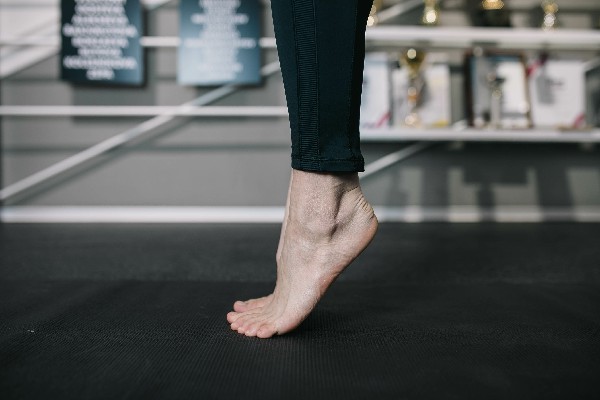
Stretching exercises for the calf muscles and plantar fascia will help reduce pain and increase mobility of the sore foot. It is generally recommended to do the exercises with both feet, even if only one foot is painful.
Stretch with a towel. Put a long towel next to your bed. Before getting up in the morning, place the towel over your foot and use it to pinch your toes together, keeping your knee straight. Repeat this three times with each leg.
wall stretching. Support yourself with your hands on a wall at shoulder height and place one foot in front of the other. Your front foot should be about 12 inches from the wall. With your back straight, bend your front leg at the knee and brace yourself against the wall until you feel tension in the calf muscles of the other leg. Relax. Repeat the exercise 10 times with one leg and then the other. Do this exercise twice a day.
Stretching on the stairs. Stand on a step facing the stairs and lean against the railing. Spread your legs slightly and let your heels hang down the stairs. Lower your heels until you feel tension in your calf muscles. Stay in this position for about 40 seconds and then return to the starting position. Repeat this exercise six times, at least twice a day.
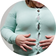
Prevent heel pain
It's not always possible to avoid heel pain, but there are steps you can take to prevent such problems in the future. It is well known that being overweight puts extra strain on the feet, especially the heels, and increases the risk of heel pain. If you are overweight, combining regular exercise with a healthy, balanced diet will help your feet lose weight and maintain a healthy weight. Calculate your body mass index (BMI) to determine if your weight is appropriate for your height and build.
Choosing the 'right' footwear is important to prevent heel disorders. Wearing heels to a party won't hurt you, but wearing them to work all week can hurt your feet, especially if you have to walk or stand a lot. It is best to choose shoes with laces and a low or medium heel that will support and protect the arch and heels. Do not wear flat-soled shoes.
Do not walk barefoot on asphalt or hard ground. Heel pain is common when you start going barefoot on vacation after having been in shoes all year. When this happens, the feet are not used to the extra pressure, resulting in heel pain.
If you lead an active lifestyle, e.g. B. running or doing other sports, you should change your running shoes regularly. Most experts recommend changing athletic shoes after running about 500 miles in them. Remember to stretch after exercise and incorporate regular strength and flexibility exercises into your workouts.
Causes and symptoms of a bone fracture
Most fractures of this type typically result from a fall from a great height, when a person falls to their feet, traffic accidents, poor bone health, osteoporosis, and excessive exercise.
Fractures of the heel bone are usually divided into several groups:
In extra-articular trauma, the heel shaft has different fracture planes and accounts for about 20 % of traumatization:
- burst injury;
- trauma to the styloid process of the heel bone;
- anterior process of the biceps ligament;
- Tear of the tuber of the calcaneus.
Intra-articular deformities occur in the joint cavity of the bone and account for approximately 80% of all calcaneal bone injuries. These injuries include:
- Fracture of the posterior lamina of the heel bone with a lingual aspect;
- Posterior lamina fracture with sagittal aspect;
- fracture of the central portion of the posterior surface mimicking a Y-shape;
- Fracture in multiple locations of the outer and middle parts of the posterior facet of the heel bone with possible 'dropping' of the middle part;
- Fragmentation fracture with multiple posterior facet injuries.
A fracture of the heel bone can be recognized by characteristic symptoms that cause discomfort. The person begins to complain about symptoms such as e.g.:
- Pains;
- Swelling;
- inability to stand on foot;
- Severe limb deformity;
- Bone splinters in the wound.
Principles of Diagnosis
If there is a suspicion of a fracture, you should see a specialist at the Sports Injury Center. The doctor performs a visual and tactile examination and assesses the condition of the skin around the heel for dislocations or skin damage. The specialist will also recommend additional examinations, such as B:
- Radiological examination. It is performed in 4 projections and helps the doctor to determine the exact location of the dislocation or the position of the fragments.
- CT scan. Allows the specialist to see and assess the extent of damage to areas of the leg.
CT scan is the gold standard for heel bone injuries.
Indications for X-ray examination of the heel
Due to its special anatomical structure and high daily loads, the heel bone is subject to frequent trauma and pathological processes. An X-ray of the heel bone is the easiest, quickest and most meaningful way to diagnose problems early on.
An appointment for an X-ray examination should be made if the following symptoms appear:
- burning or severe pain in the heel
- limited joint mobility in the foot
- Pain and discomfort when walking
- Gait changes that shift the center of gravity from the heel to the toes
- Swelling, redness and localized fever in the protruding area of the heel bone.
X-rays are taken after trauma to the foot when fractures, breaks, or dislocations are suspected. If there is bone hypertrophy in the heel area, the diagnostician can assess the development of a spur. X-rays help the doctor assess the effectiveness of treatment for inflammatory diseases, identify the features of bone fusion in fractures, and understand the causes of chronic pain.
Diagnostic radiographs are required when there is suspicion
- osteoporosis
- flat feet
- osteitis
- Achilles tendon rupture and heel sprain
- Arthritis, arthrosis, osteochondropathy.
X-ray shows signs of inflammation characteristic of bursitis, plantar fasciitis, and tendinitis. When describing the x-ray, attention should be paid to the integrity of the bony structures, the shape of the heel shaft and its position in relation to the rest of the tarsal area. If abnormalities are present, additional investigations, including a second radiograph of the foot, may be needed to make an accurate diagnosis.
Preparing for surgery
X-ray examination of the heel bone for fracture, spur or inflammation is performed without preparation. A referral for the examination can be made by a traumatologist, orthopedist, podiatrist, rheumatologist, or surgeon. It is advisable to bring the results of previous examinations so that the radiologist can follow the progression of the pathological process.
Immediately before the X-ray examination, the specialist will ask you to take off your shoes and remove socks, jewelry and foreign objects from your foot.
News


Video about the laboratory
GEMOHELP – YOUR GUIDE THROUGH THE COUNTRY OF HEALTH.
The Hemohelp Group is the largest independent central laboratory in the Nizhny Novgorod region with 74 medical departments. The laboratory is built according to European standards and is equipped with the latest generation of precision equipment, which allows automatic tests, high speed and diagnostic accuracy of the examinations carried out. The most important asset of the company is its employees. It is a well-established team of dedicated professionals.
GEMOCHELP cares about its customers and values its reputation and constantly pays attention to the quality control of the tests.
In addition to the daily internal control, the laboratory (AVK-Med Centralized Laboratory LLC) carries out an external quality assessment of the laboratory tests according to the Russian and US systems:
In July 2017, the GEMOHELP laboratory was successfully certified according to the international standard ISO 15189:2012 'Medical Laboratories. Specific requirements for quality and competence'. The certification was carried out by Haensch, a member of the certification holding company DQS-CFS (Germany).
In August 2017, the 'GEMOCHELP' laboratory underwent voluntary certification, which certifies compliance with the quality management system in relation to laboratory diagnostics, GOST R ISO 9001-2015 (ISO 9001:2015) with annual confirmation.
In August 2018, the 'GEMOHELP' laboratory was successfully certified for compliance with the requirements of GOST R ISO 15189:2015 (ISO 15189:2012) 'Medical Laboratories. Specific requirements for quality and competence' with annual confirmation.
Naise' gel
Before applying to the skin, the painful area is cleaned and thoroughly dried. A strip of gel about 3 cm long is squeezed out of the tube and applied evenly to the affected area without rubbing it in. Allow the mixture to soak in for 1-2 minutes, then cover the affected area with gauze (no closed bandages!) or leave uncovered. The treatment is repeated up to 4 times a day.
Do not use Naise for more than 10 consecutive days without consulting your doctor.
Use of Naise during pregnancy and lactation
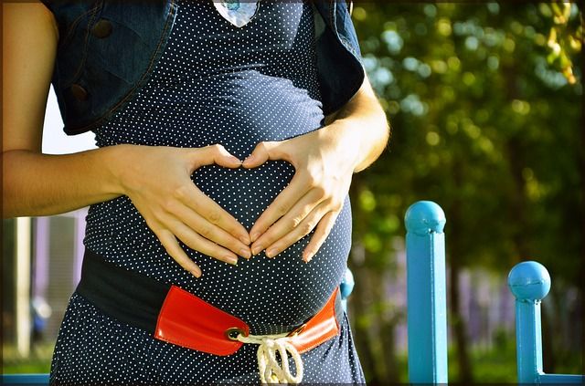
The tablet is contraindicated in pregnancy
Naise tablets are contraindicated for pregnant women and nursing mothers. It can have negative effects on the course of pregnancy and fetal development.
Nise is also contraindicated in pregnancy. Its components have been clinically proven to pass into breast milk. If taking the drug is necessary, breastfeeding should be stopped and the child switched to artificial feeding.
opinions
MP on the work of the Department of Housing and Utilities in Voronezh: 'They collect money - they do not provide services'.
'There should be a switch connected to the neural network'.
Russian paleoanthropologist, editor of the Anthropogenesis.ru website
Why don't the officers start with 'MYO!' talk about the new renovation program?
'After the transport 'reform' it became a chore to wait for and squeeze into a minibus'.
last comments
The 'Kitchen' star has been accused of slandering a Russian woman.
The 'Kitchen' star has been accused of discrediting the Russian government.
Taxi driver hits pedestrian in Voronezh (video)
MP on Voronezh management company: 'They collect money but do not provide services'.
Personal experience
Dear readers! We would like to present you our new project 'New Buildings in Voronezh'. We have marked new apartment complexes on an interactive map of the city. If you click on a complex, you will get complete information about it: location, terms of delivery, apartment plan, price per square meter, etc. Apartment in the heart of a metropolis or in a sleeping district? With or without furniture? The card answers all these questions and more. 'Novostroyki Voronezh' - offers the opportunity to quickly and comprehensively inform yourself about new residential complexes and to learn how to find your way in the variety of apartments!
The online publication MOO!
(translated as MOO! Straight Line)
Online publication registered 12/30/2014. Federal Service for Supervision of Communications, Information Technologies and Mass Media (Roskomnadzor)
EL Registration Certificate No. FS77-60431 dated 12/30/2014.
Founder: LLC Publishing House 'Svobodnaya pressa'.
Irina Bulgakova is the editor-in-chief of the editorial board of 'MYO!'.
Address of the editorial office: Address of the editorial office: 54 L. Ryabtseva Street, Voronezh, 394049
Opinions of the authors of articles published on MOO! Articles posted online, material posted in the Opinions and Popular News sections, and user comments on material posted on the Site do not necessarily reflect the opinions of the editorial staff of the MOO!
Do you have any news that interests you?
Call: (473) 267-94-00, 264-93-98. Write to: [email protected], [email protected].
For questions about advertising on the Site, call: (473) 267-94-11, 267-94-08, 267-94-07, 267-94-06, 267-94-05
- The digital newspaper 'MYO! Plus'
- About Us
- Contact
- Map
- all messages
- Attractions in Voronezh
- Useful Materials
- rules of communication
- data protection
- Structure of the heel.
- Structure of the human heel.
- heel bone injury.
- What could be the cause of the amputation?.
- Anatomy of the heel bone x-ray.
- Anatomy of the heel bone.
- The bones of the human heel.
- heel joint.