What is heel valgus? It is an inflammation of the synovial capsule, which lies between the Achilles tendon and the heel bone.
- Why does heel pain occur?
- Where does the heel hurt?
- Causes of heel pain
- Fractures and breaks
- plantar fasciitis
- Treatment of heel pain
- diagnosis
- Current treatment methods
- diagnosis
- Treatment methods for fractures depending on their type
- Indications for surgical treatment of a fracture
- Possible undesirable effects after a fracture
- Treating heel pain at home
- Peculiarities of the heel structure
- Anomalies in which the heel hurts from the outside or inside
- Classification of achillobursitis (according to various parameters)
- diagnosis
- Tarsal Tunnel Syndrome
- Sever's disease (epicondylitis of the heel bone)
- treatment of pain
- Preventive measures and therapeutic exercises
Why does heel pain occur?
A common cause of discomfort or pain is in the heel
- Trauma to the foot – ligament sprain or tear, damage to the fibrous capsule (joint capsule) and cartilage, bone fracture. Sometimes the pain is quite bearable and the affected person can walk. However, over time, an inflammatory process develops, pain increases, the load on the limbs is redistributed, and the joints are constantly displaced. Ultimately, degenerative changes occur in the tissue of the foot.
- An inflammatory process that develops as a result of infection or autoimmune disease of the joint tissue. When the immune system is weakened, pathogens can easily enter the body. A similar problem occurs when the skin is damaged. Synovitis, septic arthritis, osteoarthritis and bursitis can be caused by tuberculosis or the invasion of pus pathogens. If a malignant tumor develops in the body or a nidus of chronic infection persists over a long period of time, the immune system fails. The result is an autoimmune disease. Influenza, infectious diseases of the digestive, urinary or genital tract can trigger reactive arthritis. Other causes of inflammation of the foot tissues include fever, hypothermia, intoxication and cathartic lesions of the ENT organs.
- Metabolic problems can also cause heel pain. In gout, for example, uric acid levels rise and accumulate in the joints. This process eventually leads to arthritis. Diabetes primarily affects the nerves and arterioles – the small blood vessels. Due to the impaired blood circulation, the feet lose sensitivity and the risk of injury increases.
Where does the heel hurt?
Pain in the heel may occur if any of the following abnormalities are present:

- Fracture of the heel bone (old or recent), undetected fracture, osteoarthritis, osteochondropathy (aseptic necrosis of bone cellulose), tuberculosis, malignant tumor. If the bones adjacent to the heel are damaged, the pain may radiate into the heel.
- Damage to the joints (osteoarthritis, arthritis), consequences of wearing uncomfortable shoes, excessive stress on the feet. In gout patients, the joints on the inside of the foot (next to the talus, scaphoid and heel bone) or on the outside of the foot (between the ulna and heel bone) are affected.
- Ligamentous system – tendinitis (dystrophy or tendinitis), sprains, strains. Spurs, epiphyses, spikes – they can all put pressure on the soft tissues. If an injury occurs, the ligaments in the ankle joint can be damaged. If the Achilles tendon is damaged, the pain radiates to the back and upper part of the heel. When the joint capsule around the tendon becomes inflamed, bursitis occurs.
- Diabetes, which attacks the blood vessels and nerves. The walls of blood vessels (angioedema) or nerves (neuritis) can become inflamed after an injury. Vascular changes can also occur in immune diseases, tuberculosis and bone inflammation.
- Wounds, cracked heels, burns and fungal infections can also cause pain. When a person loses weight dramatically and significantly, the subcutaneous fat melts and the bones press against the skin, causing painful sensations.
Causes of heel pain
Typically, heel pain is caused by an inflammatory process triggered by various factors. These include trauma, endocrine disorders, infections and even autoimmune diseases. Let's take a closer look at the individual causes.
Heel pain is most often caused by an injury to the heel bone itself and inflammation of the tendons and ligaments attached to it. All of the above causes can be summarized in one statement:
- Fractures and fractures of the heel bone;
- tendon and ligament inflammation;
- deformities of the heel bone;
- inflammatory and degenerative joint diseases.
Heel pain can also be caused by various infections. Pre-existing infectious diseases can also cause heel pain, for example if an infection has triggered the development of reactive arthritis.
Complications caused by diabetes cannot be ruled out. Heel pain can be caused by trophic ulceration in diabetic foot syndrome and thrombophlebitis.
As we can see, the causes of heel pain are varied, and their nature and severity vary with each diagnosis. So let's try to understand the causes in detail.
Fractures and breaks
Statistically, fractures and fractures of the calcaneus account for up to 6 % of all skeletal fractures, and in more than 60 % of cases they are part of a combined injury, such as a lower limb injury1. They are most often caused by excessive stress on the feet. This can include a fall from a height with heel strike. A pathological fracture is possible even with mild trauma, as occurs in people with osteoporosis. The pain from a fracture is usually very severe, which is why those affected do not hesitate to see a trauma surgeon. But mild pain and swelling in the heel area are also a reason to see a doctor.
plantar fasciitis
The wide, elastic plantar fascia connects the heel bone to the toes. Inflammation of the fascia is an inflammatory condition that can occur as a result of flat feet, joint problems in the lower limbs and increased stress on the foot. The inflammation usually occurs at the point where the fascia attaches to the heel bone. If you experience severe pain, especially when taking your first steps in the morning, you should see your doctor.
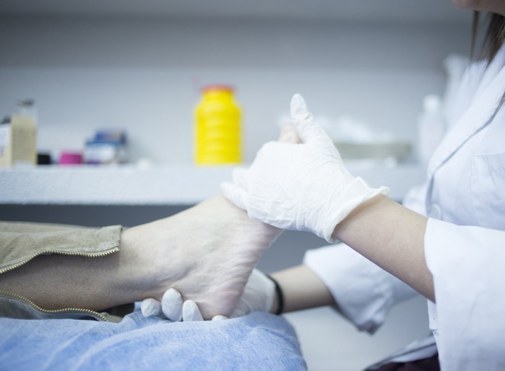
Treatment of heel pain
Before seeing a doctor, it is important to limit the stress on the painful foot. If the affected person can walk, shoes should be chosen that are spacious, comfortable and do not put additional pressure on the heel. Orthotics can help reduce pressure on the painful area. In most cases, treatment is conservative: anti-inflammatory drugs, orthoses and other aids that limit movement, therapeutic exercises, physiotherapy.
For some diagnoses, such as B. a rupture of the Achilles tendon or a fracture of the heel bone may require surgical treatment. An accurate diagnosis of heel pain is essential for determining treatment tactics.
diagnosis
If you see an orthopedist/traumatologist with heel pain, after the examination you will be referred for radiological tests such as X-rays, ultrasound, CT or MRI to confirm the diagnosis. In the case of a fracture, the diagnosis is confirmed by x-rays, but the diagnosis of plantar fasciitis is mostly based on the clinical picture. Blood tests are often required to diagnose pain and confirm systemic inflammatory diseases. Computed tomography (CT) is essential to determine the exact shape and orientation of the bone, and magnetic resonance imaging (MRI) is the best method to visualize the soft tissues and determine the condition of the bone.
Current treatment methods
Medical science does not stand still, and there are many modern methods of treatment, both surgical and conservative.
In most cases, foot pain is caused by foot deformities such as flat feet. In this case, therapeutic exercises and wearing custom-made insoles can help halt the progression of the disease.
Therapies such as shock wave therapy or platelet-rich plasma injection are very effective in treating foot tendon inflammation.
diagnosis
Treatment of a traumatic heel bone injury can be done using conservative and surgical methods. The choice of one or another tactic depends on the severity of the fracture, the patient's age and his physiological characteristics. Complications most often occur during surgical treatment.
The diagnosis, which is made after the heel fracture has been treated and which is essential for determining whether the pain has subsided, is carried out in several steps. It includes an initial examination, a differential diagnosis and the use of laboratory and instrumental procedures. For example, patients may be prescribed medications:
| diagnostic technique | Time |
|---|---|
| X-ray of the ankle | 10 mins |
| Ultrasound of the soft tissues of the foot | 30 minutes |
| electromyography | 300 minutes |
| General blood count and biochemical examination | 10 mins |
MRI is the most powerful examination method to assess the condition of the soft and bony tissues, get a three-dimensional image of the foot and understand when the pain stops after a heel fracture. The cost of the procedure is about 2500-7000 RUB.
Treatment methods for fractures depending on their type
In traumatology there are two methods of treating such injuries:
If the fractures are slightly displaced (or not), the first method is used - a plaster cast is placed on the injured leg. The fragments are aligned beforehand. For uncomplicated fractures, the ankle, hock and calf (up to the knee) are placed in a plaster cast. In the case of a club-shaped fracture, the limb is cast up to the middle of the thigh. The knee is kept in flexion.
In case of joint injuries and bone dislocations, an Ilizarov brace is applied to the patient, which is intended to stretch the skeleton: a spoke is pulled through the heel bone and a two-kilogram weight is suspended from the top of the arch of the foot. The patient wears the splint for 1.5 months, then a plaster cast is applied (without interrupting traction).
Indications for surgical treatment of a fracture
Most patients with a displaced calcaneal fracture require surgery. Objectives of the intervention:
If the hernia is closed, the operation is performed after a few days when the swelling and inflammation of the soft tissues have subsided. During this time the leg must be elevated. Open injuries require immediate intervention to clean the wound and remove the damaged tissue. This prevents infections and necrotic changes.
The main surgical method is osteosynthesis. It is carried out in two ways:
- percutaneously, where the fixation screws are inserted into the fracture site through puncture;
- Open repositioning. The surgeon makes an incision in the foot and inserts the metal structures.
The post-operative swelling subsides after two weeks and blood flow to the tissue begins again.
The operation takes place under general anesthesia and there are no age restrictions, but there are a number of contraindications to manipulation.
Possible undesirable effects after a fracture
Like any serious illness or injury, a broken heel bone can have serious, irreversible consequences. These are most often associated with a deformity of the foot.
- deforming arthritis;
- bony growths that can pinch nerve roots and affect feeling in the foot;
- difficulty walking;
- Chronic pain;
- post-traumatic arthritis;
- flat feet;
- restriction of joint mobility;
- Thrombosis;
- fracture of the heel bone;
- incomplete flexion of the ankle joint;
- nonunion;
- Torn Achilles tendon.
60 % patients develop chronic osteoarthritis after intra-articular fractures.
Postoperative complications include tendon and muscle injuries from staples and infections in the wound. These consequences are usually due to non-compliance with clinical guidelines and neglect of rehabilitation measures. They can lead to dysfunction of the entire musculoskeletal system.
Treating heel pain at home
There are many methods of traditional medicine that promise to quickly eliminate such a symptom. However, these should not be used alone as such measures can make the situation worse. However, once the heel pain and its cause have been identified, the doctor may recommend treatment at home using folk recipes. A specialist will advise you on which techniques are most effective. The most commonly recommended remedies are:
Apply a small amount of propolis to the painful area of the heel. Wrap a bandage around the foot. Leave on overnight.
Tea Mushroom Apply a small piece to the heel. Wrap in gauze or a bandage. Change the mushroom after a few hours. Use until the pain subsides.
Grate a potato on a fine grater. Apply the mushroom to the painful area and cover it with a plastic bag. Put a sock over it. Leave on for about two hours.
Peculiarities of the heel structure
This part of the leg does not look complicated, but in fact the heel area has a complex structure and does a lot of work: it bears most of the load during movement, as it is the support point of the leg. The heel is often subjected to mechanical stress when walking, jumping or running.
In addition to bones, the heel area contains:
All of these structures, as well as the skin, can be damaged by trauma or disease. This leads to pain, swelling, and other signs of discomfort. Heel pain is most common in children and teenagers who participate in strenuous sports, but older people can also suffer from heel pain.
Anomalies in which the heel hurts from the outside or inside
Various diseases can cause pain in the lateral part of the heel
- neurological;
- Dermatological;
- Orthopedic;
- vascular diseases;
- Traumatic (post-traumatic);
- degenerative-dystrophic;
- flammable.
Post-traumatic pain requires not only careful diagnosis but also long-term treatment. Even a bruise in this area can result in persistent limping and severe pain, while a broken heel bone requires crutches, medication, massage and physical therapy. Delaying treatment often results in chronic lameness, severe foot deformities, constant pain, inability to support the injured limb, and other complications. Therefore, if you develop lateral heel and foot pain after a jump, fall or bruise, you should immediately consult a traumatologist and undergo all diagnostic and therapeutic procedures recommended by your doctor.
If the foot hurts on the side above the heel without trauma, there is swelling and the skin has become hot, it is probably an inflammatory disease, e.g. B:
Inflammatory diseases can cause an increase in general body temperature. If left untreated, they can cause abscesses and tissue necrosis and, in severe cases, lead to sepsis or amputation of the foot.
Heel pain can also be accompanied by systemic pathologies, including systemic:
Arthritis and osteoarthritis can affect both large and small joints. These are degenerative-dystrophic diseases that first destroy the connective tissue and then the bone. Vessels, nerve bundles and muscles are damaged. These diseases are characterized by a slow progression. Initially they only cause mild discomfort, which increases over time and becomes chronic pain. If left untreated, the heel will gradually become deformed. The formation of osteophytes is also characteristic of osteoarthritis.
Classification of achillobursitis (according to various parameters)
- After the course:
- acute – characterized by a sudden and painful onset; usually caused by external infection as a result of various mechanical traumas;
- subacute;
- chronic – symptoms are muted and indistinct; the inflammatory process develops gradually; occurs due to the presence of underlying diseases in the body;
- recurring.
- Depending on the pathogen:
- Non-specific, as a result of macro- and micro-trauma and when infected by non-specific pathogens (staphylococci, streptococci, pneumococci, etc.);
- Specific, caused by a secondary infection with a specific infectious agent (depending on the type of primary infection: gonorrhea, brucellosis, tuberculosis, syphilis, etc.).
- Through an exudate:
- serous;
- purulent;
- hemorrhagic.
- By type of inflammation:
- purulent;
- viral;
- bacterial;
- infectious
- By localization:
- Heel;
- Heel; underfoot;
- Retachilles, also: Haglund's deformity (posterior bursitis) - inflammation of the joint capsule between the ligament and the skin;
- anterior bursitis – swelling between thick connective tissue and bone;
- Albert's disease – inflammation in the front area where the bone and tendon attach;
- subcutaneous heel bag.
- Severe throbbing pain in the heel and Achilles tendon area when walking, which may spread to the entire ankle and does not subside with rest;
- Swelling and redness in the Achilles tendon area;
- Swelling around the affected area (usually fusiform);
- Limited ankle mobility;
- Hyperthermia in the heel region (sometimes up to 39-40 ° C);
- General hyperthermia in the patient's body in the acute course of the disease;
- Audible cracking when walking and running;
- Limping, with the risk of a tendon rupture if the affected joint is constantly stressed.
diagnosis
The diagnosis of Achilles tendonitis occurs in stages. The following examinations are recommended:
To finally clarify the diagnosis, the following instrumental examinations can be ordered:
- X-rays;
- Ultrasound – to assess the condition of the tendon, musculoskeletal system and the presence of inflammation;
- MRI;
- Diagnostic puncture to collect an exudate for a bacteriological culture to determine the flora and biochemistry to determine the type of infection and sensitivity to antibiotics.
Tarsal Tunnel Syndrome
What is tarsal tunnel syndrome? It is a compression of the tibial nerve in the tarsal canal.
Causes: Swelling, cyst, arthritis, nerve nodule, benign tumor or flat foot.
Symptoms: Pain in the foot and heel, which can extend to the ankle and even the lower leg; Numbness and tingling on the inside of the foot and heel; worse at night; usually affects one leg.
Treatment: Rest, anti-inflammatory medications, orthotics, steroid injections, surgery.
Sever's disease (epicondylitis of the heel bone)
Sever's disease is the most common cause of heel pain in children.
What is heel epiphysitis? It is an inflammation of the posterior epiphysis of the heel bone.
Causes: Rapid growth of the heel bone in relation to the surrounding soft tissues on the one hand and excessive mechanical stress during prolonged standing, running or jumping on the other.
Symptoms: Pain in the back or lower heel area that occurs when walking; stiffness in the ankle joint; symptoms worsen with physical activity and resolve at rest; both feet are affected at the same time.
Treatment: Stretching and muscle strengthening exercises, rest, orthoses, medication. Symptoms subside within 2 months.
treatment of pain
After determining the cause of heel and foot pain, the doctor prescribes treatment. If all recommendations are followed, most people recover within a few months. Therapy may include:
- Taking nonsteroidal anti-inflammatory medications to reduce pain and swelling. If these medications are not effective, corticosteroid injections may help, but extreme caution should be exercised as prolonged use may cause side effects.
- Physiotherapy with exercises to stretch the plantar fascia and Achilles tendon and strengthen the shin, ankle and heel muscles.
- Aids and orthoses help to correct deficiencies in the foot and heel cushion and accelerate the healing process after an injury.
- With extracorporeal shockwave therapy, sound waves are directed into the problem area to stimulate healing. It is only recommended if other treatment methods have not been successful.
- Night splints that are attached to the calves of the feet at night to stretch the plantar fascia and Achilles tendon while you sleep.
- Surgery. This is an extreme treatment option. The surgeon partially separates the plantar fascia from the heel bone. However, there is a risk that the operation will weaken the arch of the foot.
Preventive measures and therapeutic exercises
In addition to mechanically stretching the tendon and fascia, simple exercises for the calf muscles can help:
- Sit in a chair, extend your leg in front of you and point your toes. Hold this position for at least 3-5 seconds and then relax. Repeat this 10 times for each leg.
- Stand facing the wall. Bring the affected leg behind the other leg. Keep your front knee bent and your back leg straight. Lean forward against the wall until your lower leg is supported and extended. Repeat the exercise 10 times.
Before visiting the doctor, it is important to protect your foot and avoid physical activity, especially jumping, running on hard surfaces and standing for long periods of time. A cold compress for 15 minutes will help relieve the pain. Place an ice pack wrapped in a cloth on the affected area.
It is advisable to check your footwear and only wear the most comfortable size. High heels should be avoided for the duration of the illness. To prevent pain, doctors recommend watching your weight and always warming up before exercising.
Read more:- Structure of the human heel.
- bones in the heel.
- Structure of the human ankle.
- The bones of the human heel.
- Heel bone human anatomy photo and description.
- heel bone injury.
- heel joint.
- Anatomy of the heel bone x-ray.