With enthesopathy or A pulling pain in the heel may also occur. With bunions, the pain in the heel can be cramp-like. Shooting pain is always a worrying symptom and the cause should be clarified.
- Blocking the heel spur
- How is heel spur blocking performed?
- Strain or injury to the tendon
- Infectious disease (reactive arthritis)
- CT or X-ray for a broken heel bone – which is the better choice?
- What is the best examination for a broken heel bone?
- How can the cause of heel pain be diagnosed? In what order should the examinations be carried out?
- How do you treat heel pain?
- Heel Spur Symptoms
- diagnosis
- Heel Spur Symptoms
- Treatment of heel spurs
- Medication
- physiotherapy
- indications
- heel bone
Blocking the heel spur
A heel spur is caused by a malformation of the heel bone caused by a long-term condition. This condition is very common and causes discomfort when walking. The treatment for a heel spur is a blockade, that is, an injection into the heel area under anesthesia. The anesthesia is immediate and the procedure is painful. In practice, this method is often not used. Since this method causes inconvenience during the procedure, it must be done correctly.
Heel spurs cause discomfort when wearing shoes, walking for long periods or playing sports. If the disease is advanced, pain can occur both under stress and at rest. If treatment is not started in time, the heel bone will become deformed. This leads to lameness and the inability to walk without a cane. The disease can lead to disability.
To avoid such a condition, a disease analysis is necessary to determine how the heel foot deformity arose. At the beginning of treatment, the doctor recommends resorting to simple therapies. Treatments must be carried out by a qualified doctor.
Therapeutic methods include:
- wearing individual orthopedic insoles;
- reduction in physical exertion;
- use of ointments and anti-inflammatory gels;
- carrying out all necessary treatments.
If these methods do not relieve pain, anesthetic injections are made into the heel.
How is heel spur blocking performed?
Before the procedure, the patient is informed about the possible negative effects. In addition to complications, there can also be side effects that affect various organs and systems. The doctor warns about the painfulness of the procedure, as the needle is inserted into a painful area - under the osteophytes. The medication is injected slowly so that it penetrates the plantar fasciitis and does not create pressure inside.
Due to the high density of the skin on the heel, the injection is quite painful. No anesthesia is used. Sometimes an exception is made and lidocaine is used. Before use, it is injected under the skin and a reaction is observed after 20 minutes.
Before giving the injection, the doctor will feel the entire foot to find the location of the spike. The patient lies on his stomach and places his legs so that the foot is straight and the heel hangs down. The doctor mixes an ampoule of the medication with lidocaine. It is important to know that anesthesia slows down the effects of the medication. A peculiarity of this procedure is that it is difficult to determine the correct puncture site. If the medication is not in the right place, the blockage will not be effective because the hormone only works near the tissue.
If the heel block is performed correctly, the effect will be felt immediately after the medication is injected into the heel.
To enable accurate manipulation, the injection is performed under X-ray or ultrasound control. The procedure is carried out in a private or public clinic. It cannot be done properly at home.
The block is carried out under sterile conditions to exclude infection.
Strain or injury to the tendon
This is the largest group and is usually referred to as 'heel spur'. These include tendon injuries (e.g. tear or sprain), tendon strains caused by walking in high-heeled shoes, tendon strains caused by long walking with pronounced flat feet and contusions of the heel bone with subsequent inflammation of the surrounding tissue (e.g. as a result of heel jumping).
Patients in this group most commonly complain of a burning pain under the heel, described as a 'nail' feeling, which is made worse when attempting to step on the heel.
Treatment Patients in this group require two to three weeks of complete rest and anti-inflammatory drugs (Diclofenac, Brufen, Indomethacin, Flexen, etc.) in tablet or suppository form to relieve pain and inflammation.
Sometimes laser therapy and a special 'transverse massage' with anti-inflammatory ointments can also help.
If this does not help, corticosteroid hormones (dipropane, hydrocortisone, Kenalog) can be injected directly into the heel area - sometimes one or two injections are enough to completely relieve the acute pain.
In the most severe cases, when all possible measures have failed, radiation therapy is necessary, but fortunately this is only necessary in two to three percent of patients.
Infectious disease (reactive arthritis)
Reactive arthritis of the heels is possible with certain types of infections, especially genital infections (e.g. gonorrhea, chlamydia, etc.), which sometimes occur covertly. And there are some symptoms that may indicate (or at least 'suggest') the infectious and reactive nature of a heel tendon infection. In this case, the pain in the heel area often occurs not only when walking. Patients with reactive arthritis may also experience heel pain at rest, at night. Sometimes they hurt the most at night.
In addition, heel inflammation in reactive arthritis is often accompanied by inflammation of many joints and eyes as well as discomfort in the genital area. However, all of these symptoms - including night pain, eye inflammation, joint inflammation and genital discomfort - can also be symptoms of other inflammatory diseases (see below).
In reactive arthritis, the underlying infection should first be treated with appropriate antibiotics, and anti-inflammatory medications and dimethoxide packs can be used to relieve pain.
CT or X-ray for a broken heel bone – which is the better choice?
CT and X-ray are the two main methods for diagnosing injuries to the bony structures of the heel. Radiological procedures enable a very precise assessment of the bone condition and are therefore the preferred form of diagnosis for heel fractures. Other advantages of CT and X-rays in fracture diagnosis include.
- a procedure duration of only 2-3 minutes;
- an accurate examination without surgical intervention;
- no discomfort during the examination
- no precautions for the patient;
- quick availability of test results.
For these reasons, CT and X-rays are the preferred form of examination in emergency situations where the surgeon or trauma surgeon must act quickly.
Compared to an X-ray, a CT scan of the heel is more detailed and accurate. From a diagnostic point of view, CT examination has several advantages:
- It provides a clear and volumetric three-dimensional image of the bone;
- three-dimensional bone models can be made;
- the results are immediately visible during the examination;
- The physician can assess both the bony structures and the adjacent soft tissue structures of the limb without having to overlay images of one tissue over another.
What is the best examination for a broken heel bone?
| Types of fractures | Preferred form of diagnosis |
|---|---|
| Incomplete fracture | X-ray or CT scan |
| Complete fracture | X-ray or CT scan |
| Multiple fracture | X-ray or CT scan |
| characteristics | CT SCAN | X-RAY |
|---|---|---|
| Full name | Computed Tomography | Digital or analog radiography |
| Principle of imaging | A CT scan is based on X-rays aimed at the human body. Some of the rays are retained in the tissue, while another part penetrates the body. Different tissues and organs block X-rays differently: bones block most radiation, soft tissue less, and air blocks X-rays poorly. The sensor uses this data to create a three-dimensional image of the area being examined. | X-rays are based on X-rays, where the X-rays are aimed at the human body. Some of the rays are retained in the tissue, while another part penetrates the body. Different tissues and organs stop X-rays differently: bones stop most of the rays, soft tissue stops less, and air stops the X-rays poorly. The sensor creates a two-dimensional image from this data. |
| Risks | CT scans can be dangerous due to radiation exposure and should only be done once every six months if your doctor recommends it. | X-rays can be dangerous due to radiation exposure and should be prescribed by a doctor. |
| Time | A CT scan is a quick form of diagnosis that can be performed in 2-3 minutes. Therefore, a CT scan is more commonly used in emergency care. | X-rays are a quick form of diagnosis that can be performed within 2-3 minutes. Therefore, x-rays are more commonly used in emergency care. |
How can the cause of heel pain be diagnosed? In what order should the examinations be carried out?
Meddiagnosti employees who treat heel pain.
- X-ray of the foot with the heel. It is a common misconception that an X-ray of the heel area is a universal method. However, this is true when it comes to recognizing the spur itself - a bony hypertrophy in the area of the plantar aponeurosis.
heel spur good visible on the x-ray..
The first step in making a diagnosis is usually an x-ray.
A heel spur may be visiblea during an ultrasound examination.
But it's not just heel spurs that affect you Ultrasound examination. Inflammation can also become visible.. This is the cause of the 'pain under the heel' (in the heel area). An ultrasound examination is helpful if heel pain occurs, for example. B. occurs from the side. Such pain can also be caused by abnormalities in the peripheral nervous system. A neurological examination may be necessary to diagnose this pathology.
If there is suspicion of a new growth in the foot, especially in the heel area, an MRI examination of the foot is indicated. If it is the heel area, an MRI scan of the ankle can be performed to visualize the heel bone. What’s more, heel pain can be caused by inflammation of the ankle joint – arthritis.
We use x-rays and ultrasound to diagnose pain in orthopedics and rheumatology, including heel pain. If necessary, an MRI scan may also be performed.
X-rays and ultrasound are the most conclusive methods for diagnosing heel pain. However, to truly rule out refractory (chronic) heel pain, a laboratory diagnosis may be necessary. For example in erythromelalgia.
How do you treat heel pain?
Treatment depends on the cause of the heel pain. It does not always make sense to start treatment by injecting a steroid (hormone) into the painful area of the heel. For example, in many rheumatic diseases, tumors and some other conditions, steroids are not effective or only effective for a short time. Treatment occurs after a confirmed diagnosis has been made.
The method of shock wave therapy is effective in the treatment of diseases such as calcium salt deposits or scarring after inflammation at the attachment points of ligaments, muscles and tendons in the heel area. The clinic has devices for shock wave therapy. We have 15 years of experience using this method.
However, shock wave therapy should not be used if there are visible signs of inflammation, as the likelihood of aggravation of the process is high. It makes sense to find out the cause of the inflammation and only then develop treatment tactics.
And if your heel pain persists, you need a proper diagnosis and professional treatment from us.
Complete diagnosis and treatment of heel pain takes place in one buildingAddress: Stroiteley Lane 4, Kiev, Left Bank, 250 m from Darnytsya metro station.
md
Professor at the National Medical University
Gongalsky V.
Heel Spur Symptoms
Any of the above causes can lead to the symptoms of the disease: the appearance of the so-called 'morning pain' after getting out of bed. This pain is also known as 'start-up pain'. It is attributed to the presence of inflammation in the heel bone. It is associated with plantar aponeurositis: an inflammation of the plantar fascia caused by the development of pathological changes in the area where the plantar aponeurosis attaches to the heel bone.
Achilles tendonitis (Achilles tendon pain) is an equally common pathology caused by a lesion in the area of the insertion of the Achilles tendon on the posterior surface of the heel.
In the inflamed area there is a proliferation of thick fibrous connective tissue, which thickens and becomes impregnated with calcium salts, causing irritation of the nerve endings in the periosteum and, consequently, severe heel pain.
diagnosis
To clarify the cause of pain in the foot and heel area, the Institute of Musculoskeletal Medicine and Neurology (Clinic Mediagnostica) recommends an X-ray examination of the foot with an ultrasound examination of the soft tissues and tendons of the foot.
The heel spur as well as the condition and integrity of the foot bones are visible on the x-ray. The spur may be small in size and extent, but the pain may be very pronounced.
An ultrasound scan can clarify the extent of the heel spur and reveal signs of inflammation, tissue swelling, and the integrity of tendons and ligaments.
Heel Spur Symptoms
The main symptom is pain in the heel area that occurs at the beginning of walking ('start-up pain') after sitting or lying down for a long time, e.g. B. after a long day of work while sitting, after getting out of bed after sleeping. The pain eases slightly with movement. Over time, heel pain becomes persistent and debilitating, not only with movement but also at rest, significantly limiting motor activities and reducing quality of life.
Depending on the extent of the inflammation and the degree of bone hypertrophy, the pain may be sharp and burning (patients describe it as a 'nail in the heel') or it may be mild and 'spread' throughout the heel. Due to the constant discomfort, patients try not to step on their heel, so the gait is significantly changed.
Sometimes heel spurs cause no symptoms and do not bother the sufferer at all.
Treatment of heel spurs
Medication

The following medications are used to relieve pain, reduce swelling and reduce inflammation Nonsteroidal anti-inflammatory drugs (NSAIDs) – For oral intake and topical application, but do not always achieve a significant pain-relieving effect. This is due to the high density of the soleus tissue and its poor blood circulation, which hinders the penetration of medications. Hormone injections (cortisone, hydrocortisone, dipropane, flosterone) are performed that block the heel area, but this method of treatment must be combined with physiotherapy.
physiotherapy
The physiotherapy methods for heel spurs are primarily recommended because they are very effective and, in combination with other measures (LFC, medication, orthoses), ensure that the patient maintains his motor activity.
Among these methods, extracorporeal shock wave therapy (EUVT), phonophoresis, laser therapy and magnetic therapy are currently considered the most effective for heel spurs.
EUVT Treatment is carried out once the diagnosis of heel spurs has been confirmed (radiologically). The method is based on acoustic shock waves with high amplitude and short pulse duration. The treatment is painful, but in the end there is significant pain relief, and the entire treatment leads to a significant improvement in the patient's condition, since shock wave treatment leads to the destruction of calcifications, tissue regeneration and the reduction of inflammation. Combining EWT with blocks or other physical therapy may result in increased pain.
laser therapy Laser therapy is also indicated for heel spurs. Various heads are used that emit a pulsed laser (various spectra), some of which work in drug delivery mode (Laser Fereza). High-intensity laser therapy (HIL), which is not yet widely used (due to the high cost of the equipment and the required training of personnel), is particularly effective.
It is easily absorbed through ingestion and excreted relatively quickly in the urine.
Asparkam is a source of potassium and magnesium ions, regulates metabolic processes, promotes the restoration of electrolyte balance and has an antiarrhythmic effect.
The potassium ion is involved in both the conduction of impulses along nerve fibers and in synaptic transmission, in muscle contractions and in maintaining normal cardiac function. Disturbances in potassium ion metabolism lead to changes in the excitability of nerves and muscles. Active ion transport maintains a high potassium ion gradient across the plasma membrane. In low doses, the potassium ion dilates the coronary arteries; in high doses, it narrows them. It has a negative chrono- and bathmotropic effect and, in high doses, a negative ion and dro-motropic effect, as well as a moderate diuretic effect.
The magnesium ion is a cofactor for 300 enzymatic reactions. It is an essential element in the processes that ensure the absorption and use of energy. Participates in electrolyte balance, ion transport, membrane permeability and neuromuscular excitability. Participates in the structure (pentose phosphate) of deoxyribonucleic acid. Participates in ribonucleic acid synthesis, the hereditary apparatus, cell growth and division. Limits and prevents the excessive release of catecholamines during stress, possible lipolysis and release of free fatty acids. Is a 'physiological' slow calcium channel blocker. Promotes the permeation of potassium ions into the cells.
Aspartate promotes the penetration of potassium and magnesium ions into the intracellular space and stimulates intercellular phosphate synthesis.
indications
Asparkam is used to treat heart failure, coronary artery disease, hypokalemia and cardiac arrhythmias (including myocardial infarction and overdose of cardiac glycosides).
Hypersensitivity to the drug, disorders of amino acid metabolism, hypotension, acute and chronic renal failure, hyperkalemia, hypermagnesemia, atrioventricular conduction disorders (atrioventricular block grades I-III), severe myasthenia gravis, hemolysis, adrenal insufficiency, age under 18 years (efficacy and safety not proven) .
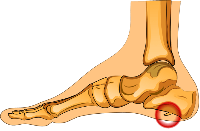
heel bone

Patients visiting their GP for heel pain are the fourth most common reason for visiting their GP. Plantar fasciitis is the most common cause of heel pain. Although there are other causes of heel problems, such as rheumatoid arthritis and gout, plantar fasciitis is the most common. The plantar fascia, a long, flat band that extends across the entire foot, provides support. It is trapezoidal in shape and tapers where it attaches to the heel bone and then becomes wider as the band continues.
archive
Ideas
- Glucosamine for osteoarthritis
- Excessive protein intake
- Minerals
- Unhealthy habits
- Osteoarthritis pain in the knee joints
- Caraway seeds
- dehydration
- Alternative medicine for gout
- Cleansing the colon
- Gout and penile function
- Curcumin benefits
- Rheumatism pronunciation
- Kava Kava Benefits
- Nutrition for arthritis
- Holisticness
- Naturopathy
- Treatment of gout infections
- Black pepper
- Gout in the big toe
- Benefits of exercise
- Symptoms of hyperacidity
- acne
- Alkaline water and gout
- Arthritis relief from knee joint pain
- Oats
We use technologies such as cookies to store and/or access information about your device. We do this to improve your browsing experience and show personalized advertising. Consent to the use of these technologies allows us to process data such as browsing behavior or unique identifiers on this website. Lack of consent or withdrawal of consent may affect certain features and functions.
- Structure of the human heel.
- Structure of the heel.
- The bones of the human heel.
- How to cure plantar fasciitis forum.
- heel joint.
- If your soles and heels hurt.
- Treatment of plantar fasciitis at home.
- Tear of the foot fascia.